National Institute of Biomedical Imaging and Bioengineering, NIH, Bethesda, MD, 20892, USA.
Gatan, Inc., 5794W. Las Positas Blvd, Pleasanton, CA, 94588, USA.
Sci Rep. 2018 Aug 28;8(1):12985. doi: 10.1038/s41598-018-31231-w.
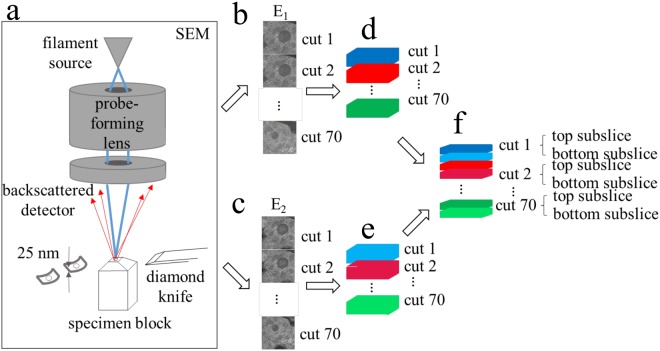
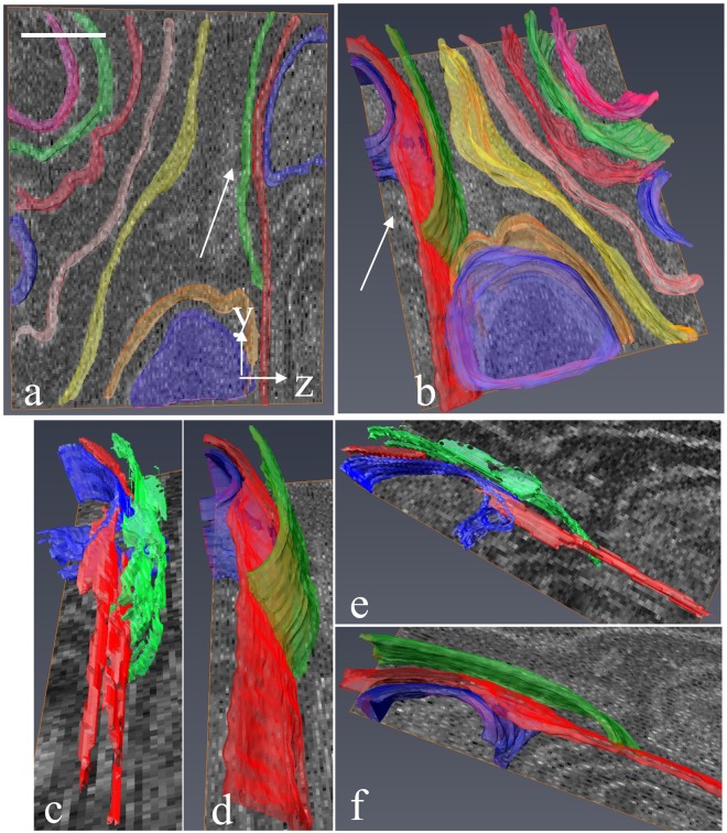
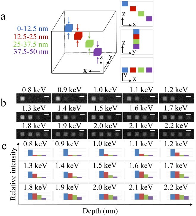
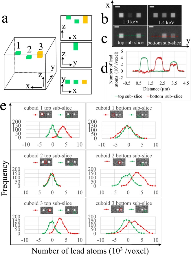
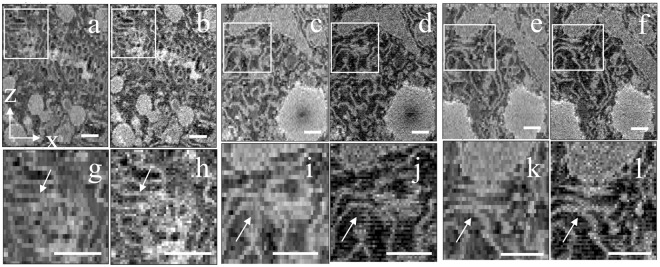
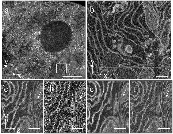
Serial block-face electron microscopy (SBEM) provides nanoscale 3D ultrastructure of embedded and stained cells and tissues in volumes of up to 10 µm. In SBEM, electrons with 1-3 keV energies are incident on a specimen block, from which backscattered electron (BSE) images are collected with x, y resolution of 5-10 nm in the block-face plane, and successive layers are removed by an in situ ultramicrotome. Spatial resolution along the z-direction, however, is limited to around 25 nm by the minimum cutting thickness. To improve the z-resolution, we have extracted depth information from BSE images acquired at dual primary beam energies, using Monte Carlo simulations of electron scattering. The relationship between depth of stain and ratio of dual-energy BSE intensities enables us to determine 3D structure with a ×2 improvement in z-resolution. We demonstrate the technique by sub-slice imaging of hepatocyte membranes in liver tissue.
连续块面电子显微镜(SBEM)可提供嵌入和染色的细胞和组织的纳米级 3D 超微结构,体积可达 10μm。在 SBEM 中,能量为 1-3keV 的电子入射到标本块上,从块表面以 x、y 方向 5-10nm 的分辨率收集背散射电子(BSE)图像,然后通过原位超微切片机去除连续的层。然而,沿 z 方向的空间分辨率由于最小切割厚度的限制,约为 25nm。为了提高 z 分辨率,我们从在双束流能量下获取的 BSE 图像中提取深度信息,使用电子散射的蒙特卡罗模拟。染色深度和双能 BSE 强度比之间的关系使我们能够以 z 分辨率提高 2 倍的方式确定 3D 结构。我们通过对肝组织中肝细胞膜的亚切片成像来演示该技术。