• 文献检索
  • 文档翻译
  • 深度研究
  • 学术资讯
  • Suppr Zotero 插件Zotero 插件
  • 邀请有礼
  • 套餐&价格
  • 历史记录
应用&插件
Suppr Zotero 插件Zotero 插件浏览器插件Mac 客户端Windows 客户端微信小程序
定价
高级版会员购买积分包购买API积分包
服务
文献检索文档翻译深度研究API 文档MCP 服务
关于我们
关于 Suppr公司介绍联系我们用户协议隐私条款
关注我们

Suppr 超能文献

核心技术专利:CN118964589B侵权必究
粤ICP备2023148730 号-1Suppr @ 2026

文献检索

告别复杂PubMed语法,用中文像聊天一样搜索,搜遍4000万医学文献。AI智能推荐,让科研检索更轻松。

立即免费搜索

文件翻译

保留排版,准确专业,支持PDF/Word/PPT等文件格式,支持 12+语言互译。

免费翻译文档

深度研究

AI帮你快速写综述,25分钟生成高质量综述,智能提取关键信息,辅助科研写作。

立即免费体验

所需的生长促进剂可推动完整脊髓损伤后的轴突再生。

Required growth facilitators propel axon regeneration across complete spinal cord injury.

机构信息

Department of Neurobiology, David Geffen School of Medicine, University of California, Los Angeles, Los Angeles, CA, USA.

Center for Neuroprosthetics and Brain Mind Institute, School of Life Sciences, Swiss Federal Institute of Technology (EPFL), Lausanne, Switzerland.

出版信息

Nature. 2018 Sep;561(7723):396-400. doi: 10.1038/s41586-018-0467-6. Epub 2018 Aug 29.

DOI:10.1038/s41586-018-0467-6
PMID:30158698
原文链接:https://pmc.ncbi.nlm.nih.gov/articles/PMC6151128/
Abstract

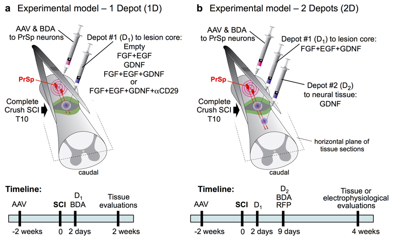
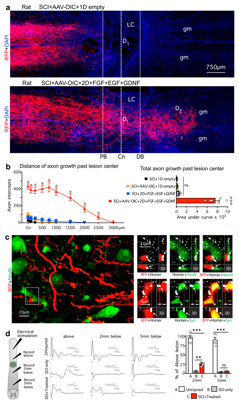
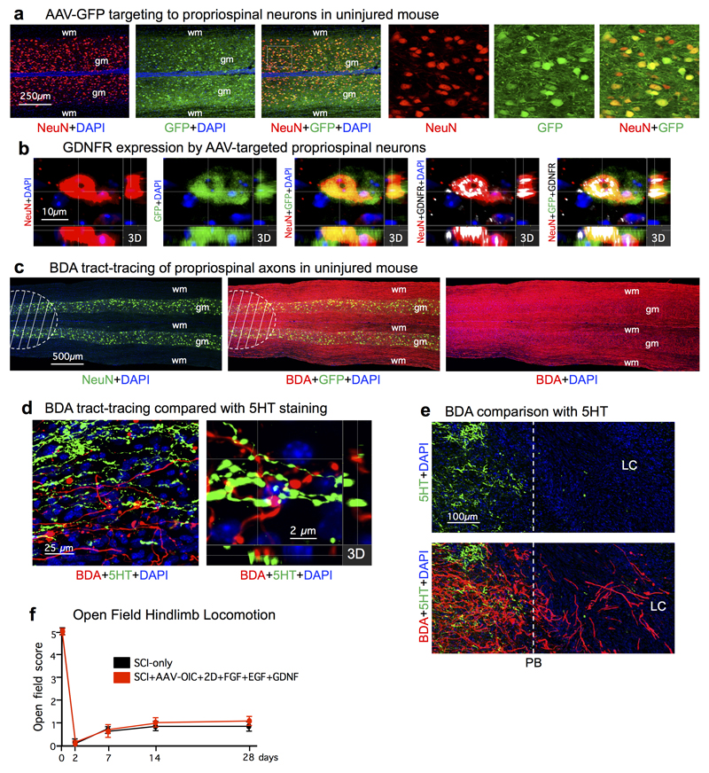
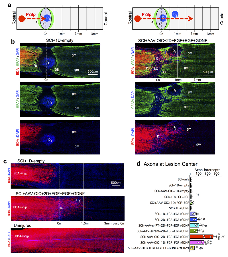
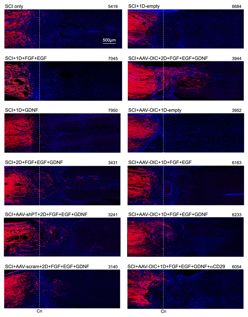
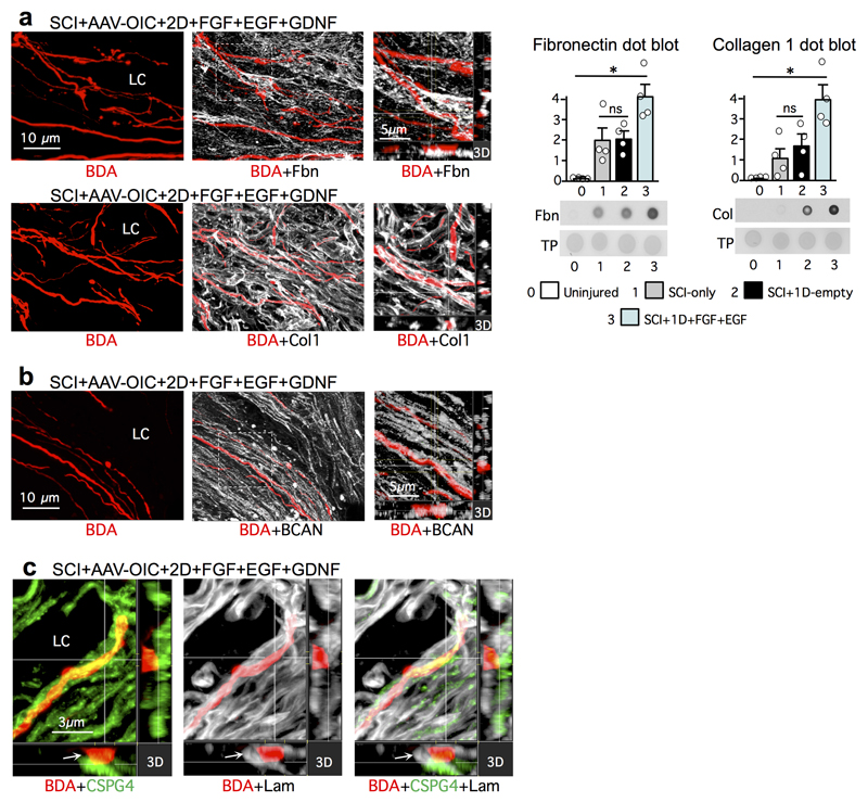
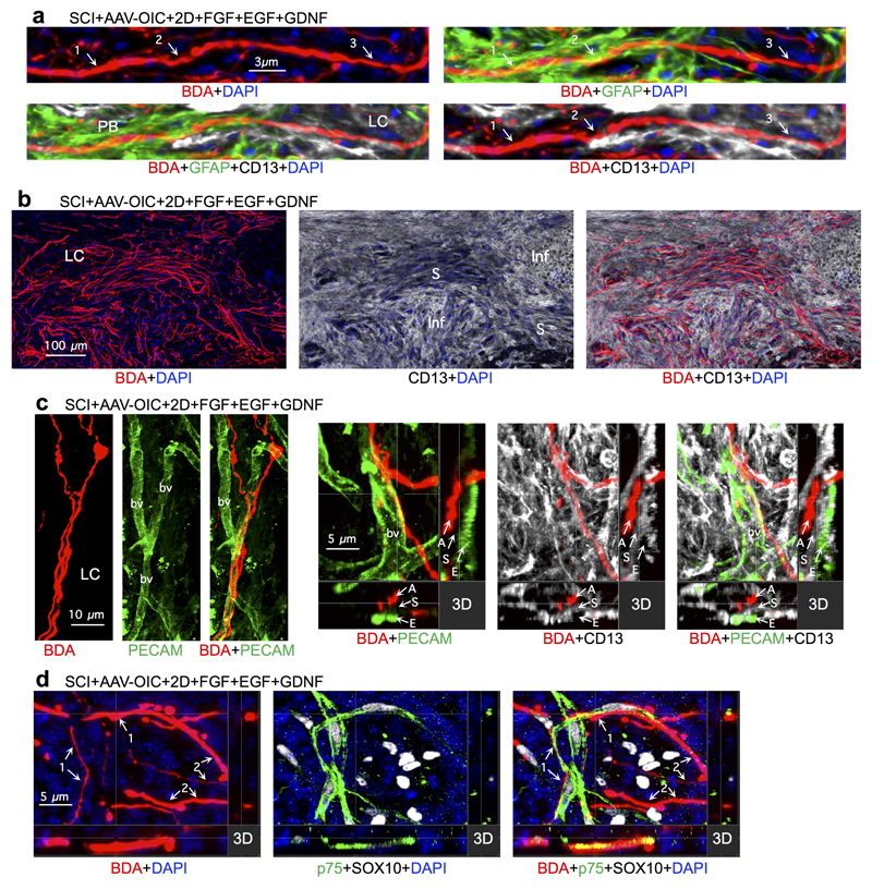
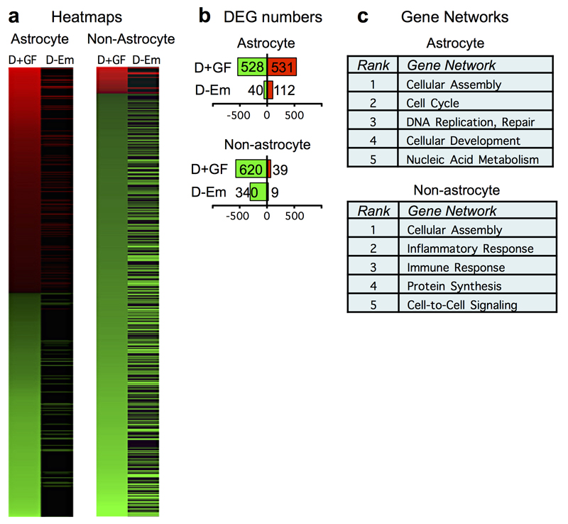
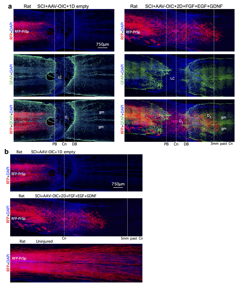
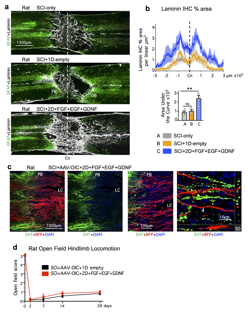
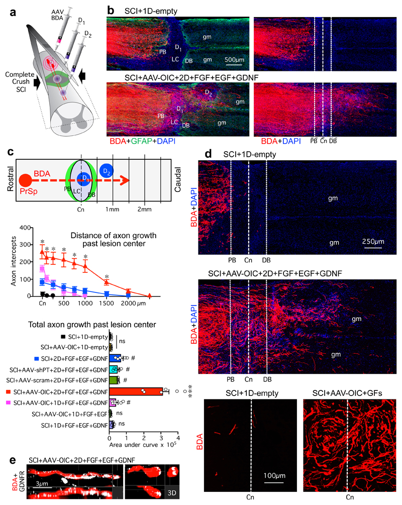
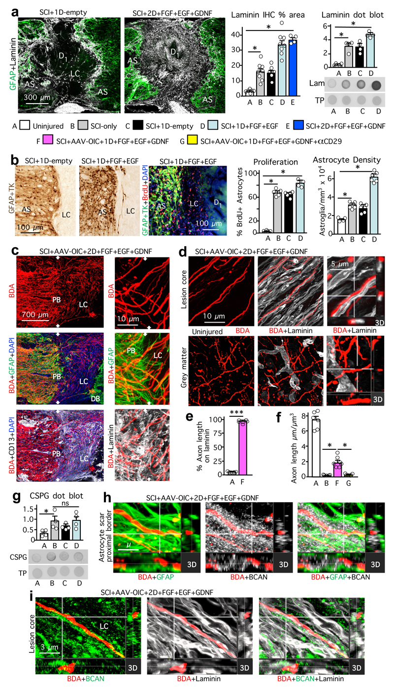
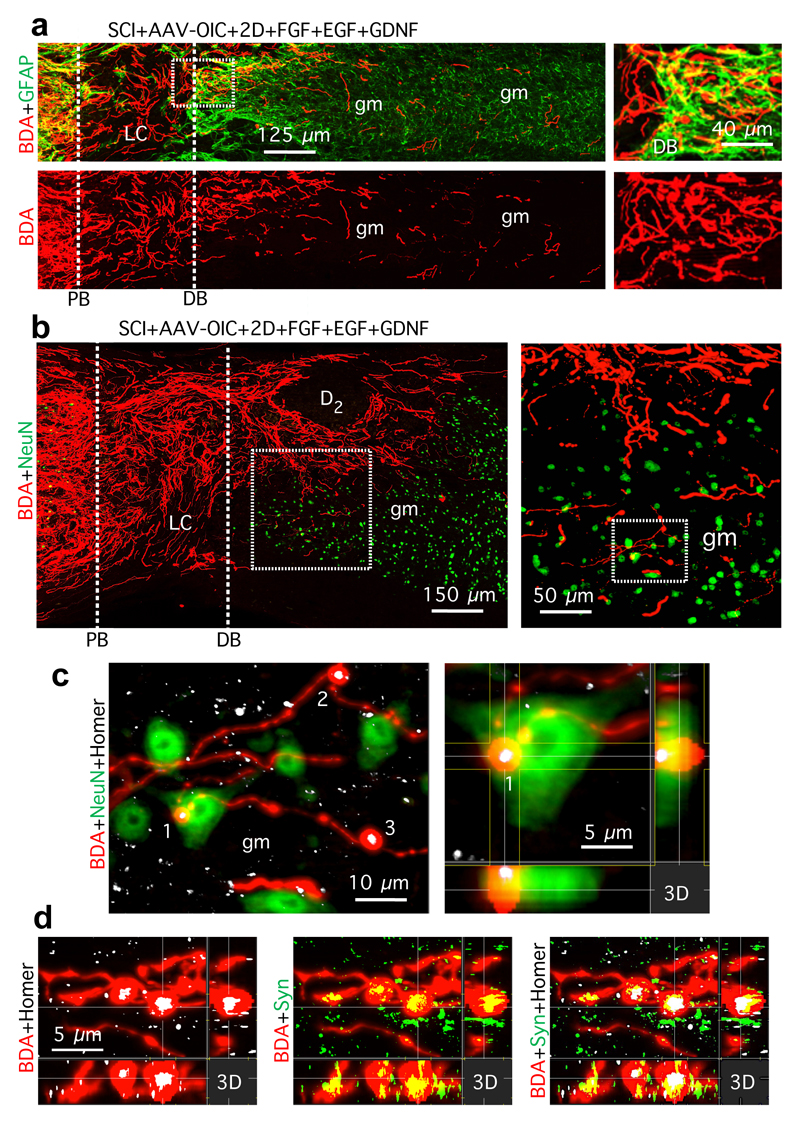
Transected axons fail to regrow across anatomically complete spinal cord injuries (SCI) in adults. Diverse molecules can partially facilitate or attenuate axon growth during development or after injury, but efficient reversal of this regrowth failure remains elusive. Here we show that three factors that are essential for axon growth during development but are attenuated or lacking in adults-(i) neuron intrinsic growth capacity, (ii) growth-supportive substrate and (iii) chemoattraction-are all individually required and, in combination, are sufficient to stimulate robust axon regrowth across anatomically complete SCI lesions in adult rodents. We reactivated the growth capacity of mature descending propriospinal neurons with osteopontin, insulin-like growth factor 1 and ciliary-derived neurotrophic factor before SCI; induced growth-supportive substrates with fibroblast growth factor 2 and epidermal growth factor; and chemoattracted propriospinal axons with glial-derived neurotrophic factor delivered via spatially and temporally controlled release from biomaterial depots, placed sequentially after SCI. We show in both mice and rats that providing these three mechanisms in combination, but not individually, stimulated robust propriospinal axon regrowth through astrocyte scar borders and across lesion cores of non-neural tissue that was over 100-fold greater than controls. Stimulated, supported and chemoattracted propriospinal axons regrew a full spinal segment beyond lesion centres, passed well into spared neural tissue, formed terminal-like contacts exhibiting synaptic markers and conveyed a significant return of electrophysiological conduction capacity across lesions. Thus, overcoming the failure of axon regrowth across anatomically complete SCI lesions after maturity required the combined sequential reinstatement of several developmentally essential mechanisms that facilitate axon growth. These findings identify a mechanism-based biological repair strategy for complete SCI lesions that could be suitable to use with rehabilitation models designed to augment the functional recovery of remodelling circuits.

摘要

轴突在成人的解剖完整的脊髓损伤(SCI)中无法再生。多种分子可以在发育过程中或损伤后部分促进或抑制轴突生长,但有效地逆转这种再生失败仍然难以捉摸。在这里,我们表明,在发育过程中对于轴突生长至关重要的三种因素-(i)神经元内在的生长能力,(ii)生长支持性基质和(iii)趋化性-在成人中均被减弱或缺乏-都是单独需要的,并且结合在一起足以刺激成年啮齿动物的解剖完整的 SCI 病变中强有力的轴突再生。我们在 SCI 之前使用骨桥蛋白、胰岛素样生长因子 1 和睫状神经营养因子重新激活成熟的下行 propriospinal 神经元的生长能力;通过成纤维细胞生长因子 2 和表皮生长因子诱导生长支持性基质;并通过生物材料库的时空控制释放来趋化 propriospinal 轴突,该方法放置在 SCI 后顺序进行。我们在小鼠和大鼠中均表明,提供这三种机制的组合而不是单独的机制,可以刺激 propriospinal 轴突通过星形胶质细胞瘢痕边界和跨越非神经组织的病变核心进行强有力的再生,其强度比对照高出 100 倍以上。刺激、支持和趋化的 propriospinal 轴突在病变中心之外再生了整整一个脊髓节段,很好地进入了剩余的神经组织中,形成了具有突触标记的末端样接触,并在病变部位传递了电生理传导能力的显著恢复。因此,克服成熟后解剖完整的 SCI 病变中轴突再生的失败需要联合重新启动几种促进轴突生长的发育必需机制。这些发现为完全性 SCI 病变确定了一种基于机制的生物学修复策略,该策略可以与旨在增强重塑回路功能恢复的康复模型一起使用。

https://cdn.ncbi.nlm.nih.gov/pmc/blobs/0d55/6151128/f4b27dc463d7/emss-78642-f004.jpg
https://cdn.ncbi.nlm.nih.gov/pmc/blobs/0d55/6151128/92a354e9c0da/emss-78642-f005.jpg
https://cdn.ncbi.nlm.nih.gov/pmc/blobs/0d55/6151128/f968479a266c/emss-78642-f006.jpg
https://cdn.ncbi.nlm.nih.gov/pmc/blobs/0d55/6151128/9626c6b387a6/emss-78642-f007.jpg
https://cdn.ncbi.nlm.nih.gov/pmc/blobs/0d55/6151128/87752fdbb01a/emss-78642-f008.jpg
https://cdn.ncbi.nlm.nih.gov/pmc/blobs/0d55/6151128/1c9ea57016dd/emss-78642-f009.jpg
https://cdn.ncbi.nlm.nih.gov/pmc/blobs/0d55/6151128/519aaf3548af/emss-78642-f010.jpg
https://cdn.ncbi.nlm.nih.gov/pmc/blobs/0d55/6151128/771d1dfd20ac/emss-78642-f011.jpg
https://cdn.ncbi.nlm.nih.gov/pmc/blobs/0d55/6151128/39d321a53ee6/emss-78642-f012.jpg
https://cdn.ncbi.nlm.nih.gov/pmc/blobs/0d55/6151128/0aaa4cf63d6a/emss-78642-f013.jpg
https://cdn.ncbi.nlm.nih.gov/pmc/blobs/0d55/6151128/2dd0a9438761/emss-78642-f001.jpg
https://cdn.ncbi.nlm.nih.gov/pmc/blobs/0d55/6151128/1557dc80d4f1/emss-78642-f002.jpg
https://cdn.ncbi.nlm.nih.gov/pmc/blobs/0d55/6151128/1e5ddf2093fe/emss-78642-f003.jpg
https://cdn.ncbi.nlm.nih.gov/pmc/blobs/0d55/6151128/f4b27dc463d7/emss-78642-f004.jpg

相似文献

1
Required growth facilitators propel axon regeneration across complete spinal cord injury.所需的生长促进剂可推动完整脊髓损伤后的轴突再生。
Nature. 2018 Sep;561(7723):396-400. doi: 10.1038/s41586-018-0467-6. Epub 2018 Aug 29.
2
Expressing Constitutively Active Rheb in Adult Neurons after a Complete Spinal Cord Injury Enhances Axonal Regeneration beyond a Chondroitinase-Treated Glial Scar.在完全性脊髓损伤后,在成年神经元中组成性表达活性Rheb可增强轴突再生,超过经软骨素酶处理的胶质瘢痕。
J Neurosci. 2015 Aug 5;35(31):11068-80. doi: 10.1523/JNEUROSCI.0719-15.2015.
3
Regeneration of Propriospinal Axons in Rat Transected Spinal Cord Injury through a Growth-Promoting Pathway Constructed by Schwann Cells Overexpressing GDNF.通过过表达 GDNF 的施万细胞构建的促生长途径促进大鼠横断脊髓损伤中的 propriospinal 轴突再生。
Cells. 2024 Jul 8;13(13):1160. doi: 10.3390/cells13131160.
4
Astrocyte scar formation aids central nervous system axon regeneration.星形胶质细胞瘢痕形成有助于中枢神经系统轴突再生。
Nature. 2016 Apr 14;532(7598):195-200. doi: 10.1038/nature17623. Epub 2016 Mar 30.
5
Proliferating NG2-Cell-Dependent Angiogenesis and Scar Formation Alter Axon Growth and Functional Recovery After Spinal Cord Injury in Mice.NG2 细胞依赖性血管生成和瘢痕形成促进小鼠脊髓损伤后的轴突生长和功能恢复。
J Neurosci. 2018 Feb 7;38(6):1366-1382. doi: 10.1523/JNEUROSCI.3953-16.2017. Epub 2017 Dec 26.
6
Astrocyte progenitor transplantation promotes regeneration of bulbospinal respiratory axons, recovery of diaphragm function, and a reduced macrophage response following cervical spinal cord injury.星形胶质细胞祖细胞移植促进延髓呼吸轴突的再生、膈肌功能的恢复以及颈脊髓损伤后巨噬细胞反应的减少。
Glia. 2019 Mar;67(3):452-466. doi: 10.1002/glia.23555. Epub 2018 Dec 11.
7
Meningeal cells and glia establish a permissive environment for axon regeneration after spinal cord injury in newts.在蝾螈的脊髓损伤后,脑膜细胞和神经胶质为轴突再生建立了一个许可的环境。
Neural Dev. 2011 Jan 4;6:1. doi: 10.1186/1749-8104-6-1.
8
Low-dose fractionated irradiation promotes axonal regeneration beyond reactive gliosis and facilitates locomotor function recovery after spinal cord injury in beagle dogs.低剂量分割照射促进轴突再生超越反应性神经胶质增生,并促进脊髓损伤后比格犬的运动功能恢复。
Eur J Neurosci. 2017 Nov;46(9):2507-2518. doi: 10.1111/ejn.13714. Epub 2017 Oct 19.
9
miR-155 Deletion in Mice Overcomes Neuron-Intrinsic and Neuron-Extrinsic Barriers to Spinal Cord Repair.小鼠体内miR-155缺失克服脊髓修复的神经元内在和外在障碍。
J Neurosci. 2016 Aug 10;36(32):8516-32. doi: 10.1523/JNEUROSCI.0735-16.2016.
10
Body weight-supported treadmill training reduces glial scar overgrowth in SCI rats by decreasing the reactivity of astrocytes during the subacute phase.体重支持的跑步机训练通过在亚急性期降低星形胶质细胞的反应性,减少脊髓损伤大鼠的胶质瘢痕过度生长。
BMC Neurosci. 2025 Apr 28;26(1):30. doi: 10.1186/s12868-025-00947-7.

引用本文的文献

1
A neuronal architecture underlying autonomic dysreflexia.自主神经反射异常的神经元结构基础。
Nature. 2025 Sep 17. doi: 10.1038/s41586-025-09487-w.
2
A laminin-based therapy for dogs with chronic spinal cord injury: promising results of a longitudinal trial.一种基于层粘连蛋白的犬慢性脊髓损伤治疗方法:一项纵向试验的可喜成果。
Front Vet Sci. 2025 Aug 13;12:1592687. doi: 10.3389/fvets.2025.1592687. eCollection 2025.
3
Integrative analysis and experimental validation identify the role of CD44 and Nucleolin in regulating gliogenesis following spinal cord injury.

本文引用的文献

1
Dissecting spinal cord regeneration.解析脊髓再生。
Nature. 2018 May;557(7705):343-350. doi: 10.1038/s41586-018-0068-4. Epub 2018 May 16.
2
Cortico-reticulo-spinal circuit reorganization enables functional recovery after severe spinal cord contusion.皮质-网状脊髓环路重建使严重脊髓挫伤后功能恢复成为可能。
Nat Neurosci. 2018 Apr;21(4):576-588. doi: 10.1038/s41593-018-0093-5. Epub 2018 Mar 19.
3
Cell biology of spinal cord injury and repair.脊髓损伤与修复的细胞生物学
综合分析与实验验证确定了CD44和核仁素在脊髓损伤后调节神经胶质生成中的作用。
Cell Regen. 2025 Aug 13;14(1):35. doi: 10.1186/s13619-025-00253-x.
4
Precise rewiring of corticospinal axons and spinal interneurons via near-infrared optogenetics for spinal cord injury treatment.通过近红外光遗传学精确重连皮质脊髓轴突和脊髓中间神经元以治疗脊髓损伤
Sci Adv. 2025 Aug;11(31):eads4938. doi: 10.1126/sciadv.ads4938. Epub 2025 Aug 1.
5
Identifying pyroptosis- and inflammation-related genes in spinal cord injury based on bioinformatics analysis.基于生物信息学分析鉴定脊髓损伤中与焦亡和炎症相关的基因。
Sci Rep. 2025 Jul 14;15(1):25424. doi: 10.1038/s41598-025-10541-w.
6
Advances in Neuromodulation and Digital Brain-Spinal Cord Interfaces for Spinal Cord Injury.用于脊髓损伤的神经调节和数字脑脊髓接口的进展
Int J Mol Sci. 2025 Jun 23;26(13):6021. doi: 10.3390/ijms26136021.
7
Dual-Responsive Multi-Functional Silica Nanoparticles With Repaired Mitochondrial Functions for Efficient Alleviation of Spinal Cord Injury.具有修复线粒体功能的双响应多功能二氧化硅纳米颗粒用于有效减轻脊髓损伤
Exploration (Beijing). 2025 Feb 6;5(3):270012. doi: 10.1002/EXP.70012. eCollection 2025 Jun.
8
Transcriptomics of Various Diseases Reveals the Core Role of Immune System Pathways in Retinal Damage Repair and Nerve Regeneration.多种疾病的转录组学揭示了免疫系统通路在视网膜损伤修复和神经再生中的核心作用。
Mol Neurobiol. 2025 Apr 17. doi: 10.1007/s12035-025-04929-y.
9
The diversity and plasticity of descending motor pathways rewired after stroke and trauma in rodents.啮齿动物中风和创伤后重新布线的下行运动通路的多样性和可塑性。
Front Neural Circuits. 2025 Mar 21;19:1566562. doi: 10.3389/fncir.2025.1566562. eCollection 2025.
10
The Glial Scar: To Penetrate or Not for Motor Pathway Restoration?胶质瘢痕:穿透与否以恢复运动通路?
Cell Transplant. 2025 Jan-Dec;34:9636897251315271. doi: 10.1177/09636897251315271. Epub 2025 Mar 28.
J Clin Invest. 2017 Sep 1;127(9):3259-3270. doi: 10.1172/JCI90608. Epub 2017 Jul 24.
4
The Calcium Channel Subunit Alpha2delta2 Suppresses Axon Regeneration in the Adult CNS.钙通道亚基 α2δ2 抑制成年中枢神经系统中的轴突再生。
Neuron. 2016 Oct 19;92(2):419-434. doi: 10.1016/j.neuron.2016.09.026. Epub 2016 Oct 6.
5
Intrinsic Control of Axon Regeneration.轴突再生的内在控制。
Neuron. 2016 May 4;90(3):437-51. doi: 10.1016/j.neuron.2016.04.022.
6
Evidence for an Age-Dependent Decline in Axon Regeneration in the Adult Mammalian Central Nervous System.成年哺乳动物中枢神经系统中轴突再生随年龄下降的证据。
Cell Rep. 2016 Apr 12;15(2):238-46. doi: 10.1016/j.celrep.2016.03.028. Epub 2016 Mar 31.
7
Astrocyte scar formation aids central nervous system axon regeneration.星形胶质细胞瘢痕形成有助于中枢神经系统轴突再生。
Nature. 2016 Apr 14;532(7598):195-200. doi: 10.1038/nature17623. Epub 2016 Mar 30.
8
Restoration of Visual Function by Enhancing Conduction in Regenerated Axons.通过增强再生轴突的传导来恢复视觉功能
Cell. 2016 Jan 14;164(1-2):219-232. doi: 10.1016/j.cell.2015.11.036.
9
FGF22 signaling regulates synapse formation during post-injury remodeling of the spinal cord.成纤维细胞生长因子22(FGF22)信号通路在脊髓损伤后重塑过程中调节突触形成。
EMBO J. 2015 May 5;34(9):1231-43. doi: 10.15252/embj.201490578. Epub 2015 Mar 12.
10
Subtype-specific regeneration of retinal ganglion cells following axotomy: effects of osteopontin and mTOR signaling.轴突切断后视网膜神经节细胞的亚型特异性再生:骨桥蛋白和mTOR信号通路的作用
Neuron. 2015 Mar 18;85(6):1244-56. doi: 10.1016/j.neuron.2015.02.017. Epub 2015 Mar 5.