China Astronaut Researching and Training Center, Beijing, China.
Biomed Eng Online. 2018 Aug 30;17(1):116. doi: 10.1186/s12938-018-0548-7.
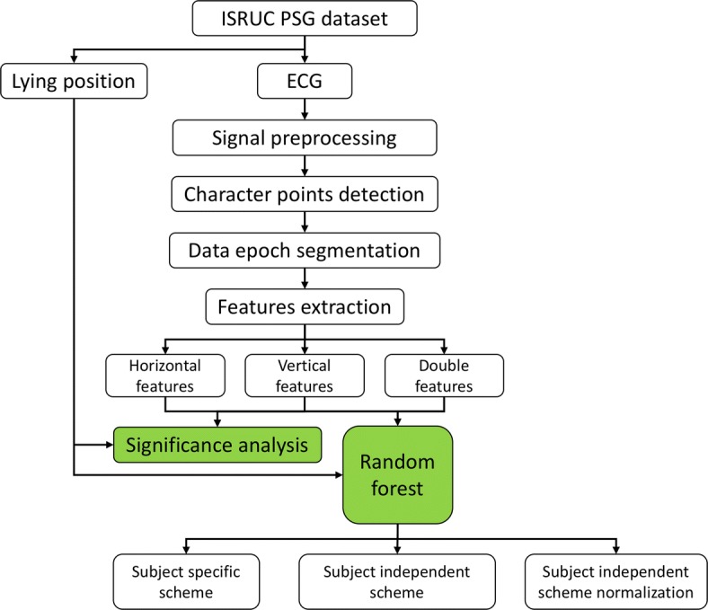
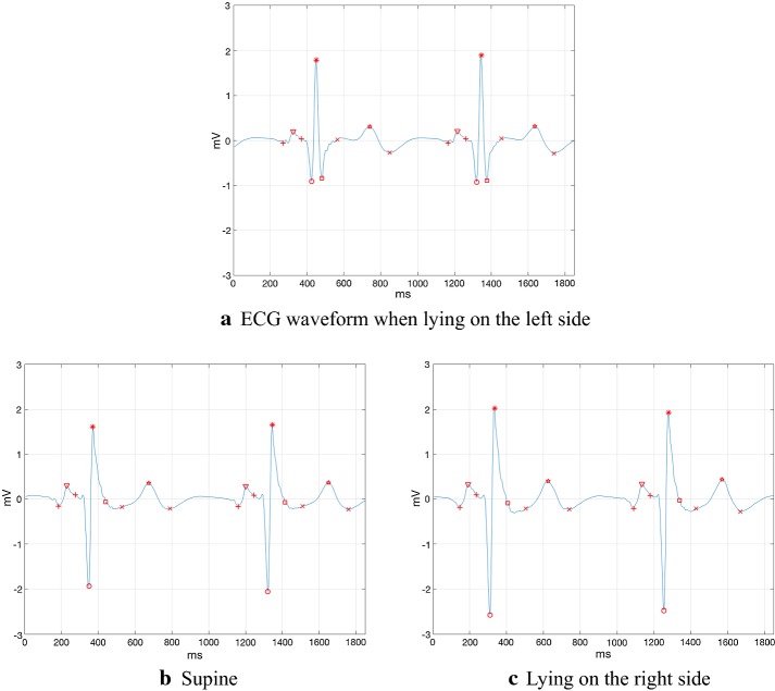
Several different lying positions, such as lying on the left side, supine, lying on the right side and prone position, existed when healthy people fell asleep. This article explored the influence of lying positions on the shape of ECG (electrocardiograph) waveform during sleep, and then lying position classification based on ECG waveform features and random forest was achieved.
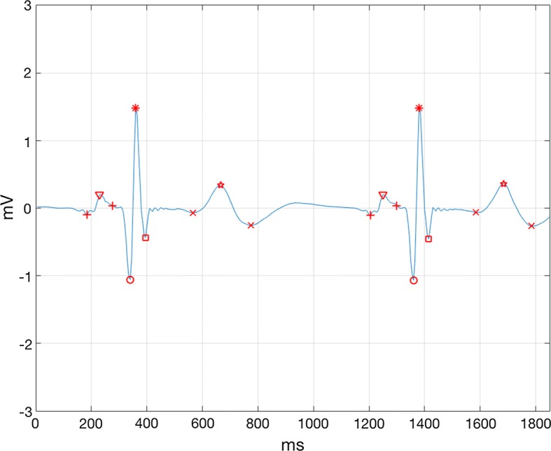
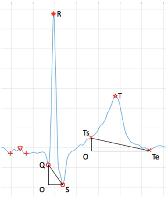
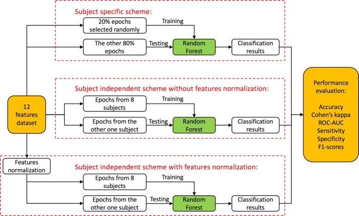
By means of de-noising the overnight sleep ECG data from ISRUC website dataset, as well as extracting the waveform features, we calculated a total of 30 ECG waveform features, including 2 newly proposed features, S/R and ∠QSR. The means and significant difference level of these features within different lying positions were calculated, respectively. Then 12 features were selected for three kinds of classification schemes.
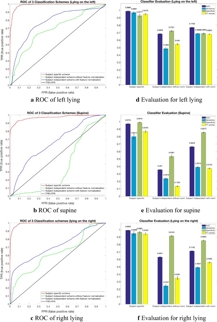
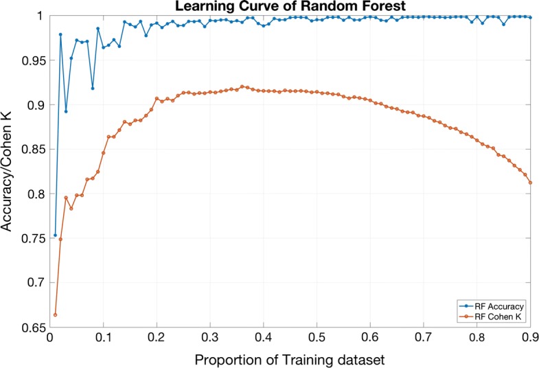
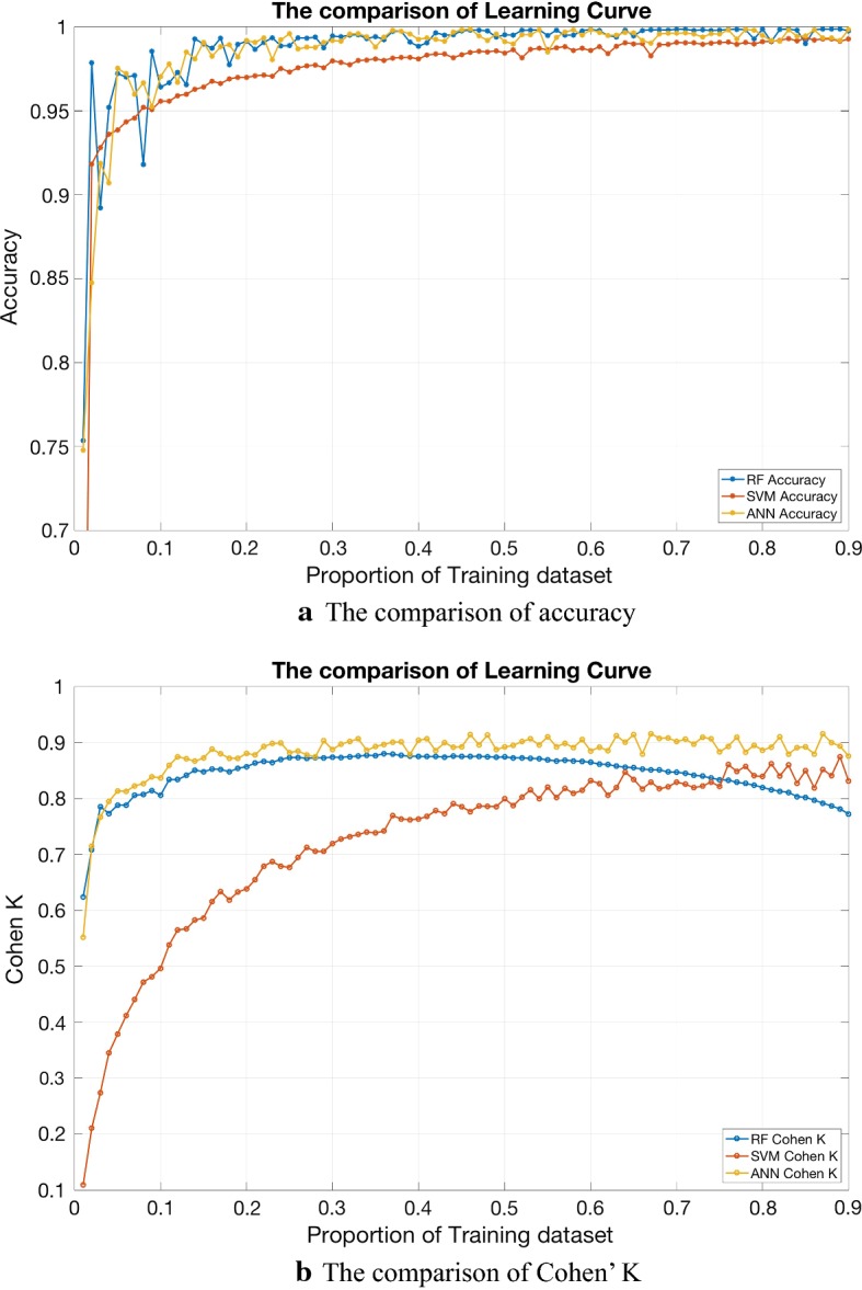
The lying positions had comparatively less effect on time-limit features. QT interval and RR interval were significantly lower than that in supine ([Formula: see text]). Significant differences appeared in most of the amplitude and double-direction features. When lying on the left side, the height of P wave and T wave, QRS area and T area, the QR potential difference and ∠QSR were significantly lower than those in supine ([Formula: see text]). However, S/R was significantly greater on left than those in supine ([Formula: see text]) and on right ([Formula: see text]). The height of T wave and area under T wave were significantly higher in supine than those on right ([Formula: see text]). For the subject specific classifier, a mean accuracy of 97.17% with Cohen's kappa statistic κ of 0.91, and AUC > 0.97 were achieved. While the accuracy and κ dropped to 63.87% and 0.32, AUC > 0.66, respectively when the subject independent classifier was considered.
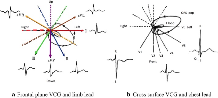
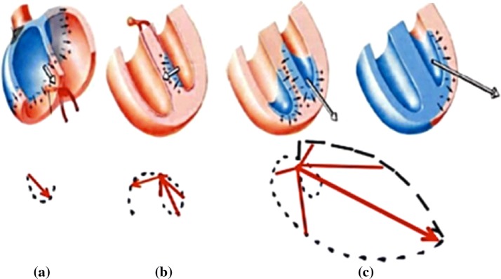
When subjects were lying on the left side during sleep, due to the effect of gravity on heart, the position of heart changed, for example, turned and rotated, causing changes in the vectorcardiogram of frontal plane and horizontal plane, which lead to a change in ECG. When lying on the right side, the heart was upheld by the mediastinum, so that the degree of freedom was poor, and the ECG waveform was almost unchanged. The proposed method could be used as a technique for convenient lying position classification.
健康人入睡时会采取左侧卧位、仰卧位、右侧卧位和俯卧位等不同的卧位。本文探讨了卧位对睡眠时心电图(ECG)波形形态的影响,并基于 ECG 波形特征和随机森林实现了卧位分类。
通过对 ISRUC 网站数据集的整夜睡眠 ECG 数据进行去噪以及提取波形特征,计算了总共 30 个 ECG 波形特征,包括 2 个新提出的特征 S/R 和 ∠QSR。分别计算了不同卧位下这些特征的均值和显著差异水平。然后,为三种分类方案选择了 12 个特征。
卧位对时限特征的影响相对较小。QT 间期和 RR 间期明显低于仰卧位([公式:见正文])。大多数幅度和双向特征都存在显著差异。左侧卧位时,P 波和 T 波高度、QRS 面积和 T 波面积、QR 位差和 ∠QSR 明显低于仰卧位([公式:见正文])。然而,S/R 在左侧卧位时明显大于仰卧位([公式:见正文])和右侧卧位([公式:见正文])。仰卧位时 T 波高度和 T 波下面积明显高于右侧卧位([公式:见正文])。对于个体特异性分类器,平均准确率为 97.17%,Cohen's kappa 统计量 κ 为 0.91,AUC>0.97。而当考虑个体独立分类器时,准确率和 κ 分别降至 63.87%和 0.32,AUC>0.66。
当受试者在睡眠中左侧卧位时,由于重力对心脏的影响,心脏位置发生改变,例如转动和旋转,导致额面和水平面的心向量图发生变化,从而导致 ECG 发生变化。右侧卧位时,心脏被纵隔托起,自由度较差,心电图波形几乎不变。该方法可作为一种方便的卧位分类技术。