Department of Radiology, Xinhua Hospital affiliated to Shanghai Jiaotong University School of Medicine, 1665 Kongjiang Road, Shanghai, 200092, China.
Department of Radiology, Jinshan Hospital, Fudan University, 1508 Longhang Road, Shanghai, 201508, China.
J Ovarian Res. 2018 Aug 30;11(1):73. doi: 10.1186/s13048-018-0444-6.
To investigate MRI for differentiating benign from malignant sex cord-stromal tumors of the ovary (SCSTs) emphasizing on the value of diffusion-weighted (DW) magnetic resonance (MR) imaging.
This retrospective study included 29 benign SCSTs in 28 patients and 13 malignant SCSTs in 13 patients. DW imaging as well as conventional MR imaging was performed. Signal intensity on DW imaging was assessed and apparent diffusion coefficient (ADC) value was measured. In addition, T2 signal intensity and contrast enhancement pattern were also assessed and compared between benign and malignant SCSTs.
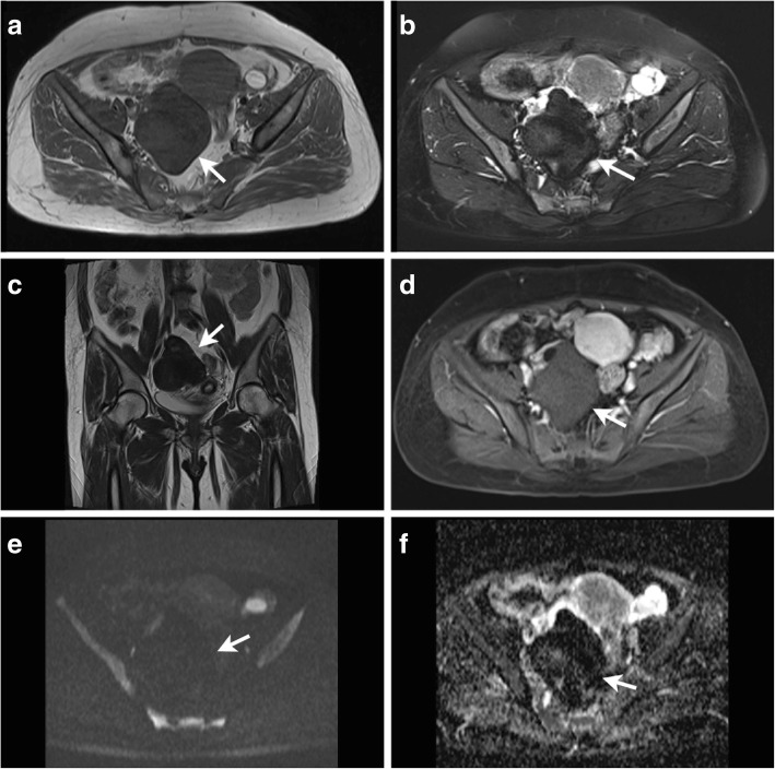
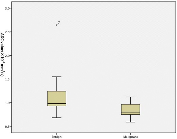
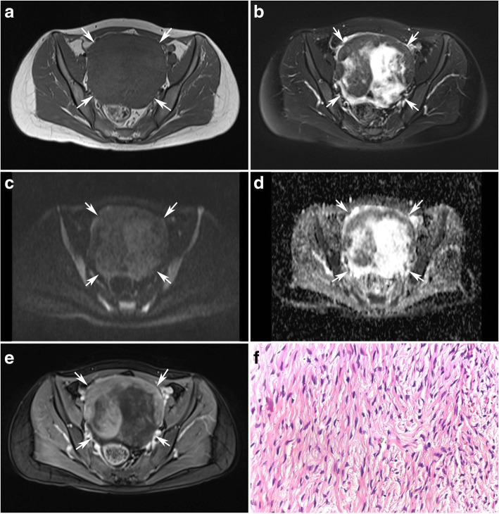
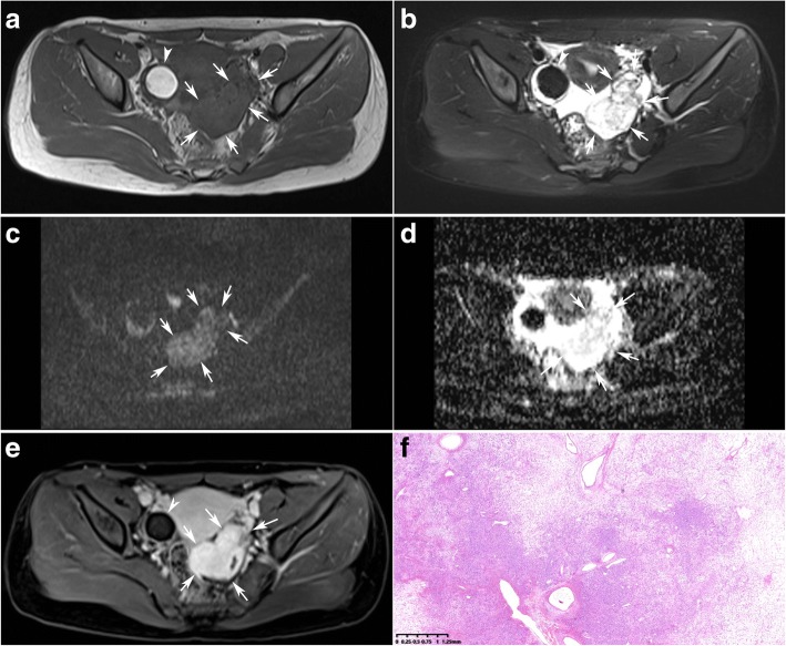
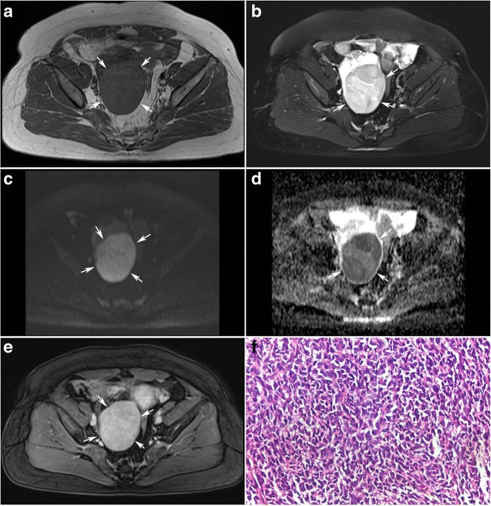
Both of the T2 hypointensity and mild enhancement were specific to benign SCSTs. The majority of malignant SCSTs showed high signal intensity on DW imaging, whereas most benign SCSTs showed low or moderate signal intensity (p = 0.000). Fibromas were the tumors with the lowest observed ADC value (0.470 × 10 mm/s). Sclerosing stromal tumors were the tumors with the highest observed ADC value (2.291 × 10 mm/s). ADC value of solid component was significantly lower in malignant SCSTs (0.825 ± 0.129 × 10 mm/s) than in benign SCSTs (1.343 ± 0.528 × 10 mm/s) when fibromas were excluded (p = 0.024). T2, DCE and DW imaging has a limited value on the differential diagnosis of the benign and malignant SCSTs with an accuracy of 69.0%,71.4% and 78.1% respectively. Combination of T2, DCE and DW imaging permitted the distinction with an accuracy of 88.0%.
It is more helpful for distinction of the benign and malignant SCSTs by combining of T2, DCE and DW imaging than using each of the three sequences independently.
探讨磁共振成像(MRI)在鉴别卵巢性索-间质肿瘤(SCST)良恶性中的作用,重点评估弥散加权(DW)成像的价值。
本回顾性研究纳入 28 例 29 个良性 SCST 和 13 例 13 个恶性 SCST。所有患者均行常规 MRI 及 DW 成像,评估 DW 图像信号强度并测量表观弥散系数(ADC)值,同时评估并比较良恶性 SCST 的 T2 信号强度和增强模式。
T2 低信号和轻度强化均为良性 SCST 的特征。恶性 SCST 多表现为 DW 高信号,而良性 SCST 多为低或中等信号(p=0.000)。纤维瘤的 ADC 值最低(0.470×10 mm/s),而硬化性间质瘤的 ADC 值最高(2.291×10 mm/s)。排除纤维瘤后,恶性 SCST 的实性成分 ADC 值[0.825±0.129×10 mm/s]显著低于良性 SCST[1.343±0.528×10 mm/s](p=0.024)。T2、DCE 和 DW 成像对良恶性 SCST 的鉴别诊断效能有限,其准确性分别为 69.0%、71.4%和 78.1%,三者联合应用的准确性为 88.0%。
与单独使用 T2、DCE 或 DW 序列相比,联合应用 T2、DCE 和 DW 序列更有助于鉴别卵巢良恶性 SCST。