• 文献检索
  • 文档翻译
  • 深度研究
  • 学术资讯
  • Suppr Zotero 插件Zotero 插件
  • 邀请有礼
  • 套餐&价格
  • 历史记录
应用&插件
Suppr Zotero 插件Zotero 插件浏览器插件Mac 客户端Windows 客户端微信小程序
定价
高级版会员购买积分包购买API积分包
服务
文献检索文档翻译深度研究API 文档MCP 服务
关于我们
关于 Suppr公司介绍联系我们用户协议隐私条款
关注我们

Suppr 超能文献

核心技术专利:CN118964589B侵权必究
粤ICP备2023148730 号-1Suppr @ 2026

文献检索

告别复杂PubMed语法,用中文像聊天一样搜索,搜遍4000万医学文献。AI智能推荐,让科研检索更轻松。

立即免费搜索

文件翻译

保留排版,准确专业,支持PDF/Word/PPT等文件格式,支持 12+语言互译。

免费翻译文档

深度研究

AI帮你快速写综述,25分钟生成高质量综述,智能提取关键信息,辅助科研写作。

立即免费体验

基于常规 MRI 的分析和纹理分析在鉴别卵巢卵泡膜细胞瘤-纤维瘤组与卵巢颗粒细胞瘤中的诊断效用。

Diagnostic utility of a conventional MRI-based analysis and texture analysis for discriminating between ovarian thecoma-fibroma groups and ovarian granulosa cell tumors.

机构信息

Department of Radiology, Saitama Medical University, 38 Morohongou, Moroyama-machi, Iruma-gun, Saitama, Japan.

Department of Radiology, Tokyo Metropolitan Cancer and Infectious Diseases Center Komagome Hospital, 3-18-22 Honkomagome, Bunkyo-ku, Tokyo, Japan.

出版信息

J Ovarian Res. 2022 May 25;15(1):65. doi: 10.1186/s13048-022-00989-z.

DOI:10.1186/s13048-022-00989-z
PMID:35610706
原文链接:https://pmc.ncbi.nlm.nih.gov/articles/PMC9131674/
Abstract

OBJECTIVE

To evaluate the diagnostic utility of conventional magnetic resonance imaging (MRI)-based characteristics and a texture analysis (TA) for discriminating between ovarian thecoma-fibroma groups (OTFGs) and ovarian granulosa cell tumors (OGCTs).

METHODS

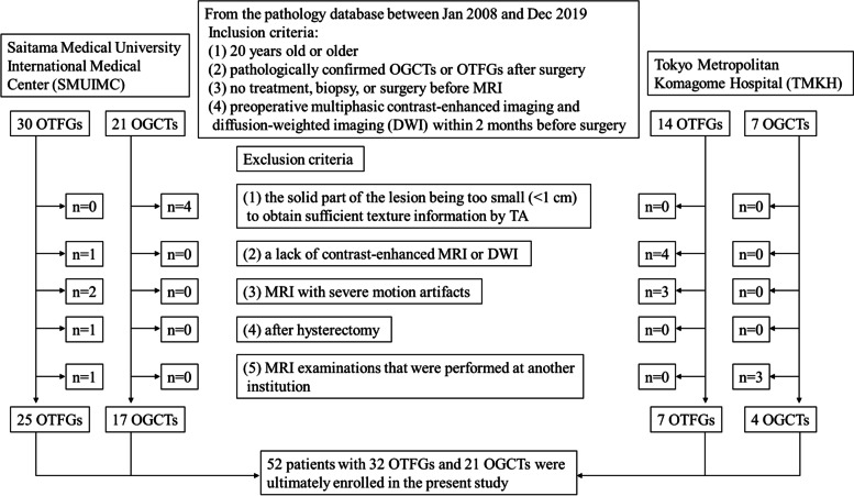
This retrospective multicenter study enrolled 52 patients with 32 OGCTs and 21 OTFGs, which were dissected and pathologically diagnosed between January 2008 and December 2019. MRI-based features (MBFs) and texture features (TFs) were evaluated and compared between OTFGs and OGCTs. A least absolute shrinkage and selection operator (LASSO) regression analysis was performed to select features and construct the discriminating model. ROC analyses were conducted on MBFs, TFs, and their combination to discriminate between the two diseases.

RESULTS

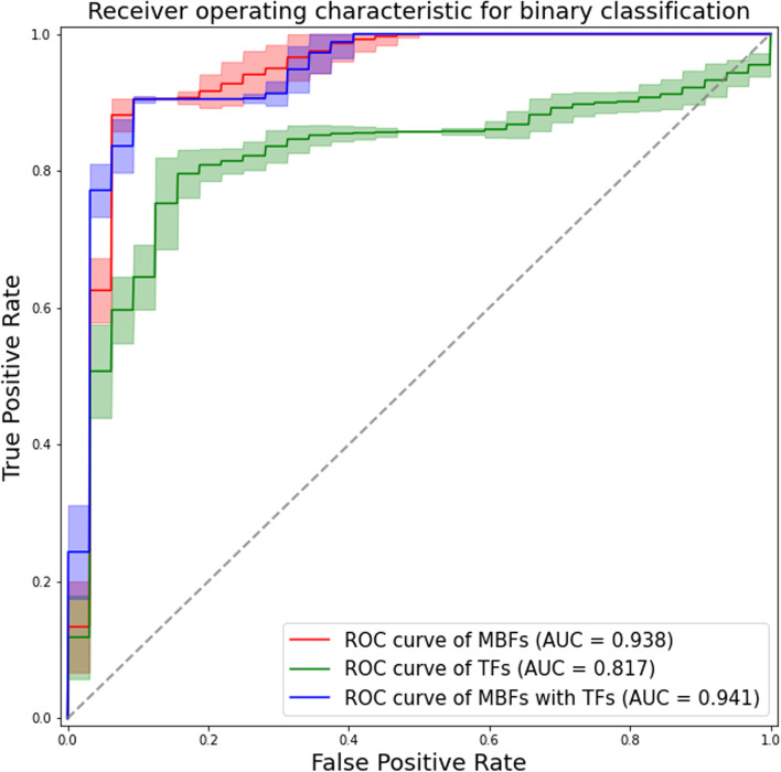
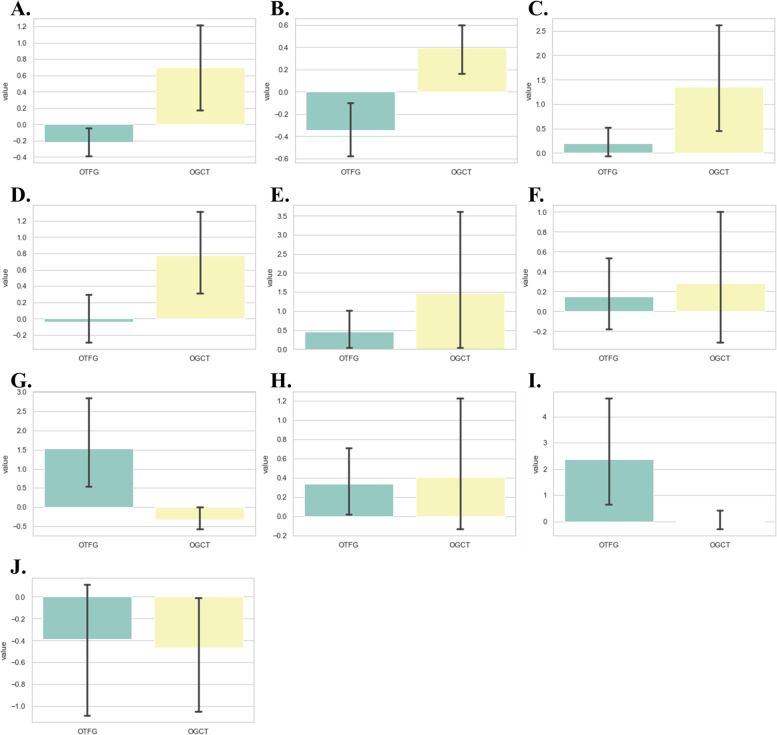
We selected 3 features with the highest absolute value of the LASSO regression coefficient for each model: the apparent diffusion coefficient (ADC), peripheral cystic area, and contrast enhancement in the venous phase (VCE) for the MRI-based model; the 10th percentile, difference variance, and maximal correlation coefficient for the TA-based model; and ADC, VCE, and the difference variance for the combination model. The areas under the curves of the constructed models were 0.938, 0.817, and 0.941, respectively. The diagnostic performance of the MRI-based and combination models was similar (p = 0.38), but significantly better than that of the TA-based model (p < 0.05).

CONCLUSIONS

The conventional MRI-based analysis has potential as a method to differentiate OTFGs from OGCTs. TA did not appear to be of any additional benefit. Further studies are needed on the use of these methods for a preoperative differential diagnosis of these two diseases.

摘要

目的

评估基于常规磁共振成像(MRI)的特征和纹理分析(TA)在鉴别卵巢卵泡膜细胞瘤-纤维瘤组(OTFG)和卵巢颗粒细胞瘤(OGCT)中的诊断效用。

方法

本回顾性多中心研究纳入了 2008 年 1 月至 2019 年 12 月期间经手术切除和病理诊断为 32 例 OGCT 和 21 例 OTFG 的 52 例患者。评估并比较了 OTFG 和 OGCT 之间的 MRI 特征(MBFs)和纹理特征(TFs)。使用最小绝对收缩和选择算子(LASSO)回归分析来选择特征并构建鉴别模型。对 MBFs、TFs 及其组合进行 ROC 分析,以鉴别这两种疾病。

结果

我们为每个模型选择了 3 个具有最高 LASSO 回归系数绝对值的特征:基于 MRI 的模型中的表观扩散系数(ADC)、周围囊腔区和静脉期增强(VCE);基于 TA 的模型中的 10 百分位数、差异方差和最大相关系数;以及组合模型中的 ADC、VCE 和差异方差。构建模型的曲线下面积分别为 0.938、0.817 和 0.941。基于 MRI 的和组合模型的诊断性能相似(p=0.38),但明显优于基于 TA 的模型(p<0.05)。

结论

基于常规 MRI 的分析有可能成为鉴别 OTFG 和 OGCT 的方法。TA 似乎没有任何额外的益处。需要进一步研究这些方法在这两种疾病术前鉴别诊断中的应用。

https://cdn.ncbi.nlm.nih.gov/pmc/blobs/8c39/9131674/17e8a26a80da/13048_2022_989_Fig5_HTML.jpg
https://cdn.ncbi.nlm.nih.gov/pmc/blobs/8c39/9131674/706c6df9522c/13048_2022_989_Fig1_HTML.jpg
https://cdn.ncbi.nlm.nih.gov/pmc/blobs/8c39/9131674/a32c9c859bbb/13048_2022_989_Fig2_HTML.jpg
https://cdn.ncbi.nlm.nih.gov/pmc/blobs/8c39/9131674/c13fc83f6763/13048_2022_989_Fig3_HTML.jpg
https://cdn.ncbi.nlm.nih.gov/pmc/blobs/8c39/9131674/41b9ff6581e1/13048_2022_989_Fig4_HTML.jpg
https://cdn.ncbi.nlm.nih.gov/pmc/blobs/8c39/9131674/17e8a26a80da/13048_2022_989_Fig5_HTML.jpg

相似文献

1
Diagnostic utility of a conventional MRI-based analysis and texture analysis for discriminating between ovarian thecoma-fibroma groups and ovarian granulosa cell tumors.基于常规 MRI 的分析和纹理分析在鉴别卵巢卵泡膜细胞瘤-纤维瘤组与卵巢颗粒细胞瘤中的诊断效用。
J Ovarian Res. 2022 May 25;15(1):65. doi: 10.1186/s13048-022-00989-z.
2
The Value of MRI Findings Combined With Texture Analysis in the Differential Diagnosis of Primary Ovarian Granulosa Cell Tumors and Ovarian Thecoma-Fibrothecoma.MRI表现联合纹理分析在原发性卵巢颗粒细胞瘤与卵巢泡膜-纤维瘤鉴别诊断中的价值
Front Oncol. 2021 Oct 27;11:758036. doi: 10.3389/fonc.2021.758036. eCollection 2021.
3
MRI combined with clinical features to differentiate ovarian thecoma-fibroma with cystic degeneration from ovary adenofibroma.MRI 结合临床特征鉴别卵巢卵泡膜细胞瘤-纤维瘤伴囊性变与卵巢性腺纤维瘤。
Br J Radiol. 2024 May 7;97(1157):1057-1065. doi: 10.1093/bjr/tqae046.
4
Magnetic resonance imaging-based radiomics analysis of the differential diagnosis of ovarian clear cell carcinoma and endometrioid carcinoma: a retrospective study.基于磁共振成像的影像组学分析在卵巢透明细胞癌与子宫内膜样癌鉴别诊断中的应用:一项回顾性研究。
Jpn J Radiol. 2024 Jul;42(7):731-743. doi: 10.1007/s11604-024-01545-z. Epub 2024 Mar 12.
5
Combination of clinical and MRI features in diagnosing ovarian granulosa cell tumor: A comparison with other ovarian sex cord-gonadal stromal tumors.结合临床和 MRI 特征诊断卵巢颗粒细胞瘤:与其他卵巢性索-性腺间质肿瘤的比较。
Eur J Radiol. 2023 Jan;158:110593. doi: 10.1016/j.ejrad.2022.110593. Epub 2022 Nov 5.
6
Application of convolutional neural network for differentiating ovarian thecoma-fibroma and solid ovarian cancer based on MRI.基于 MRI 的卷积神经网络在鉴别卵巢卵泡膜细胞瘤-纤维瘤与卵巢癌实性成分中的应用。
Acta Radiol. 2024 Jul;65(7):860-868. doi: 10.1177/02841851241252951. Epub 2024 May 15.
7
Value of diffusion-weighted imaging combined with conventional magnetic resonance imaging in the diagnosis of thecomas/fibrothecomas and their differential diagnosis with malignant pelvic solid tumors.扩散加权成像联合传统磁共振成像在诊断卵巢纤维瘤/卵泡膜细胞瘤及其与盆腔恶性实性肿瘤鉴别诊断中的价值。
World J Surg Oncol. 2016 Jan 8;14(1):5. doi: 10.1186/s12957-015-0760-x.
8
MR findings of primary ovarian granulosa cell tumor with focus on the differentiation with other ovarian sex cord-stromal tumors.卵巢原发性颗粒细胞瘤的磁共振表现,重点在于与其他卵巢性索-间质肿瘤的鉴别。
J Ovarian Res. 2018 Jun 5;11(1):46. doi: 10.1186/s13048-018-0416-x.
9
Value of diffusion-weighted imaging combined with conventional magnetic resonance imaging in the diagnosis of thecomas and their differential diagnosis with adult granulosa cell tumors.弥散加权成像联合传统磁共振成像在昏迷诊断及其与成人颗粒细胞瘤鉴别诊断中的价值。
Acta Radiol. 2019 Nov;60(11):1532-1542. doi: 10.1177/0284185119830280. Epub 2019 Feb 18.
10
Diagnostic Performance of MR Imaging-based Features and Texture Analysis in the Differential Diagnosis of Ovarian Thecomas/Fibrothecomas and Uterine Fibroids in the Adnexal Area.基于 MRI 的特征和纹理分析在附件区卵巢性索间质瘤/纤维瘤与子宫肌瘤鉴别诊断中的诊断性能。
Acad Radiol. 2020 Oct;27(10):1406-1415. doi: 10.1016/j.acra.2019.12.025. Epub 2020 Feb 5.

引用本文的文献

1
Findings on conventional ultrasonography and contrast-enhanced ultrasonography in different histopathological subtypes of ovarian thecoma-fibroma group.卵巢纤维瘤-卵泡膜细胞瘤组不同组织病理学亚型的传统超声和超声造影检查结果
BMC Med Imaging. 2025 May 20;25(1):175. doi: 10.1186/s12880-025-01693-2.
2
Conventional ultrasound and high-frame-rate contrast-enhanced ultrasound characteristics of ovarian thecoma-fibroma groups.卵巢纤维瘤-卵泡膜细胞瘤组的传统超声及高帧率超声造影特征
Quant Imaging Med Surg. 2025 May 1;15(5):3875-3890. doi: 10.21037/qims-24-2200. Epub 2025 Apr 17.
3
Performance of radiomics analysis in ultrasound imaging for differentiating benign from malignant adnexal masses: A systematic review and meta-analysis.

本文引用的文献

1
The Value of MRI Findings Combined With Texture Analysis in the Differential Diagnosis of Primary Ovarian Granulosa Cell Tumors and Ovarian Thecoma-Fibrothecoma.MRI表现联合纹理分析在原发性卵巢颗粒细胞瘤与卵巢泡膜-纤维瘤鉴别诊断中的价值
Front Oncol. 2021 Oct 27;11:758036. doi: 10.3389/fonc.2021.758036. eCollection 2021.
2
Texture analysis of muscle MRI: machine learning-based classifications in idiopathic inflammatory myopathies.肌肉 MRI 的纹理分析:基于机器学习的特发性炎性肌病分类。
Sci Rep. 2021 May 10;11(1):9821. doi: 10.1038/s41598-021-89311-3.
3
Analysis of magnetic resonance imaging features of ovarian thecoma.
超声成像中影像组学分析在鉴别附件肿块良恶性中的性能:一项系统评价和荟萃分析。
Acta Obstet Gynecol Scand. 2025 May 1. doi: 10.1111/aogs.15146.
4
Evaluation of Uterine Carcinosarcoma and Uterine Endometrial Carcinoma Using Magnetic Resonance Imaging Findings and Texture Features.利用磁共振成像结果和纹理特征评估子宫癌肉瘤和子宫内膜癌
Cureus. 2024 Mar 10;16(3):e55916. doi: 10.7759/cureus.55916. eCollection 2024 Mar.
5
Magnetic resonance imaging-based radiomics analysis of the differential diagnosis of ovarian clear cell carcinoma and endometrioid carcinoma: a retrospective study.基于磁共振成像的影像组学分析在卵巢透明细胞癌与子宫内膜样癌鉴别诊断中的应用:一项回顾性研究。
Jpn J Radiol. 2024 Jul;42(7):731-743. doi: 10.1007/s11604-024-01545-z. Epub 2024 Mar 12.
6
Hyper α2,6-Sialylation Promotes CD4 T-Cell Activation and Induces the Occurrence of Ulcerative Colitis.高 α2,6-唾液酸化促进 CD4 T 细胞激活并诱导溃疡性结肠炎的发生。
Adv Sci (Weinh). 2023 Sep;10(26):e2302607. doi: 10.1002/advs.202302607. Epub 2023 Jul 9.
7
A systematic review and meta-analysis of CT and MRI radiomics in ovarian cancer: methodological issues and clinical utility.卵巢癌CT和MRI影像组学的系统评价与Meta分析:方法学问题与临床应用
Insights Imaging. 2023 Jul 3;14(1):117. doi: 10.1186/s13244-023-01464-z.
8
Serum vascular endothelial growth factor associated with the progression of granulosa cell tumor: a report of two cases.血清血管内皮生长因子与颗粒细胞瘤的进展相关:两例报告。
J Ovarian Res. 2023 Jun 14;16(1):112. doi: 10.1186/s13048-023-01197-z.
9
Normalization Strategies in Multi-Center Radiomics Abdominal MRI: Systematic Review and Meta-Analyses.多中心腹部MRI影像组学中的标准化策略:系统评价与荟萃分析
IEEE Open J Eng Med Biol. 2023 Apr 28;4:67-76. doi: 10.1109/OJEMB.2023.3271455. eCollection 2023.
卵巢纤维瘤的磁共振成像特征分析
Medicine (Baltimore). 2020 May 22;99(21):e20358. doi: 10.1097/MD.0000000000020358.
4
Diagnostic Performance of MR Imaging-based Features and Texture Analysis in the Differential Diagnosis of Ovarian Thecomas/Fibrothecomas and Uterine Fibroids in the Adnexal Area.基于 MRI 的特征和纹理分析在附件区卵巢性索间质瘤/纤维瘤与子宫肌瘤鉴别诊断中的诊断性能。
Acad Radiol. 2020 Oct;27(10):1406-1415. doi: 10.1016/j.acra.2019.12.025. Epub 2020 Feb 5.
5
Value of diffusion-weighted imaging combined with conventional magnetic resonance imaging in the diagnosis of thecomas and their differential diagnosis with adult granulosa cell tumors.弥散加权成像联合传统磁共振成像在昏迷诊断及其与成人颗粒细胞瘤鉴别诊断中的价值。
Acta Radiol. 2019 Nov;60(11):1532-1542. doi: 10.1177/0284185119830280. Epub 2019 Feb 18.
6
Radiomics: the facts and the challenges of image analysis.放射组学:图像分析的现状与挑战
Eur Radiol Exp. 2018 Nov 14;2(1):36. doi: 10.1186/s41747-018-0068-z.
7
Clear Cell Renal Cell Carcinoma: Machine Learning-Based Quantitative Computed Tomography Texture Analysis for Prediction of Fuhrman Nuclear Grade.透明细胞肾细胞癌:基于机器学习的定量 CT 纹理分析预测 Fuhrman 核分级。
Eur Radiol. 2019 Mar;29(3):1153-1163. doi: 10.1007/s00330-018-5698-2. Epub 2018 Aug 30.
8
The value of MRI for differentiating benign from malignant sex cord-stromal tumors of the ovary: emphasis on diffusion-weighted MR imaging.MRI 对卵巢性索-间质肿瘤良恶性鉴别的价值:着重于弥散加权 MR 成像。
J Ovarian Res. 2018 Aug 30;11(1):73. doi: 10.1186/s13048-018-0444-6.
9
MR findings of primary ovarian granulosa cell tumor with focus on the differentiation with other ovarian sex cord-stromal tumors.卵巢原发性颗粒细胞瘤的磁共振表现,重点在于与其他卵巢性索-间质肿瘤的鉴别。
J Ovarian Res. 2018 Jun 5;11(1):46. doi: 10.1186/s13048-018-0416-x.
10
Texture analysis of paraspinal musculature in MRI of the lumbar spine: analysis of the lumbar stenosis outcome study (LSOS) data.腰椎MRI中椎旁肌肉组织的纹理分析:腰椎管狭窄症结局研究(LSOS)数据分析
Skeletal Radiol. 2018 Jul;47(7):947-954. doi: 10.1007/s00256-018-2919-3. Epub 2018 Mar 1.