• 文献检索
  • 文档翻译
  • 深度研究
  • 学术资讯
  • Suppr Zotero 插件Zotero 插件
  • 邀请有礼
  • 套餐&价格
  • 历史记录
应用&插件
Suppr Zotero 插件Zotero 插件浏览器插件Mac 客户端Windows 客户端微信小程序
定价
高级版会员购买积分包购买API积分包
服务
文献检索文档翻译深度研究API 文档MCP 服务
关于我们
关于 Suppr公司介绍联系我们用户协议隐私条款
关注我们

Suppr 超能文献

核心技术专利:CN118964589B侵权必究
粤ICP备2023148730 号-1Suppr @ 2026

文献检索

告别复杂PubMed语法,用中文像聊天一样搜索,搜遍4000万医学文献。AI智能推荐,让科研检索更轻松。

立即免费搜索

文件翻译

保留排版,准确专业,支持PDF/Word/PPT等文件格式,支持 12+语言互译。

免费翻译文档

深度研究

AI帮你快速写综述,25分钟生成高质量综述,智能提取关键信息,辅助科研写作。

立即免费体验

光学相干断层扫描血管造影参数与糖尿病视网膜病变严重程度相关的统计模型。

Statistical Model of Optical Coherence Tomography Angiography Parameters That Correlate With Severity of Diabetic Retinopathy.

机构信息

Department of Ophthalmology, Feinberg School of Medicine, Northwestern University, Chicago, Illinois, United States.

Department of Ophthalmology, Alexandria Faculty of Medicine, Alexandria, Egypt.

出版信息

Invest Ophthalmol Vis Sci. 2018 Aug 1;59(10):4292-4298. doi: 10.1167/iovs.18-24142.

DOI:10.1167/iovs.18-24142
PMID:30167660
原文链接:https://pmc.ncbi.nlm.nih.gov/articles/PMC6110573/
Abstract

PURPOSE

To determine whether combining quantitative optical coherence tomography angiography (OCTA) parameters can achieve high sensitivity and specificity to distinguish eyes with nonproliferative diabetic retinopathy (NPDR) from those with proliferative diabetic retinopathy (PDR) as well as eyes with diabetes and no DR (NoDR) from those with clinical DR (any DR).

METHODS

This cross-sectional study included 28 eyes (17 patients) with NoDR, 54 eyes (34 patients) with NPDR, and 56 eyes (36 patients) with PDR. OCTA images were processed to quantify the foveal avascular zone (FAZ) area, acircularity, vessel density, skeletonized vessel density, fractal dimension, and intersections and average vessel diameter for the superficial (SCP) and the deep capillary plexus (DCP). Binary logistic regression models were used to identify the OCTA parameters that best distinguished DR severity groups. The area (AUC) under the receiver operating characteristic (ROC) curves, and sensitivity and specificity were calculated for each model.

RESULTS

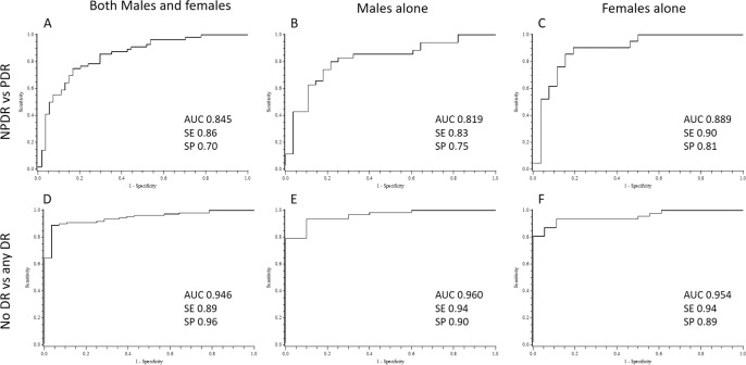
The regression model identified the SCP FAZ area, DCP vessel density, and acircularity as parameters that best distinguished between DR severity groups. ROC curves for NPDR versus PDR had an AUC of 0.845 (P < 0.001) and sensitivity and specificity of 86% and 70%, respectively. ROC curves for NoDR versus any DR showed an AUC of 0.946 (P < 0.001) with sensitivity of 89% and specificity of 96%, with comparable results when explored in males and females separately.

CONCLUSIONS

We identified a set of OCTA parameters with high sensitivity and specificity for distinguishing between groups based on DR severity, suggesting potential clinical application for OCTA as a screening tool for DR.

摘要

目的

确定定量光学相干断层扫描血管造影(OCTA)参数的组合是否可以实现高灵敏度和特异性,以区分非增殖性糖尿病视网膜病变(NPDR)和增殖性糖尿病视网膜病变(PDR)的眼睛,以及糖尿病无DR(NoDR)和有临床 DR(任何 DR)的眼睛。

方法

本横断面研究纳入了 28 只眼(17 例患者)的 NoDR、54 只眼(34 例患者)的 NPDR 和 56 只眼(36 例患者)的 PDR。对 OCTA 图像进行处理,以定量测量黄斑无血管区(FAZ)面积、非圆度、血管密度、骨架血管密度、分形维数以及浅层毛细血管丛(SCP)和深层毛细血管丛(DCP)的交叉点和平均血管直径。采用二元逻辑回归模型确定区分 DR 严重程度组的最佳 OCTA 参数。计算每个模型的接收者操作特征(ROC)曲线下面积(AUC)以及灵敏度和特异性。

结果

回归模型确定 SCP FAZ 面积、DCP 血管密度和非圆度是区分 DR 严重程度组的最佳参数。NPDR 与 PDR 的 ROC 曲线 AUC 为 0.845(P<0.001),灵敏度和特异性分别为 86%和 70%。NoDR 与任何 DR 的 ROC 曲线 AUC 为 0.946(P<0.001),灵敏度为 89%,特异性为 96%,分别在男性和女性中进行探索时,结果相似。

结论

我们确定了一组 OCTA 参数,具有区分 DR 严重程度组的高灵敏度和特异性,提示 OCTA 作为 DR 筛查工具具有潜在的临床应用价值。

https://cdn.ncbi.nlm.nih.gov/pmc/blobs/2bd0/6110573/65e758c6a51d/i1552-5783-59-10-4292-f01.jpg

相似文献

1
Statistical Model of Optical Coherence Tomography Angiography Parameters That Correlate With Severity of Diabetic Retinopathy.光学相干断层扫描血管造影参数与糖尿病视网膜病变严重程度相关的统计模型。
Invest Ophthalmol Vis Sci. 2018 Aug 1;59(10):4292-4298. doi: 10.1167/iovs.18-24142.
2
Macular Capillary Perfusion in Chinese Patients With Diabetic Retinopathy Obtained With Optical Coherence Tomography Angiography.光学相干断层扫描血管造影术检测中国糖尿病视网膜病变患者的黄斑区毛细血管灌注情况
Ophthalmic Surg Lasers Imaging Retina. 2019 Apr 1;50(4):e88-e95. doi: 10.3928/23258160-20190401-12.
3
Linking Retinal Microvasculature Features With Severity of Diabetic Retinopathy Using Optical Coherence Tomography Angiography.使用光学相干断层扫描血管造影术将视网膜微血管特征与糖尿病性视网膜病变的严重程度相联系。
Invest Ophthalmol Vis Sci. 2016 Jul 1;57(9):OCT519-25. doi: 10.1167/iovs.15-18901.
4
QUANTIFICATION OF RETINAL VESSEL TORTUOSITY IN DIABETIC RETINOPATHY USING OPTICAL COHERENCE TOMOGRAPHY ANGIOGRAPHY.应用光学相干断层扫描血管造影术定量分析糖尿病性视网膜病变中的视网膜血管迂曲。
Retina. 2018 May;38(5):976-985. doi: 10.1097/IAE.0000000000001618.
5
Interaction Between the Distribution of Diabetic Retinopathy Lesions and the Association of Optical Coherence Tomography Angiography Scans With Diabetic Retinopathy Severity.糖尿病视网膜病变病变分布与光学相干断层扫描血管造影扫描与糖尿病视网膜病变严重程度的相关性分析。
JAMA Ophthalmol. 2020 Dec 1;138(12):1291-1297. doi: 10.1001/jamaophthalmol.2020.4516.
6
OCT Angiography Assessment of Retinal Microvascular Changes in Diabetic Eyes in an Urban Safety-Net Hospital.城市安全网医院中糖尿病眼病视网膜微血管变化的光学相干断层扫描血管造影评估
Ophthalmol Retina. 2020 Apr;4(4):425-432. doi: 10.1016/j.oret.2019.11.008. Epub 2019 Nov 15.
7
Deep Capillary Geometric Perfusion Deficits on OCT Angiography Detect Clinically Referable Eyes with Diabetic Retinopathy.OCT 血管造影上的深层毛细血管几何灌注不足可检测出有临床意义的糖尿病视网膜病变眼。
Ophthalmol Retina. 2022 Dec;6(12):1194-1205. doi: 10.1016/j.oret.2022.05.028. Epub 2022 Jun 2.
8
Superficial capillary perfusion on optical coherence tomography angiography differentiates moderate and severe nonproliferative diabetic retinopathy.光学相干断层扫描血管造影的浅层毛细血管灌注可区分中度和重度非增殖性糖尿病性视网膜病变。
PLoS One. 2020 Oct 22;15(10):e0240064. doi: 10.1371/journal.pone.0240064. eCollection 2020.
9
Detection of the Microvascular Changes of Diabetic Retinopathy Progression Using Optical Coherence Tomography Angiography.利用光相干断层扫描血管造影术检测糖尿病视网膜病变进展的微血管变化。
Transl Vis Sci Technol. 2021 Jun 1;10(7):31. doi: 10.1167/tvst.10.7.31.
10
Changes of blood flow in macular zone of patients with diabetic retinopathy at different stages evaluated by optical coherence tomography angiography.应用光学相干断层扫描血管造影评估不同分期糖尿病视网膜病变患者黄斑区血流变化。
J Fr Ophtalmol. 2022 Sep;45(7):728-734. doi: 10.1016/j.jfo.2022.01.010. Epub 2022 May 13.

引用本文的文献

1
The Impact of Hypertension on Macular Perfusion in Patients With Referable Diabetic Retinopathy: An OCTA Analysis.高血压对可转诊糖尿病视网膜病变患者黄斑灌注的影响:一项光学相干断层扫描血管造影分析
Transl Vis Sci Technol. 2025 Jul 1;14(7):2. doi: 10.1167/tvst.14.7.2.
2
Diabetic foveal neovascularization is associated with diminished subfoveal choroidal flow on optical coherence tomography angiography.糖尿病性黄斑新生血管形成与光学相干断层扫描血管造影显示的黄斑下脉络膜血流减少有关。
Eye (Lond). 2025 May 6. doi: 10.1038/s41433-025-03806-1.
3
Diagnostic value of choroidal vascular density in predicting the progression of diabetic retinopathy.

本文引用的文献

1
Importance of Considering the Middle Capillary Plexus on OCT Angiography in Diabetic Retinopathy.重视 OCT 血管造影中糖尿病视网膜病变的中层毛细血管丛
Invest Ophthalmol Vis Sci. 2018 Apr 1;59(5):2167-2176. doi: 10.1167/iovs.17-23304.
2
Automated diabetic retinopathy detection using optical coherence tomography angiography: a pilot study.基于光相干断层扫描血管造影的自动糖尿病性视网膜病变检测:一项初步研究。
Br J Ophthalmol. 2018 Nov;102(11):1564-1569. doi: 10.1136/bjophthalmol-2017-311489. Epub 2018 Jan 23.
3
Optical coherence tomography angiography analysis of foveal microvascular changes and inner retinal layer thinning in patients with diabetes.
脉络膜血管密度在预测糖尿病视网膜病变进展中的诊断价值
Sci Rep. 2025 May 5;15(1):15671. doi: 10.1038/s41598-025-00528-y.
4
Diabetic Retinopathy (DR) nomogram construction based on optical coherence tomography angiography parameters: a preliminary exploration of DR prediction.基于光学相干断层扫描血管造影参数构建糖尿病视网膜病变(DR)列线图:DR预测的初步探索
Graefes Arch Clin Exp Ophthalmol. 2025 Jul;263(7):1867-1876. doi: 10.1007/s00417-025-06824-7. Epub 2025 Apr 8.
5
Visual Deficits in Type 2 Diabetes Mellitus Without Retinopathy: From Retinal Structure to Higher-Level Visual Functions.无视网膜病变的2型糖尿病患者的视觉缺陷:从视网膜结构到高级视觉功能
Transl Vis Sci Technol. 2025 Mar 3;14(3):10. doi: 10.1167/tvst.14.3.10.
6
Visual Function Measurements in Eyes With Diabetic Retinopathy: An Expert Opinion on Available Measures.糖尿病视网膜病变患者的视觉功能测量:关于现有测量方法的专家意见
Ophthalmol Sci. 2024 Apr 6;4(5):100519. doi: 10.1016/j.xops.2024.100519. eCollection 2024 Sep-Oct.
7
Resilience to diabetic retinopathy.糖尿病视网膜病变的抵抗力。
Prog Retin Eye Res. 2024 Jul;101:101271. doi: 10.1016/j.preteyeres.2024.101271. Epub 2024 May 11.
8
Retinal vasculature of different diameters and plexuses exhibit distinct vulnerability in varying severity of diabetic retinopathy.不同直径和丛的视网膜血管在不同严重程度的糖尿病视网膜病变中表现出明显的脆弱性。
Eye (Lond). 2024 Jun;38(9):1762-1769. doi: 10.1038/s41433-024-03021-4. Epub 2024 Mar 21.
9
Optical Coherence Tomography Angiography as a Diagnostic Tool for Diabetic Retinopathy.光学相干断层扫描血管造影术作为糖尿病视网膜病变的诊断工具
Diagnostics (Basel). 2024 Feb 2;14(3):326. doi: 10.3390/diagnostics14030326.
10
Is There a Nonperfusion Threshold on OCT Angiography Associated With New Vessels Detected on Ultra-Wide-Field Imaging in Diabetic Retinopathy?OCT 血管造影与超广角成像中新血管检测相关的非灌注阈值是否存在于糖尿病视网膜病变中?
Transl Vis Sci Technol. 2023 Sep 1;12(9):15. doi: 10.1167/tvst.12.9.15.
糖尿病患者黄斑区微血管改变及内层视网膜变薄的光相干断层扫描血管造影分析。
Br J Ophthalmol. 2018 Sep;102(9):1226-1231. doi: 10.1136/bjophthalmol-2017-311149. Epub 2017 Dec 19.
4
Quantitative Analysis of Three Distinct Retinal Capillary Plexuses in Healthy Eyes Using Optical Coherence Tomography Angiography.使用光学相干断层扫描血管造影术对健康眼睛中三个不同视网膜毛细血管丛进行定量分析。
Invest Ophthalmol Vis Sci. 2017 Oct 1;58(12):5548-5555. doi: 10.1167/iovs.17-22036.
5
Foveal avascular zone area and parafoveal vessel density measurements in different stages of diabetic retinopathy by optical coherence tomography angiography.利用光学相干断层扫描血管造影术测量糖尿病视网膜病变不同阶段的黄斑无血管区面积和黄斑旁血管密度
Int J Ophthalmol. 2017 Oct 18;10(10):1545-1551. doi: 10.18240/ijo.2017.10.11. eCollection 2017.
6
Quantifying Microvascular Abnormalities With Increasing Severity of Diabetic Retinopathy Using Optical Coherence Tomography Angiography.使用光学相干断层扫描血管造影术量化糖尿病视网膜病变严重程度增加时的微血管异常。
Invest Ophthalmol Vis Sci. 2017 May 1;58(6):BIO307-BIO315. doi: 10.1167/iovs.17-21787.
7
Deep capillary plexus impairment in patients with type 1 diabetes mellitus with no signs of diabetic retinopathy revealed using optical coherence tomography angiography.使用光学相干断层扫描血管造影术发现,1型糖尿病且无糖尿病视网膜病变迹象患者的深部毛细血管丛受损。
Acta Ophthalmol. 2018 Mar;96(2):e264-e265. doi: 10.1111/aos.13510. Epub 2017 Sep 9.
8
Macular Vascular Fractal Dimension in the Deep Capillary Layer as an Early Indicator of Microvascular Loss for Retinopathy in Type 2 Diabetic Patients.2型糖尿病患者视网膜病变中深层毛细血管层黄斑血管分形维数作为微血管损失的早期指标
Invest Ophthalmol Vis Sci. 2017 Jul 1;58(9):3785-3794. doi: 10.1167/iovs.17-21461.
9
VALUE OF FRACTAL ANALYSIS OF OPTICAL COHERENCE TOMOGRAPHY ANGIOGRAPHY IN VARIOUS STAGES OF DIABETIC RETINOPATHY.光学相干断层扫描血管造影术在糖尿病视网膜病变不同分期中的分形分析价值。
Retina. 2018 Sep;38(9):1816-1823. doi: 10.1097/IAE.0000000000001774.
10
Assessing the Accuracy of Foveal Avascular Zone Measurements Using Optical Coherence Tomography Angiography: Segmentation and Scaling.使用光学相干断层扫描血管造影术评估黄斑无血管区测量的准确性:分割与缩放
Transl Vis Sci Technol. 2017 Jun 9;6(3):16. doi: 10.1167/tvst.6.3.16. eCollection 2017 Jun.