Waters Corporation, Stamford Avenue, Altricham Road, Wilmslow, SK9 4AX, UK.
Waters Research Centre, Záhony utca., C ép., 1. Em., Budapest, 1031, Hungary.
J Am Soc Mass Spectrom. 2018 Dec;29(12):2456-2466. doi: 10.1007/s13361-018-2049-0. Epub 2018 Aug 30.
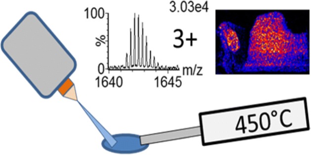
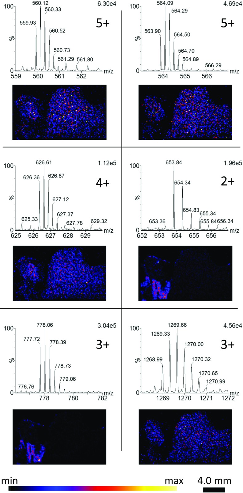
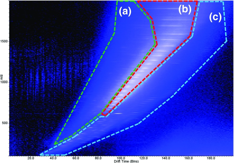
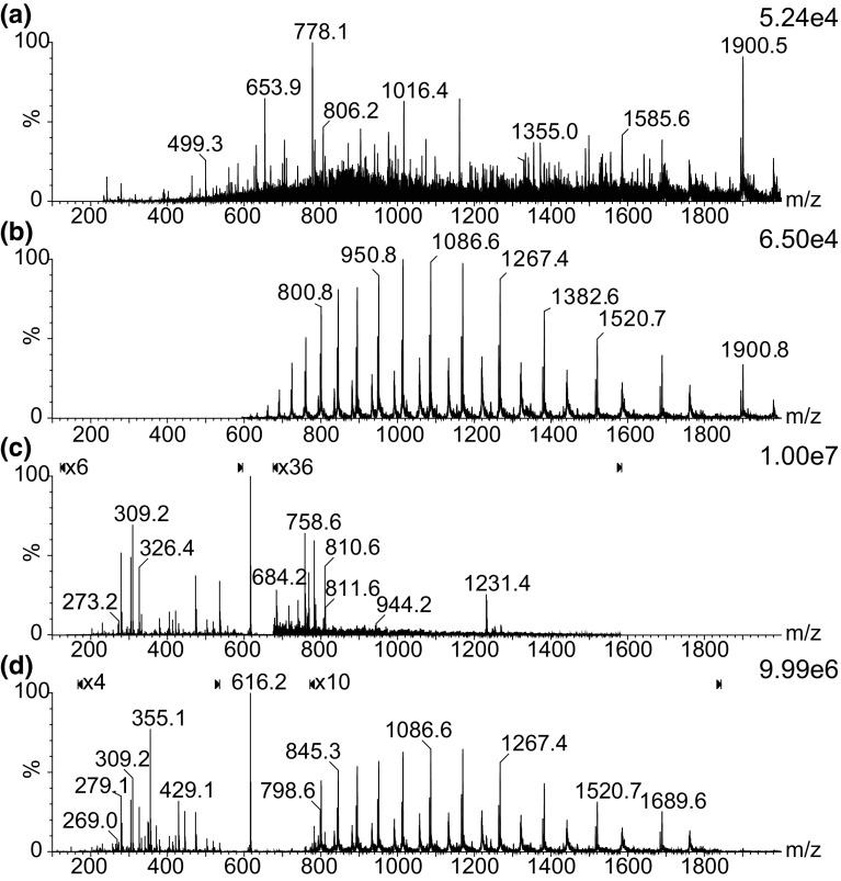
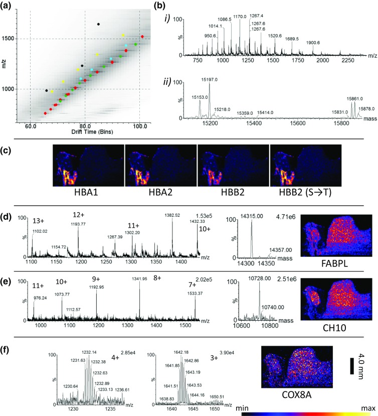
Desorption electrospray ionisation mass spectrometry imaging (DESI-MSI) is typically known for the ionisation of small molecules such as lipids and metabolites, in singly charged form. Here we present a method that allows the direct detection of proteins and peptides in multiply charged forms directly from tissue sections by DESI. Utilising a heated mass spectrometer inlet capillary, combined with ion mobility separation (IMS), the conditions with regard to solvent composition, nebulising gas flow, and solvent flow rate have been explored and optimised. Without the use of ion mobility separation prior to mass spectrometry analysis, only the most abundant charge series were observed. In addition to the dominant haemoglobin subunit(s) related trend line in the m/z vs drift time (DT) 2D plot, trend lines were found relating to background solvent peaks, residual lipids and, more importantly, small proteins/large peptides of lower abundance. These small proteins/peptides were observed with charge states from 1+ to 12+, the majority of which could only be resolved from the background when using IMS. By extracting charge series from the 2D m/z vs DT plot, a number of proteins could be tentatively assigned by accurate mass. Tissue images were acquired with a pixel size of 150 μm showing a marked improvement in protein image resolution compared to other liquid-based ambient imaging techniques such as liquid extraction surface analysis (LESA) and continuous-flow liquid microjunction surface sampling probe (LMJ-SSP) imaging. Graphical Abstract ᅟ.
解吸电喷雾电离质谱成像(DESI-MSI)通常以单电荷形式电离小分子,如脂质和代谢物。在这里,我们提出了一种方法,通过 DESI 可以直接从组织切片中直接检测到多电荷形式的蛋白质和肽。利用加热的质谱仪进样毛细管,结合离子淌度分离(IMS),我们已经探索和优化了有关溶剂组成、雾化气流和溶剂流速的条件。在进行质谱分析之前不使用离子淌度分离,仅观察到最丰富的电荷系列。除了 m/z 与漂移时间(DT)二维图中与血红蛋白亚基相关的主导趋势线外,还发现了与背景溶剂峰、残留脂质有关的趋势线,更重要的是,还发现了低丰度的小蛋白质/大肽的趋势线。这些小蛋白质/肽的电荷状态从 1+到 12+,其中大多数在使用 IMS 时只能从背景中分辨出来。通过从二维 m/z 与 DT 图中提取电荷系列,可以通过精确质量初步鉴定出一些蛋白质。以 150 μm 的像素大小获取组织图像,与其他基于液体的环境成像技术(如液体萃取表面分析(LESA)和连续流动液体微连接表面采样探针(LMJ-SSP)成像)相比,蛋白质图像分辨率得到显著提高。