Blander J Magarian
The Jill Roberts Institute for Research in Inflammatory Bowel Disease.
Gastroenterology and Hepatology Division, Joan and Sanford I. Weill Department of Medicine.
Curr Opin Gastroenterol. 2018 Nov;34(6):413-419. doi: 10.1097/MOG.0000000000000481.
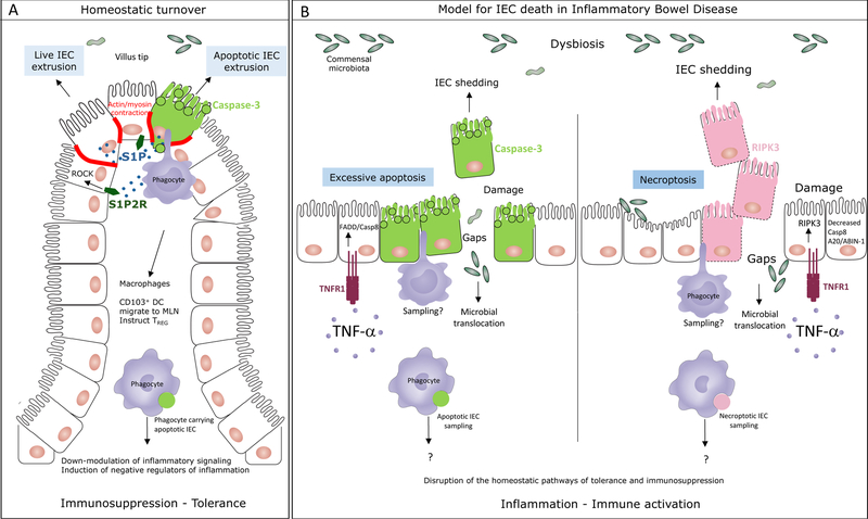
Both apoptotic and nonapoptotic cell extrusion preserve the barrier functions of epithelia. Live cell extrusion is the paradigm for homeostatic renewal of intestinal epithelial cells (IEC). By extension, as extruded cells are not apoptotic, this form of cell shedding is thought to be largely ignored by lamina propria phagocytes and without immune consequence.
Visualization of apoptotic IEC inside distinct subsets of intestinal phagocytes during homeostasis has highlighted apoptosis as a normal component of the natural turnover of the intestinal epithelium. Analysis of phagocytes with or without apoptotic IEC corpses has shown how apoptotic IEC constrain inflammatory pathways within phagocytes and induce immunosuppressive regulatory CD4 T-cell differentiation. Many of the genes involved overlap with susceptibility genes for inflammatory bowel disease (IBD).
Excessive IEC death and loss-of-barrier function is characteristic of IBD. As regulatory and tolerogenic mechanisms are broken in IBD, a molecular understanding of the precise triggers and modes of IEC death as well as their consequences on intestinal inflammation is necessary. This characterization should guide new therapies that restore homeostatic apoptosis, along with its associated programs of immune tolerance and immunosuppression, to achieve mucosal healing and long-term remission.
凋亡性和非凋亡性细胞挤出均能维持上皮的屏障功能。活细胞挤出是肠上皮细胞(IEC)稳态更新的范例。由此推断,由于挤出的细胞并非凋亡细胞,这种细胞脱落形式被认为在很大程度上会被固有层吞噬细胞忽略,且不会产生免疫后果。
在稳态过程中,对肠道不同亚群吞噬细胞内凋亡IEC的可视化研究突出了凋亡是肠上皮自然更新的正常组成部分。对含有或不含有凋亡IEC尸体的吞噬细胞分析表明,凋亡IEC如何限制吞噬细胞内的炎症途径并诱导免疫抑制性调节性CD4 T细胞分化。许多相关基因与炎症性肠病(IBD)的易感基因重叠。
IEC过度死亡和屏障功能丧失是IBD的特征。由于IBD中调节和耐受机制被破坏,因此有必要从分子层面精确了解IEC死亡的触发因素和模式及其对肠道炎症的影响。这一特征描述应指导新的治疗方法,恢复稳态凋亡及其相关的免疫耐受和免疫抑制程序,以实现黏膜愈合和长期缓解。