• 文献检索
  • 文档翻译
  • 深度研究
  • 学术资讯
  • Suppr Zotero 插件Zotero 插件
  • 邀请有礼
  • 套餐&价格
  • 历史记录
应用&插件
Suppr Zotero 插件Zotero 插件浏览器插件Mac 客户端Windows 客户端微信小程序
定价
高级版会员购买积分包购买API积分包
服务
文献检索文档翻译深度研究API 文档MCP 服务
关于我们
关于 Suppr公司介绍联系我们用户协议隐私条款
关注我们

Suppr 超能文献

核心技术专利:CN118964589B侵权必究
粤ICP备2023148730 号-1Suppr @ 2026

文献检索

告别复杂PubMed语法,用中文像聊天一样搜索,搜遍4000万医学文献。AI智能推荐,让科研检索更轻松。

立即免费搜索

文件翻译

保留排版,准确专业,支持PDF/Word/PPT等文件格式,支持 12+语言互译。

免费翻译文档

深度研究

AI帮你快速写综述,25分钟生成高质量综述,智能提取关键信息,辅助科研写作。

立即免费体验

磁共振成像与乳腺癌肿瘤浸润淋巴细胞相关的分子特征。

Magnetic resonance imaging and molecular features associated with tumor-infiltrating lymphocytes in breast cancer.

机构信息

Department of Radiation Oncology, Stanford University School of Medicine, 1070 Arastradero Road, Stanford, CA, 94305, USA.

Department of Pathology, First Affiliated Hospital of Zhejiang University, Hangzhou, 310058, Zhejiang, China.

出版信息

Breast Cancer Res. 2018 Sep 3;20(1):101. doi: 10.1186/s13058-018-1039-2.

DOI:10.1186/s13058-018-1039-2
PMID:30176944
原文链接:https://pmc.ncbi.nlm.nih.gov/articles/PMC6122724/
Abstract

BACKGROUND

We sought to investigate associations between dynamic contrast-enhanced (DCE) magnetic resonance imaging (MRI) features and tumor-infiltrating lymphocytes (TILs) in breast cancer, as well as to study if MRI features are complementary to molecular markers of TILs.

METHODS

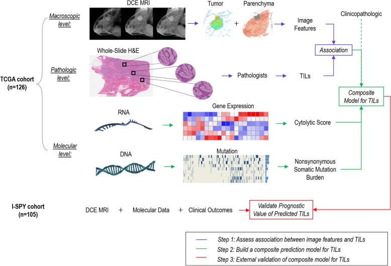
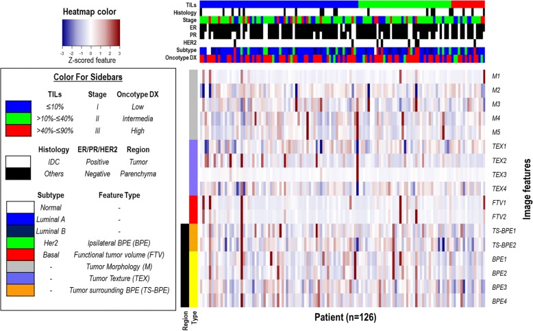
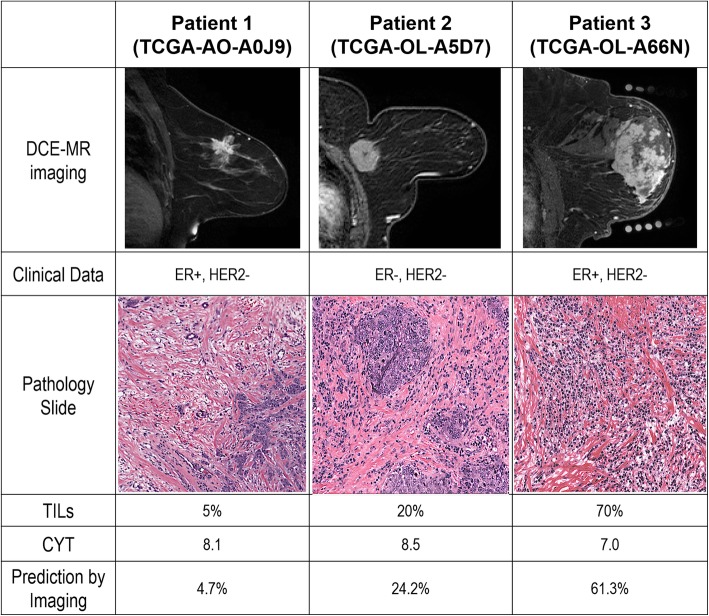
In this retrospective study, we extracted 17 computational DCE-MRI features to characterize tumor and parenchyma in The Cancer Genome Atlas cohort (n = 126). The percentage of stromal TILs was evaluated on H&E-stained histological whole-tumor sections. We first evaluated associations between individual imaging features and TILs. Multiple-hypothesis testing was corrected by the Benjamini-Hochberg method using false discovery rate (FDR). Second, we implemented LASSO (least absolute shrinkage and selection operator) and linear regression nested with tenfold cross-validation to develop an imaging signature for TILs. Next, we built a composite prediction model for TILs by combining imaging signature with molecular features. Finally, we tested the prognostic significance of the TIL model in an independent cohort (I-SPY 1; n = 106).

RESULTS

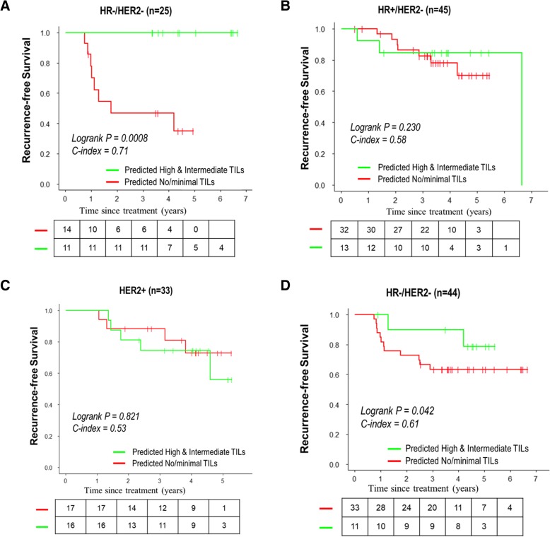
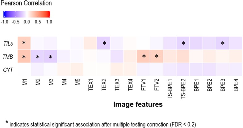
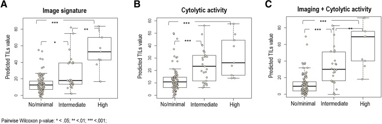
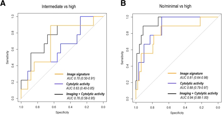
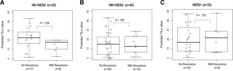
Four imaging features were significantly associated with TILs (P < 0.05 and FDR < 0.2), including tumor volume, cluster shade of signal enhancement ratio (SER), mean SER of tumor-surrounding background parenchymal enhancement (BPE), and proportion of BPE. Among molecular and clinicopathological factors, only cytolytic score was correlated with TILs (ρ = 0.51; 95% CI, 0.36-0.63; P = 1.6E-9). An imaging signature that linearly combines five features showed correlation with TILs (ρ = 0.40; 95% CI, 0.24-0.54; P = 4.2E-6). A composite model combining the imaging signature and cytolytic score improved correlation with TILs (ρ = 0.62; 95% CI, 0.50-0.72; P = 9.7E-15). The composite model successfully distinguished low vs high, intermediate vs high, and low vs intermediate TIL groups, with AUCs of 0.94, 0.76, and 0.79, respectively. During validation (I-SPY 1), the predicted TILs from the imaging signature separated patients into two groups with distinct recurrence-free survival (RFS), with log-rank P = 0.042 among triple-negative breast cancer (TNBC). The composite model further improved stratification of patients with distinct RFS (log-rank P = 0.0008), where TNBC with no/minimal TILs had a worse prognosis.

CONCLUSIONS

Specific MRI features of tumor and parenchyma are associated with TILs in breast cancer, and imaging may play an important role in the evaluation of TILs by providing key complementary information in equivocal cases or situations that are prone to sampling bias.

摘要

背景

本研究旨在探讨动态对比增强(DCE)磁共振成像(MRI)特征与乳腺癌肿瘤浸润淋巴细胞(TILs)之间的关联,并研究 MRI 特征是否与 TILs 的分子标志物互补。

方法

本回顾性研究从癌症基因组图谱(TCGA)队列(n=126)中提取了 17 种计算 DCE-MRI 特征来描述肿瘤和实质。在 H&E 染色的全肿瘤切片上评估间质 TILs 的百分比。我们首先评估了个体影像学特征与 TILs 的相关性。采用错误发现率(FDR)对多重假设检验进行 Benjamini-Hochberg 校正。其次,我们采用 LASSO(最小绝对收缩和选择算子)和嵌套十折交叉验证的线性回归来开发 TILs 的影像学特征。接下来,我们通过结合影像学特征和分子特征构建了 TILs 的复合预测模型。最后,我们在独立队列(I-SPY 1;n=106)中测试了 TIL 模型的预后意义。

结果

四个影像学特征与 TILs 显著相关(P<0.05, FDR<0.2),包括肿瘤体积、信号增强比值(SER)簇的色调、肿瘤周围背景实质增强(BPE)的平均 SER 和 BPE 比例。在分子和临床病理因素中,只有细胞毒性评分与 TILs 相关(ρ=0.51;95%CI,0.36-0.63;P=1.6E-9)。一个线性组合五个特征的影像学特征与 TILs 相关(ρ=0.40;95%CI,0.24-0.54;P=4.2E-6)。结合影像学特征和细胞毒性评分的复合模型改善了与 TILs 的相关性(ρ=0.62;95%CI,0.50-0.72;P=9.7E-15)。该复合模型成功区分了低 vs 高、中 vs 高和低 vs 中 TIL 组,AUC 分别为 0.94、0.76 和 0.79。在验证(I-SPY 1)中,来自影像学特征的预测 TILs 将患者分为两组,两组患者的无复发生存(RFS)有明显差异,三阴乳腺癌(TNBC)患者的对数秩 P=0.042。复合模型进一步改善了具有明显 RFS 的患者的分层(对数秩 P=0.0008),其中 TILs 无/最小的 TNBC 患者预后更差。

结论

乳腺癌肿瘤和实质的特定 MRI 特征与 TILs 相关,影像学可能通过在有疑问的情况下或在容易发生取样偏倚的情况下提供关键的补充信息,在 TILs 的评估中发挥重要作用。

https://cdn.ncbi.nlm.nih.gov/pmc/blobs/630f/6122724/ac8862a42bcd/13058_2018_1039_Fig8_HTML.jpg
https://cdn.ncbi.nlm.nih.gov/pmc/blobs/630f/6122724/44ed5ffaf40c/13058_2018_1039_Fig1_HTML.jpg
https://cdn.ncbi.nlm.nih.gov/pmc/blobs/630f/6122724/ef99eeeddd43/13058_2018_1039_Fig2_HTML.jpg
https://cdn.ncbi.nlm.nih.gov/pmc/blobs/630f/6122724/f31913740a97/13058_2018_1039_Fig3_HTML.jpg
https://cdn.ncbi.nlm.nih.gov/pmc/blobs/630f/6122724/34b16930c2c5/13058_2018_1039_Fig4_HTML.jpg
https://cdn.ncbi.nlm.nih.gov/pmc/blobs/630f/6122724/2668bf46cf1e/13058_2018_1039_Fig5_HTML.jpg
https://cdn.ncbi.nlm.nih.gov/pmc/blobs/630f/6122724/2b8cf8c9814f/13058_2018_1039_Fig6_HTML.jpg
https://cdn.ncbi.nlm.nih.gov/pmc/blobs/630f/6122724/3204eb2f421c/13058_2018_1039_Fig7_HTML.jpg
https://cdn.ncbi.nlm.nih.gov/pmc/blobs/630f/6122724/ac8862a42bcd/13058_2018_1039_Fig8_HTML.jpg

相似文献

1
Magnetic resonance imaging and molecular features associated with tumor-infiltrating lymphocytes in breast cancer.磁共振成像与乳腺癌肿瘤浸润淋巴细胞相关的分子特征。
Breast Cancer Res. 2018 Sep 3;20(1):101. doi: 10.1186/s13058-018-1039-2.
2
Tumor-infiltrating lymphocytes are significantly associated with better overall survival and disease-free survival in triple-negative but not estrogen receptor-positive breast cancers.肿瘤浸润淋巴细胞与三阴性乳腺癌而非雌激素受体阳性乳腺癌的更好的总生存期和无病生存期显著相关。
Hum Pathol. 2017 Jun;64:7-12. doi: 10.1016/j.humpath.2017.01.004. Epub 2017 Jan 30.
3
Tumour-infiltrating lymphocytes as a prognostic and tamoxifen predictive marker in premenopausal breast cancer: data from a randomised trial with long-term follow-up.肿瘤浸润淋巴细胞作为绝经前乳腺癌的预后和他莫昔芬预测标志物:来自一项具有长期随访的随机试验的数据。
Breast Cancer Res. 2020 Dec 23;22(1):140. doi: 10.1186/s13058-020-01364-w.
4
Diffuse distribution of tumor-infiltrating lymphocytes is a marker for better prognosis and chemotherapeutic effect in triple-negative breast cancer.肿瘤浸润淋巴细胞弥漫分布是三阴性乳腺癌预后较好和化疗效果较好的标志物。
Breast Cancer Res Treat. 2019 Nov;178(2):283-294. doi: 10.1007/s10549-019-05390-x. Epub 2019 Aug 12.
5
Tumour-infiltrating lymphocytes and prognosis in different subtypes of breast cancer: a pooled analysis of 3771 patients treated with neoadjuvant therapy.浸润淋巴细胞与不同亚型乳腺癌患者预后的关系:新辅助化疗治疗 3771 例患者的汇总分析
Lancet Oncol. 2018 Jan;19(1):40-50. doi: 10.1016/S1470-2045(17)30904-X. Epub 2017 Dec 7.
6
Abundant tumor infiltrating lymphocytes after primary systemic chemotherapy predicts poor prognosis in estrogen receptor-positive/HER2-negative breast cancers.原发性全身化疗后丰富的肿瘤浸润淋巴细胞预示着雌激素受体阳性/HER2 阴性乳腺癌预后不良。
Breast Cancer Res Treat. 2018 Feb;168(1):135-145. doi: 10.1007/s10549-017-4575-z. Epub 2017 Nov 22.
7
Heterogeneity of tumour-infiltrating lymphocytes in breast cancer and its prognostic significance.乳腺癌肿瘤浸润淋巴细胞的异质性及其预后意义。
Histopathology. 2018 Dec;73(6):887-896. doi: 10.1111/his.13695. Epub 2018 Oct 9.
8
Heterogeneous Enhancement Patterns of Tumor-adjacent Parenchyma at MR Imaging Are Associated with Dysregulated Signaling Pathways and Poor Survival in Breast Cancer.磁共振成像中肿瘤邻近实质的异质性强化模式与乳腺癌信号通路失调及生存预后不良相关。
Radiology. 2017 Nov;285(2):401-413. doi: 10.1148/radiol.2017162823. Epub 2017 Jul 14.
9
Identifying relations between imaging phenotypes and molecular subtypes of breast cancer: Model discovery and external validation.鉴定乳腺癌影像学表型与分子亚型之间的关系:模型发现与外部验证。
J Magn Reson Imaging. 2017 Oct;46(4):1017-1027. doi: 10.1002/jmri.25661. Epub 2017 Feb 8.
10
Predicting the level of tumor-infiltrating lymphocytes in patients with triple-negative breast cancer: Usefulness of breast MRI computer-aided detection and diagnosis.预测三阴性乳腺癌患者肿瘤浸润淋巴细胞的水平:乳腺 MRI 计算机辅助检测和诊断的作用。
J Magn Reson Imaging. 2018 Mar;47(3):760-766. doi: 10.1002/jmri.25802. Epub 2017 Jul 4.

引用本文的文献

1
Role of Preoperative Breast MRI in Predicting Tumor-Infiltrating Lymphocytes in Breast Cancer: Is There an Association with Tumor Biological Subtypes?术前乳腺磁共振成像在预测乳腺癌肿瘤浸润淋巴细胞中的作用:与肿瘤生物学亚型是否存在关联?
Biomedicines. 2025 Jun 2;13(6):1364. doi: 10.3390/biomedicines13061364.
2
Radiologic imaging biomarkers in triple-negative breast cancer: a literature review about the role of artificial intelligence and the way forward.三阴性乳腺癌的放射影像学生物标志物:关于人工智能作用及未来发展方向的文献综述
BJR Artif Intell. 2024 Nov 13;1(1):ubae016. doi: 10.1093/bjrai/ubae016. eCollection 2024 Jan.
3

本文引用的文献

1
Current Applications and Future Impact of Machine Learning in Radiology.机器学习在放射学中的当前应用和未来影响。
Radiology. 2018 Aug;288(2):318-328. doi: 10.1148/radiol.2018171820. Epub 2018 Jun 26.
2
Precision Medicine and Radiogenomics in Breast Cancer: New Approaches toward Diagnosis and Treatment.精准医学与乳腺癌放射组学:诊断与治疗的新方法。
Radiology. 2018 Jun;287(3):732-747. doi: 10.1148/radiol.2018172171.
3
Intratumoral Spatial Heterogeneity at Perfusion MR Imaging Predicts Recurrence-free Survival in Locally Advanced Breast Cancer Treated with Neoadjuvant Chemotherapy.
Radiomics-based biomarker for PD-1 status and prognosis analysis in patients with HCC.
基于影像组学的生物标志物用于肝癌患者PD-1状态及预后分析
Front Immunol. 2025 Jan 29;16:1435668. doi: 10.3389/fimmu.2025.1435668. eCollection 2025.
4
Radiological Features for Predicting the Status of CD8-Positive Lymphocytes in HER2 Positive Breast Cancer.预测HER2阳性乳腺癌中CD8阳性淋巴细胞状态的放射学特征
Balkan Med J. 2024 May 3;41(3):213-221. doi: 10.4274/balkanmedj.galenos.2024.2024-2-64.
5
Comprehensive assessment of immune context and immunotherapy response via noninvasive imaging in gastric cancer.通过无创成像技术对胃癌的免疫微环境和免疫治疗反应进行全面评估。
J Clin Invest. 2024 Jan 25;134(6):e175834. doi: 10.1172/JCI175834.
6
Magnetic Resonance Imaging Features Associated with a High and Low Expression of Tumor-Infiltrating Lymphocytes: A Stratified Analysis According to Molecular Subtypes.与肿瘤浸润淋巴细胞高表达和低表达相关的磁共振成像特征:根据分子亚型的分层分析
Cancers (Basel). 2023 Nov 30;15(23):5672. doi: 10.3390/cancers15235672.
7
Radiogenomic analysis of cellular tumor-stroma heterogeneity as a prognostic predictor in breast cancer.基于肿瘤细胞-基质异质性的放射基因组分析在乳腺癌中的预后预测价值。
J Transl Med. 2023 Nov 25;21(1):851. doi: 10.1186/s12967-023-04748-6.
8
Associations of Multiparametric Breast MRI Features, Tumor-Infiltrating Lymphocytes, and Immune Gene Signature Scores Following a Single Dose of Trastuzumab in HER2-Positive Early-Stage Breast Cancer.HER2阳性早期乳腺癌单剂量曲妥珠单抗治疗后多参数乳腺MRI特征、肿瘤浸润淋巴细胞和免疫基因特征评分的相关性
Cancers (Basel). 2023 Aug 30;15(17):4337. doi: 10.3390/cancers15174337.
9
Non-invasive tumor microenvironment evaluation and treatment response prediction in gastric cancer using deep learning radiomics.利用深度学习放射组学评估胃癌无创肿瘤微环境和预测治疗反应。
Cell Rep Med. 2023 Aug 15;4(8):101146. doi: 10.1016/j.xcrm.2023.101146. Epub 2023 Aug 8.
10
Association between radiomics features of DCE-MRI and CD8 and CD4 TILs in advanced gastric cancer.动态对比增强磁共振成像的放射组学特征与晚期胃癌中 CD8 和 CD4 TILs 的相关性。
Pathol Oncol Res. 2023 Jun 5;29:1611001. doi: 10.3389/pore.2023.1611001. eCollection 2023.
瘤内灌注磁共振成像的空间异质性预测新辅助化疗治疗局部晚期乳腺癌的无复发生存。
Radiology. 2018 Jul;288(1):26-35. doi: 10.1148/radiol.2018172462. Epub 2018 May 1.
4
High-Risk Breast Lesions: A Machine Learning Model to Predict Pathologic Upgrade and Reduce Unnecessary Surgical Excision.高危乳腺病变:一种预测病理升级和减少不必要手术切除的机器学习模型。
Radiology. 2018 Mar;286(3):810-818. doi: 10.1148/radiol.2017170549. Epub 2017 Oct 17.
5
Breast Cancer Immunotherapy: Facts and Hopes.乳腺癌免疫治疗:现状与展望。
Clin Cancer Res. 2018 Feb 1;24(3):511-520. doi: 10.1158/1078-0432.CCR-16-3001. Epub 2017 Aug 11.
6
The immune contexture in cancer prognosis and treatment.癌症预后和治疗中的免疫结构。
Nat Rev Clin Oncol. 2017 Dec;14(12):717-734. doi: 10.1038/nrclinonc.2017.101. Epub 2017 Jul 25.
7
Heterogeneous Enhancement Patterns of Tumor-adjacent Parenchyma at MR Imaging Are Associated with Dysregulated Signaling Pathways and Poor Survival in Breast Cancer.磁共振成像中肿瘤邻近实质的异质性强化模式与乳腺癌信号通路失调及生存预后不良相关。
Radiology. 2017 Nov;285(2):401-413. doi: 10.1148/radiol.2017162823. Epub 2017 Jul 14.
8
Intratumoral and peritumoral radiomics for the pretreatment prediction of pathological complete response to neoadjuvant chemotherapy based on breast DCE-MRI.基于乳腺动态对比增强磁共振成像的瘤内和瘤周影像组学对新辅助化疗病理完全缓解的治疗前预测
Breast Cancer Res. 2017 May 18;19(1):57. doi: 10.1186/s13058-017-0846-1.
9
Identifying relations between imaging phenotypes and molecular subtypes of breast cancer: Model discovery and external validation.鉴定乳腺癌影像学表型与分子亚型之间的关系:模型发现与外部验证。
J Magn Reson Imaging. 2017 Oct;46(4):1017-1027. doi: 10.1002/jmri.25661. Epub 2017 Feb 8.
10
Unsupervised Clustering of Quantitative Image Phenotypes Reveals Breast Cancer Subtypes with Distinct Prognoses and Molecular Pathways.无监督的定量图像表型聚类揭示了具有不同预后和分子途径的乳腺癌亚型。
Clin Cancer Res. 2017 Jul 1;23(13):3334-3342. doi: 10.1158/1078-0432.CCR-16-2415. Epub 2017 Jan 10.