INSERM UMR 1186, Integrative Tumor Immunology and Genetic Oncology, Gustave Roussy, EPHE, PSL, Faculté de Médecine, University Paris-Sud, Université Paris-Saclay, 39, rue Camille Desmoulins, F-94805, Villejuif, France.
INSERM U970, Universite Paris Descartes, Paris, France.
J Immunother Cancer. 2018 Sep 4;6(1):87. doi: 10.1186/s40425-018-0399-6.
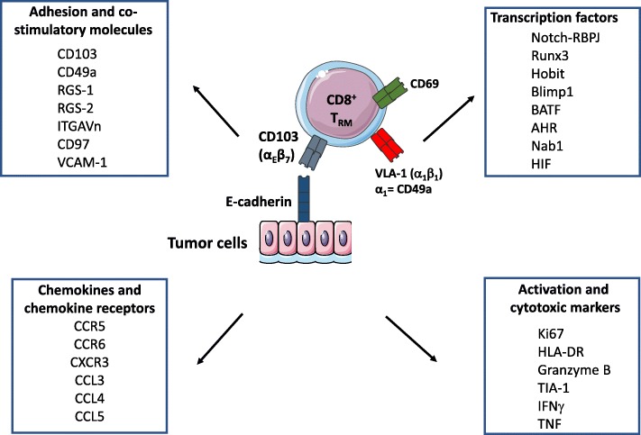
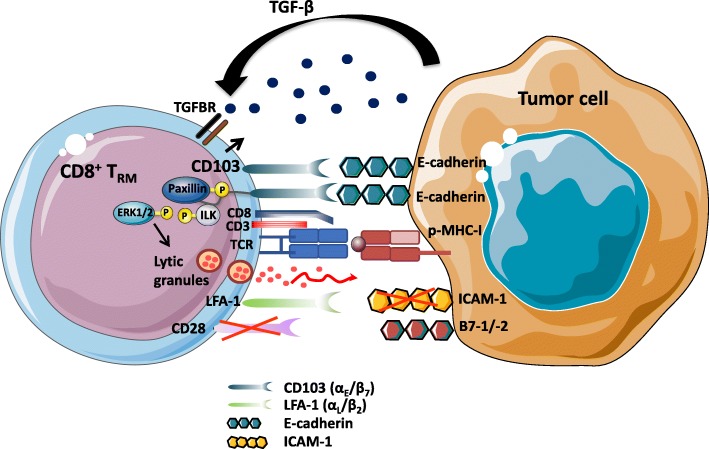
CD8 T lymphocytes are the major anti-tumor effector cells. Most cancer immunotherapeutic approaches seek to amplify cytotoxic T lymphocytes (CTL) specific to malignant cells. A recently identified subpopulation of memory CD8 T cells, named tissue-resident memory T (T) cells, persists in peripheral tissues and does not recirculate. This T-cell subset is considered an independent memory T-cell lineage with a specific profile of transcription factors, including Runx3, Notch, Hobit, Blimp1, BATF, AHR, EOMES and Tbet. It is defined by expression of CD103 (α(CD103)β) and CD49a (VLA-1 or αβ) integrins and C-type lectin CD69, which are most likely involved in retention of T cells in non-lymphoid tissues, including solid tumors. CD103 binds to the epithelial cell marker E-cadherin, thereby favoring the location and retention of T in epithelial tumor regions in close contact with malignant cells. The CD103-E-cadherin interaction is required for polarized exocytosis of lytic granules, in particular, when ICAM-1 expression on cancer cells is missing, leading to target cell death. T cells also express high levels of granzyme B, IFNγ and TNFα, supporting their cytotoxic features. Moreover, the local route of immunization targeting tissue dendritic cells (DC), and the presence of environmental factors (i.e. TGF-β, IL-33 and IL-15), promote differentiation of this T-cell subtype. In both spontaneous tumor models and engrafted tumors, natural T cells or cancer-vaccine-induced T directly control tumor growth. In line with these results, T infiltration into various human cancers, including lung cancer, are correlated with better clinical outcome in both univariate and multivariate analyses independently of CD8 T cells. T cells also predominantly express checkpoint receptors such as PD-1, CTLA-4 and Tim-3. Blockade of PD-1 with neutralizing antibodies on T cells isolated from human lung cancer promotes cytolytic activity toward autologous tumor cells. Thus, T cells appear to represent important components in tumor immune surveillance. Their induction by cancer vaccines or other immunotherapeutic approaches may be critical for the success of these treatments. Several arguments, such as their close contact with tumor cells, dominant expression of checkpoint receptors and their recognition of cancer cells, strongly suggest that they may be involved in the success of immune checkpoint inhibitors in various cancers.
CD8 T 淋巴细胞是主要的抗肿瘤效应细胞。大多数癌症免疫治疗方法都试图扩增针对恶性细胞的细胞毒性 T 淋巴细胞 (CTL)。最近发现的记忆 CD8 T 细胞亚群,称为组织驻留记忆 T (T) 细胞,存在于外周组织中,不会再循环。这种 T 细胞亚群被认为是一种独立的记忆 T 细胞谱系,具有特定的转录因子特征,包括 Runx3、Notch、Hobit、Blimp1、BATF、AHR、EOMES 和 Tbet。它通过表达 CD103(α(CD103)β)和 CD49a(VLA-1 或 αβ)整合素和 C 型凝集素 CD69 来定义,这些可能参与了非淋巴组织中 T 细胞的保留,包括实体瘤。CD103 与上皮细胞标志物 E-钙粘蛋白结合,从而有利于 T 细胞在与恶性细胞紧密接触的上皮肿瘤区域的位置和保留。CD103-E-钙粘蛋白相互作用对于裂解颗粒的极化胞吐作用是必需的,特别是当癌细胞上的 ICAM-1 表达缺失时,导致靶细胞死亡。T 细胞还表达高水平的 granzyme B、IFNγ 和 TNFα,支持其细胞毒性特征。此外,针对组织树突状细胞 (DC) 的局部免疫接种途径和环境因素(即 TGF-β、IL-33 和 IL-15)的存在促进了这种 T 细胞亚型的分化。在自发肿瘤模型和移植瘤中,天然 T 细胞或癌症疫苗诱导的 T 细胞直接控制肿瘤生长。与这些结果一致,T 细胞浸润到各种人类癌症中,包括肺癌,与单因素和多因素分析中独立于 CD8 T 细胞的更好的临床结果相关。T 细胞还主要表达检查点受体,如 PD-1、CTLA-4 和 Tim-3。用中和抗体阻断从人类肺癌分离的 T 细胞上的 PD-1 可促进对自体肿瘤细胞的细胞溶解活性。因此,T 细胞似乎是肿瘤免疫监视的重要组成部分。它们通过癌症疫苗或其他免疫治疗方法的诱导可能对这些治疗的成功至关重要。一些论点,如它们与肿瘤细胞的密切接触、检查点受体的优势表达及其对癌细胞的识别,强烈表明它们可能参与了各种癌症中免疫检查点抑制剂的成功。