• 文献检索
  • 文档翻译
  • 深度研究
  • 学术资讯
  • Suppr Zotero 插件Zotero 插件
  • 邀请有礼
  • 套餐&价格
  • 历史记录
应用&插件
Suppr Zotero 插件Zotero 插件浏览器插件Mac 客户端Windows 客户端微信小程序
定价
高级版会员购买积分包购买API积分包
服务
文献检索文档翻译深度研究API 文档MCP 服务
关于我们
关于 Suppr公司介绍联系我们用户协议隐私条款
关注我们

Suppr 超能文献

核心技术专利:CN118964589B侵权必究
粤ICP备2023148730 号-1Suppr @ 2026

文献检索

告别复杂PubMed语法,用中文像聊天一样搜索,搜遍4000万医学文献。AI智能推荐,让科研检索更轻松。

立即免费搜索

文件翻译

保留排版,准确专业,支持PDF/Word/PPT等文件格式,支持 12+语言互译。

免费翻译文档

深度研究

AI帮你快速写综述,25分钟生成高质量综述,智能提取关键信息,辅助科研写作。

立即免费体验

使用扫频源光学相干断层扫描技术对后角膜进行定量分析。

Quantification of the Posterior Cornea Using Swept Source Optical Coherence Tomography.

作者信息

Wahlig Stephen, Yam Gary Hin-Fai, Chong Wesley, Seah Xin-Yi, Kocaba Viridiana, Ang Marcus, Htoon Hla Myint, Tun Tin A, Ong Hon Shing, Mehta Jodhbir S

机构信息

Singapore Eye Research Institute (SERI), Singapore.

Duke University School of Medicine, Durham, NC, USA.

出版信息

Transl Vis Sci Technol. 2018 Sep 4;7(5):2. doi: 10.1167/tvst.7.5.2. eCollection 2018.

DOI:10.1167/tvst.7.5.2
PMID:30197834
原文链接:https://pmc.ncbi.nlm.nih.gov/articles/PMC6126962/
Abstract

PURPOSE

We define optical coherence tomography (OCT) measurement parameters of the corneal endothelium/Descemet's membrane (DM) complex and peripheral transition zone (TZ) and describe these measurements in an ethnically Chinese population.

METHODS

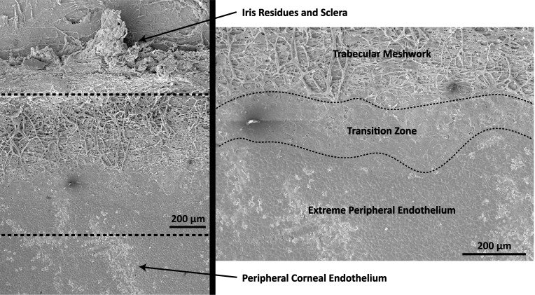
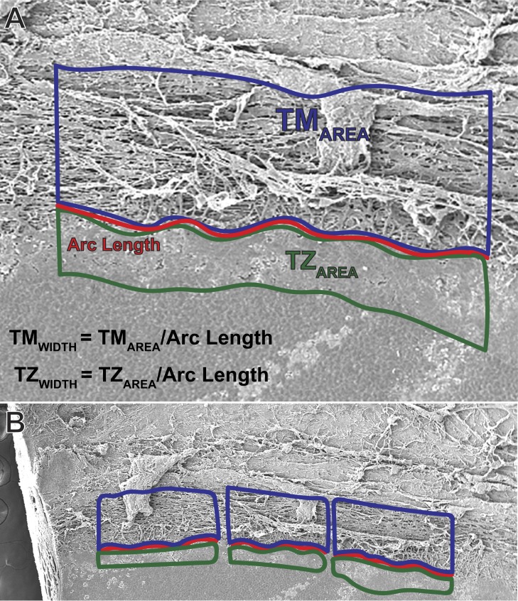
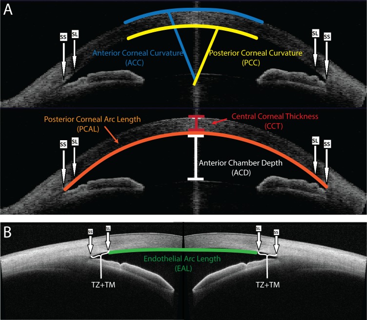
OCT images of the anterior segment and iridocorneal angle were obtained from 129 healthy Chinese subjects (129 eyes), aged 40 to 81 years. The scleral spur (SS) and Schwalbe's line (SL) were identified in each image. Endothelium/DM diameter, referred to as endothelial arc length (EAL), is the SL-to-SL distance. The SS-to-SL distance encompasses the TZ and trabecular meshwork (TM). Since the TZ cannot be visualized by OCT, a ratio of TZ-to-TZ+TM width was calculated from scanning electron microscopy (SEM) images obtained from 5 cadaveric corneas. The SS-to-SL distance was multiplied by this ratio to approximate in vivo TZ width.

RESULTS

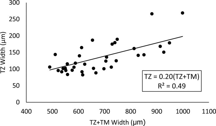
From SEM measurements, the relationship TZ = 0.20*(TZ+TM) was determined. From OCT measurements, mean EAL was 12.15 ± 0.58 mm and mean TZ width was 156 ± 20 μm. For eyes with horizontal and vertical images, vertical EAL was significantly greater than horizontal EAL ( = 0.03).

CONCLUSIONS

Corneal endothelium/DM diameter and TZ width can be obtained from OCT images. Although only combined TZ+TM is visualized on OCT, TZ width can be reasonably approximated.

TRANSLATIONAL RELEVANCE

Emerging procedures, like endothelial cell injection and DM transplantation (DMT), require accurate measurements of endothelium/DM size for preoperative planning. Size of the TZ, which may contain progenitor cells, also could contribute to endothelial regeneration in these procedures.

摘要

目的

我们定义角膜内皮/后弹力层(DM)复合体及周边移行区(TZ)的光学相干断层扫描(OCT)测量参数,并在华裔人群中描述这些测量值。

方法

获取129名年龄在40至81岁的健康华裔受试者(129只眼)眼前节和虹膜角膜角的OCT图像。在每张图像中识别巩膜突(SS)和施瓦贝线(SL)。内皮/DM直径,称为内皮弧长(EAL),是SL到SL的距离。SS到SL的距离包括TZ和小梁网(TM)。由于TZ无法通过OCT可视化,因此从5个尸体角膜获得的扫描电子显微镜(SEM)图像计算TZ与TZ+TM宽度的比值。将SS到SL的距离乘以该比值以近似体内TZ宽度。

结果

通过SEM测量,确定了TZ = 0.20×(TZ+TM)的关系。通过OCT测量,平均EAL为12.15±0.58mm,平均TZ宽度为156±20μm。对于有水平和垂直图像的眼睛,垂直EAL显著大于水平EAL(P = 0.03)。

结论

角膜内皮/DM直径和TZ宽度可从OCT图像中获得。虽然在OCT上仅能看到合并的TZ+TM,但TZ宽度可以合理地近似。

转化相关性

新兴手术,如内皮细胞注射和后弹力层移植(DMT),术前规划需要准确测量内皮/DM大小。可能含有祖细胞的TZ大小也可能有助于这些手术中的内皮再生。

https://cdn.ncbi.nlm.nih.gov/pmc/blobs/b260/6126962/a69722db5fd6/i2164-2591-7-5-2-f04.jpg
https://cdn.ncbi.nlm.nih.gov/pmc/blobs/b260/6126962/70dc68ad15e0/i2164-2591-7-5-2-f01.jpg
https://cdn.ncbi.nlm.nih.gov/pmc/blobs/b260/6126962/358faec4ed7d/i2164-2591-7-5-2-f02.jpg
https://cdn.ncbi.nlm.nih.gov/pmc/blobs/b260/6126962/358fa3c5f3f8/i2164-2591-7-5-2-f03.jpg
https://cdn.ncbi.nlm.nih.gov/pmc/blobs/b260/6126962/a69722db5fd6/i2164-2591-7-5-2-f04.jpg

相似文献

1
Quantification of the Posterior Cornea Using Swept Source Optical Coherence Tomography.使用扫频源光学相干断层扫描技术对后角膜进行定量分析。
Transl Vis Sci Technol. 2018 Sep 4;7(5):2. doi: 10.1167/tvst.7.5.2. eCollection 2018.
2
Assessment of trabecular meshwork width using swept source optical coherence tomography.应用扫频源光学相干断层扫描评估小梁网宽度。
Graefes Arch Clin Exp Ophthalmol. 2013 Jun;251(6):1587-92. doi: 10.1007/s00417-013-2285-8. Epub 2013 Feb 23.
3
Anterior chamber angle measurements using Schwalbe's line with high-resolution fourier-domain optical coherence tomography.应用高分辨率傅里叶域光学相干断层扫描技术的 Schwalbe 线前房角测量。
J Glaucoma. 2013 Dec;22(9):684-8. doi: 10.1097/IJG.0b013e318264b921.
4
Comparison of spectral domain and swept source optical coherence tomography for angle assessment of Chinese elderly subjects.光谱域光学相干断层扫描与扫频源光学相干断层扫描用于中国老年受试者房角评估的比较
BMC Ophthalmol. 2019 Jul 8;19(1):142. doi: 10.1186/s12886-019-1145-7.
5
Imaging of the iridocorneal angle with the RTVue spectral domain optical coherence tomography.应用 RTVue 谱域光学相干断层扫描仪对眼前房角成像。
Invest Ophthalmol Vis Sci. 2012 Apr 2;53(4):1710-3. doi: 10.1167/iovs.11-8159.
6
Anterior chamber angle imaging with swept-source optical coherence tomography: detecting the scleral spur, Schwalbe's Line, and Schlemm's Canal.前房角扫频源光学相干断层扫描成像:检测巩膜突、Schwalbe 线和 Schlemm 管。
J Glaucoma. 2013 Aug;22(6):468-72. doi: 10.1097/IJG.0b013e31824485fa.
7
Comparison of the trabecular meshwork length between open and closed angle with evaluation of the scleral spur location.比较开角和闭角的小梁网长度,并评估巩膜突的位置。
Sci Rep. 2019 May 2;9(1):6857. doi: 10.1038/s41598-019-43315-2.
8
Characterization of Human Transition Zone Reveals a Putative Progenitor-Enriched Niche of Corneal Endothelium.人眼过渡区细胞的特征揭示了角膜内皮细胞中富含前体细胞的潜在龛位
Cells. 2019 Oct 12;8(10):1244. doi: 10.3390/cells8101244.
9
Identification of iridocorneal angle structures assessed by Fourier domain optical coherence tomography.傅里叶域光学相干断层扫描评估的虹膜角膜角结构的识别
Arch Soc Esp Oftalmol. 2016 Feb;91(2):74-80. doi: 10.1016/j.oftal.2015.10.007. Epub 2015 Dec 2.
10
Stem cell markers in the human posterior limbus and corneal endothelium of unwounded and wounded corneas.未受伤和受伤角膜的人后弹力层及角膜内皮中的干细胞标志物
Mol Vis. 2007 Oct 18;13:1984-2000.

引用本文的文献

1
Anatomic Relationship Among Descemet's Membrane, Trabecular Meshwork, Scleral Spur, and Ciliary Muscle.Descemet膜、小梁网、巩膜突和睫状肌之间的解剖关系。
Invest Ophthalmol Vis Sci. 2025 Feb 3;66(2):8. doi: 10.1167/iovs.66.2.8.
2
Posterior corneoscleral limbus: Architecture, stem cells, and clinical implications.后角巩膜缘:结构、干细胞及临床意义。
Prog Retin Eye Res. 2023 Sep;96:101192. doi: 10.1016/j.preteyeres.2023.101192. Epub 2023 Jun 29.
3
Regenerative capacity of the corneal transition zone for endothelial cell therapy.

本文引用的文献

1
Regenerative Therapy for Fuchs Endothelial Corneal Dystrophy.富克斯内皮性角膜营养不良的再生治疗
Cornea. 2018 Apr;37(4):523-527. doi: 10.1097/ICO.0000000000001518.
2
Evolving therapies for Fuchs' endothelial dystrophy.针对富克斯角膜内皮营养不良的不断发展的治疗方法。
Regen Med. 2018 Jan;13(1):97-115. doi: 10.2217/rme-2017-0081. Epub 2018 Jan 23.
3
Feasibility of cell-based therapy combined with descemetorhexis for treating Fuchs endothelial corneal dystrophy in rabbit model.基于细胞的疗法联合后弹力层撕除术治疗兔模型中富克斯内皮性角膜营养不良的可行性。
角膜过渡区的内皮细胞治疗再生能力。
Stem Cell Res Ther. 2020 Dec 4;11(1):523. doi: 10.1186/s13287-020-02046-2.
4
Characterization of Human Transition Zone Reveals a Putative Progenitor-Enriched Niche of Corneal Endothelium.人眼过渡区细胞的特征揭示了角膜内皮细胞中富含前体细胞的潜在龛位
Cells. 2019 Oct 12;8(10):1244. doi: 10.3390/cells8101244.
PLoS One. 2018 Jan 16;13(1):e0191306. doi: 10.1371/journal.pone.0191306. eCollection 2018.
4
Wide-field contact specular microscopy analysis of corneal endothelium post trabeculectomy.小梁切除术后角膜内皮细胞的广角接触式镜面显微镜分析
Graefes Arch Clin Exp Ophthalmol. 2018 Apr;256(4):751-757. doi: 10.1007/s00417-017-3889-1. Epub 2018 Jan 4.
5
Directed differentiation of periocular mesenchyme from human embryonic stem cells.诱导人眼周间质细胞从人胚胎干细胞分化。
Differentiation. 2018 Jan-Feb;99:62-69. doi: 10.1016/j.diff.2017.11.003. Epub 2017 Nov 16.
6
Regulatory Compliant Tissue-Engineered Human Corneal Endothelial Grafts Restore Corneal Function of Rabbits with Bullous Keratopathy.经监管批准的组织工程化人眼角膜内皮移植物可恢复大泡性角膜病变兔的角膜功能。
Sci Rep. 2017 Oct 26;7(1):14149. doi: 10.1038/s41598-017-14723-z.
7
Predictive Factors for Corneal Clearance After Descemetorhexis Without Endothelial Keratoplasty.无内皮角膜移植的后弹力层撕除术后角膜透明性的预测因素
Cornea. 2018 Feb;37(2):137-140. doi: 10.1097/ICO.0000000000001427.
8
Trabecular Meshwork Height in Primary Open-Angle Glaucoma Versus Primary Angle-Closure Glaucoma.原发性开角型青光眼与原发性闭角型青光眼的小梁网高度
Am J Ophthalmol. 2017 Nov;183:42-47. doi: 10.1016/j.ajo.2017.08.018. Epub 2017 Sep 6.
9
Allogeneic Descemet's Membrane Transplantation Enhances Corneal Endothelial Monolayer Formation and Restores Functional Integrity Following Descemet's Stripping.同种异体Descemet膜移植可促进角膜内皮单层形成,并在Descemet膜剥除后恢复功能完整性。
Invest Ophthalmol Vis Sci. 2017 Aug 1;58(10):4249-4260. doi: 10.1167/iovs.17-22106.
10
Application of Rho Kinase Inhibitors for the Treatment of Corneal Endothelial Diseases.Rho激酶抑制剂在角膜内皮疾病治疗中的应用。
J Ophthalmol. 2017;2017:2646904. doi: 10.1155/2017/2646904. Epub 2017 Jul 2.