• 文献检索
  • 文档翻译
  • 深度研究
  • 学术资讯
  • Suppr Zotero 插件Zotero 插件
  • 邀请有礼
  • 套餐&价格
  • 历史记录
应用&插件
Suppr Zotero 插件Zotero 插件浏览器插件Mac 客户端Windows 客户端微信小程序
定价
高级版会员购买积分包购买API积分包
服务
文献检索文档翻译深度研究API 文档MCP 服务
关于我们
关于 Suppr公司介绍联系我们用户协议隐私条款
关注我们

Suppr 超能文献

核心技术专利:CN118964589B侵权必究
粤ICP备2023148730 号-1Suppr @ 2026

文献检索

告别复杂PubMed语法,用中文像聊天一样搜索,搜遍4000万医学文献。AI智能推荐,让科研检索更轻松。

立即免费搜索

文件翻译

保留排版,准确专业,支持PDF/Word/PPT等文件格式,支持 12+语言互译。

免费翻译文档

深度研究

AI帮你快速写综述,25分钟生成高质量综述,智能提取关键信息,辅助科研写作。

立即免费体验

角膜过渡区的内皮细胞治疗再生能力。

Regenerative capacity of the corneal transition zone for endothelial cell therapy.

机构信息

Tissue Engineering and Cell Therapy Group, Singapore Eye Research Institute, 20 College Road, The Academia, Discovery Tower Level 6, Singapore, 169856, Singapore.

Singapore National Eye Centre, Singapore, 168751, Singapore.

出版信息

Stem Cell Res Ther. 2020 Dec 4;11(1):523. doi: 10.1186/s13287-020-02046-2.

DOI:10.1186/s13287-020-02046-2
PMID:33276809
原文链接:https://pmc.ncbi.nlm.nih.gov/articles/PMC7716425/
Abstract

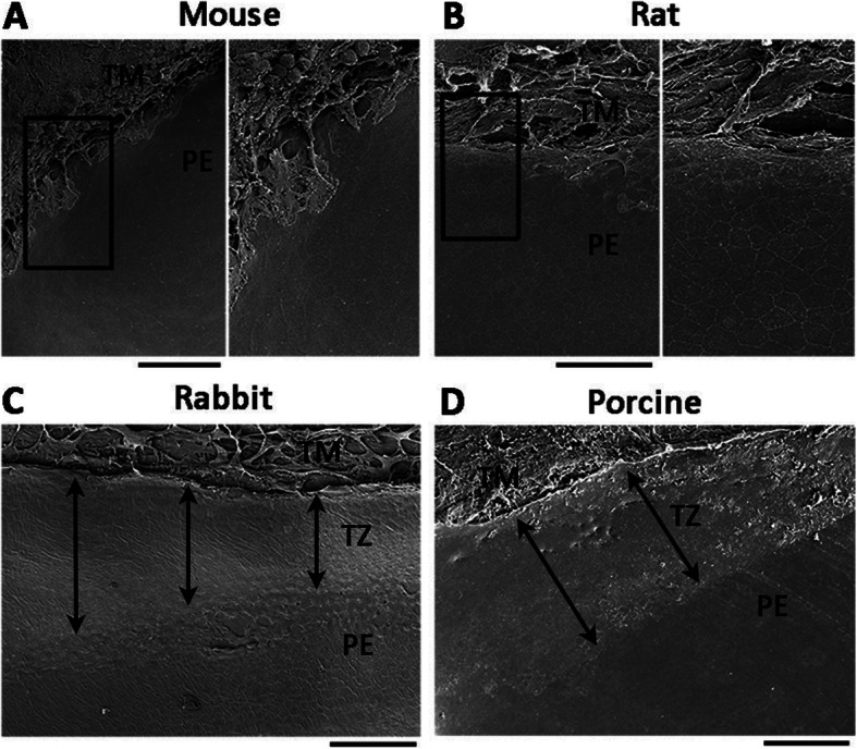
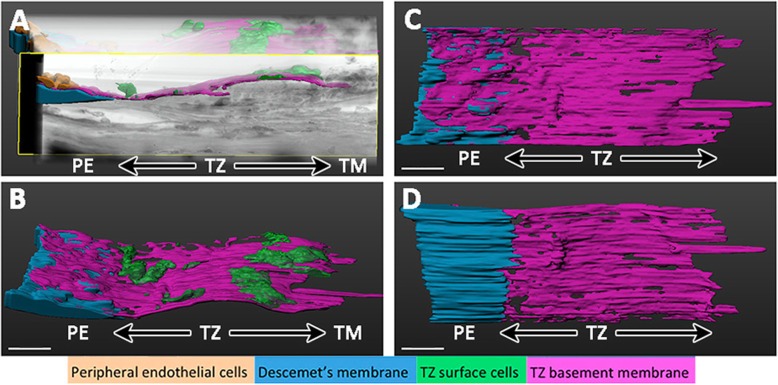
The corneal endothelium located on the posterior corneal surface is responsible for regulating stromal hydration. This is contributed by a monolayer of corneal endothelial cells (CECs), which are metabolically active in a continuous fluid-coupled efflux of ions from the corneal stroma into the aqueous humor, preventing stromal over-hydration and preserving the orderly arrangement of stromal collagen fibrils, which is essential for corneal transparency. Mature CECs do not have regenerative capacity and cell loss due to aging and diseases results in irreversible stromal edema and a loss of corneal clarity. The current gold standard of treatment for this worldwide blindness caused by corneal endothelial failure is the corneal transplantation using cadaveric donor corneas. The top indication is Fuchs corneal endothelial dystrophy/degeneration, which represents 39% of all corneal transplants performed. However, the global shortage of transplantable donor corneas has restricted the treatment outcomes, hence instigating a need to research for alternative therapies. One such avenue is the CEC regeneration from endothelial progenitors, which have been identified in the peripheral endothelium and the adjacent transition zone. This review examines the evidence supporting the existence of endothelial progenitors in the posterior limbus and summarizes the existing knowledge on the microanatomy of the transitional zone. We give an overview of the isolation and ex vivo propagation of human endothelial progenitors in the transition zone, and their growth and differentiation capacity to the corneal endothelium. Transplanting these bioengineered constructs into in vivo models of corneal endothelial degeneration will prove the efficacy and viability, and the long-term maintenance of functional endothelium. This will develop a novel regenerative therapy for the management of corneal endothelial diseases.

摘要

角膜内皮位于后角膜表面,负责调节基质的水化。这是由一层角膜内皮细胞(CEC)完成的,这些细胞在代谢上是活跃的,它们将离子从角膜基质连续不断地泵出到房水中,从而防止基质过度水化,并维持基质胶原纤维的有序排列,这对角膜透明性至关重要。成熟的 CEC 没有再生能力,由于衰老和疾病导致的细胞丢失会导致不可逆的基质水肿和角膜清晰度丧失。目前,针对由角膜内皮功能衰竭引起的这种全球失明的治疗金标准是使用尸体供体角膜进行角膜移植。最主要的适应证是 Fuchs 角膜内皮营养不良/变性,占所有角膜移植的 39%。然而,全球可用于移植的供体角膜短缺限制了治疗效果,因此需要研究替代疗法。其中一种方法是从内皮祖细胞中再生 CEC,这些内皮祖细胞已在周边内皮和相邻的过渡区中被发现。这篇综述检查了支持后缘部存在内皮祖细胞的证据,并总结了过渡区的微观解剖学的现有知识。我们概述了人内皮祖细胞在过渡区的分离和体外培养,以及它们向角膜内皮生长和分化的能力。将这些生物工程构建体移植到角膜内皮变性的体内模型中,将证明其功效和可行性,以及功能性内皮的长期维持。这将为角膜内皮疾病的管理开发一种新的再生治疗方法。

https://cdn.ncbi.nlm.nih.gov/pmc/blobs/9fef/7716425/70d9dcfe42ee/13287_2020_2046_Fig3_HTML.jpg
https://cdn.ncbi.nlm.nih.gov/pmc/blobs/9fef/7716425/2570758cfccb/13287_2020_2046_Fig1_HTML.jpg
https://cdn.ncbi.nlm.nih.gov/pmc/blobs/9fef/7716425/47e75c77172f/13287_2020_2046_Fig2_HTML.jpg
https://cdn.ncbi.nlm.nih.gov/pmc/blobs/9fef/7716425/70d9dcfe42ee/13287_2020_2046_Fig3_HTML.jpg
https://cdn.ncbi.nlm.nih.gov/pmc/blobs/9fef/7716425/2570758cfccb/13287_2020_2046_Fig1_HTML.jpg
https://cdn.ncbi.nlm.nih.gov/pmc/blobs/9fef/7716425/47e75c77172f/13287_2020_2046_Fig2_HTML.jpg
https://cdn.ncbi.nlm.nih.gov/pmc/blobs/9fef/7716425/70d9dcfe42ee/13287_2020_2046_Fig3_HTML.jpg

相似文献

1
Regenerative capacity of the corneal transition zone for endothelial cell therapy.角膜过渡区的内皮细胞治疗再生能力。
Stem Cell Res Ther. 2020 Dec 4;11(1):523. doi: 10.1186/s13287-020-02046-2.
2
Specular Microscopy镜面显微镜检查
3
Stem cell markers in the human posterior limbus and corneal endothelium of unwounded and wounded corneas.未受伤和受伤角膜的人后弹力层及角膜内皮中的干细胞标志物
Mol Vis. 2007 Oct 18;13:1984-2000.
4
Characterization of Human Transition Zone Reveals a Putative Progenitor-Enriched Niche of Corneal Endothelium.人眼过渡区细胞的特征揭示了角膜内皮细胞中富含前体细胞的潜在龛位
Cells. 2019 Oct 12;8(10):1244. doi: 10.3390/cells8101244.
5
Proliferative capacity of the corneal endothelium.角膜内皮细胞的增殖能力。
Prog Retin Eye Res. 2003 May;22(3):359-89. doi: 10.1016/s1350-9462(02)00065-4.
6
In Vitro Expansion of Corneal Endothelial Cells for Transplantation.体外扩增角膜内皮细胞用于移植。
Methods Mol Biol. 2020;2145:17-27. doi: 10.1007/978-1-0716-0599-8_2.
7
[Transplantation of corneal endothelial cells].[角膜内皮细胞移植]
Nippon Ganka Gakkai Zasshi. 2002 Dec;106(12):805-35; discussion 836.
8
Generation and Feasibility Assessment of a New Vehicle for Cell-Based Therapy for Treating Corneal Endothelial Dysfunction.用于治疗角膜内皮功能障碍的基于细胞疗法的新型载体的生成及可行性评估
PLoS One. 2016 Jun 29;11(6):e0158427. doi: 10.1371/journal.pone.0158427. eCollection 2016.
9
Characterization and Prospective of Human Corneal Endothelial Progenitors.人角膜内皮祖细胞的特性与前景
Int J Med Sci. 2017 Jun 30;14(8):705-710. doi: 10.7150/ijms.19018. eCollection 2017.
10
[Endothelial cell transplantation and growth behavior of the human corneal endothelium].[人角膜内皮细胞移植与生长行为]
Ophthalmologe. 1999 Sep;96(9):555-62. doi: 10.1007/s003470050452.

引用本文的文献

1
Decoding the Cornea-Glaucoma Association: Evidence From Mendelian Randomization.解码角膜-青光眼关联:孟德尔随机化研究证据
Invest Ophthalmol Vis Sci. 2025 Jul 1;66(9):22. doi: 10.1167/iovs.66.9.22.
2
Current concepts and recent trends in endothelial keratoplasty.内皮角膜移植术的当前概念与近期趋势
World J Transplant. 2025 Jun 18;15(2):102507. doi: 10.5500/wjt.v15.i2.102507.
3
Anatomic Relationship Among Descemet's Membrane, Trabecular Meshwork, Scleral Spur, and Ciliary Muscle.Descemet膜、小梁网、巩膜突和睫状肌之间的解剖关系。

本文引用的文献

1
Organogenesis and distribution of the ocular lymphatic vessels in the anterior eye.眼前部眼淋巴血管的发生和分布。
JCI Insight. 2020 Jul 9;5(13):135121. doi: 10.1172/jci.insight.135121.
2
Phenotypic and functional characterization of corneal endothelial cells during in vitro expansion.体外扩增过程中角膜内皮细胞的表型和功能特征。
Sci Rep. 2020 May 4;10(1):7402. doi: 10.1038/s41598-020-64311-x.
3
Successful culture of human transition zone cells.成功培养人过渡区细胞。
Invest Ophthalmol Vis Sci. 2025 Feb 3;66(2):8. doi: 10.1167/iovs.66.2.8.
4
Biomedical Application of MSCs in Corneal Regeneration and Repair.间充质干细胞在角膜再生与修复中的生物医学应用
Int J Mol Sci. 2025 Jan 15;26(2):695. doi: 10.3390/ijms26020695.
5
The Role of Mesenchymal Stem Cells for Corneal Endothelial Regeneration: A Systematic Review.间充质干细胞在角膜内皮再生中的作用:一项系统综述。
Rambam Maimonides Med J. 2024 Oct 28;15(4):e0017. doi: 10.5041/RMMJ.10531.
6
Isolation, Culture, and Quality Assessment of Clinical-Grade Corneal Stromal Stem Cells.临床级角膜基质干细胞的分离、培养和质量评估。
Methods Mol Biol. 2025;2848:3-23. doi: 10.1007/978-1-0716-4087-6_1.
7
Increased Risk of Glaucoma in Fuchs Endothelial Corneal Dystrophy Is Independent of TCF4 Trinucleotide Repeat Expansion.富克斯内皮性角膜营养不良患者青光眼风险增加与TCF4三核苷酸重复序列扩增无关。
Cornea. 2024 Sep 3;44(8):961-964. doi: 10.1097/ICO.0000000000003695.
8
Safety of intraoperative autologous plasma incubation of corneal grafts for reducing endothelial cell loss: a pilot study.术中自体血浆孵育角膜移植片以减少内皮细胞丢失的安全性:一项初步研究。
Front Med (Lausanne). 2024 Aug 12;11:1368117. doi: 10.3389/fmed.2024.1368117. eCollection 2024.
9
Microstructure of the corneal endothelial transition zone in different laboratory animals.不同实验动物角膜内皮过渡区的微观结构。
Mol Vis. 2024 Mar 17;30:107-113. eCollection 2024.
10
Absence of the Klotho Function Causes Cornea Degeneration with Specific Features Resembling Fuchs Endothelial Corneal Dystrophy and Bullous Keratopathy.Klotho功能缺失导致角膜变性,具有类似Fuchs内皮角膜营养不良和大疱性角膜病变的特定特征。
Biology (Basel). 2024 Feb 20;13(3):133. doi: 10.3390/biology13030133.
Clin Exp Ophthalmol. 2020 Jul;48(5):689-700. doi: 10.1111/ceo.13756. Epub 2020 Apr 28.
4
Optimisation of Storage and Transportation Conditions of Cultured Corneal Endothelial Cells for Cell Replacement Therapy.优化培养的角膜内皮细胞的储存和运输条件用于细胞替代治疗。
Sci Rep. 2020 Feb 3;10(1):1681. doi: 10.1038/s41598-020-58700-5.
5
Replacing what's lost: a new era of stem cell therapy for Parkinson's disease.替代所失之物:帕金森病干细胞治疗的新时代。
Transl Neurodegener. 2020 Jan 7;9:2. doi: 10.1186/s40035-019-0180-x. eCollection 2020.
6
Göttingen Minipig is not a Suitable Animal Model for Testing of Tissue-Engineered Corneal Endothelial Cell-Carrier Sheets and for Endothelial Keratoplasty.哥廷根小型猪不适合作为组织工程角膜内皮细胞-载体片的测试和内皮角膜移植的动物模型。
Curr Eye Res. 2020 Aug;45(8):945-949. doi: 10.1080/02713683.2019.1706747. Epub 2020 Jan 6.
7
Research and therapy with induced pluripotent stem cells (iPSCs): social, legal, and ethical considerations.诱导多能干细胞(iPSCs)的研究和治疗:社会、法律和伦理方面的考虑。
Stem Cell Res Ther. 2019 Nov 21;10(1):341. doi: 10.1186/s13287-019-1455-y.
8
Treatment of corneal endothelial damage in a rabbit model with a bioengineered graft using human decellularized corneal lamina and cultured human corneal endothelium.使用人去细胞角膜板层和培养的人角膜内皮细胞构建的生物工程移植物治疗兔角膜内皮损伤模型。
PLoS One. 2019 Nov 21;14(11):e0225480. doi: 10.1371/journal.pone.0225480. eCollection 2019.
9
Coexistence of Congenital Hereditary Endothelial Dystrophy and Fuchs Endothelial Corneal Dystrophy Associated With SLC4A11 Mutations in Affected Families.先天性遗传性血管内皮营养不良与 Fuchs 内皮角膜营养不良共存,并与 SLC4A11 基因突变相关,在受影响的家族中。
Cornea. 2020 Mar;39(3):354-357. doi: 10.1097/ICO.0000000000002183.
10
Characterization of Human Transition Zone Reveals a Putative Progenitor-Enriched Niche of Corneal Endothelium.人眼过渡区细胞的特征揭示了角膜内皮细胞中富含前体细胞的潜在龛位
Cells. 2019 Oct 12;8(10):1244. doi: 10.3390/cells8101244.