• 文献检索
  • 文档翻译
  • 深度研究
  • 学术资讯
  • Suppr Zotero 插件Zotero 插件
  • 邀请有礼
  • 套餐&价格
  • 历史记录
应用&插件
Suppr Zotero 插件Zotero 插件浏览器插件Mac 客户端Windows 客户端微信小程序
定价
高级版会员购买积分包购买API积分包
服务
文献检索文档翻译深度研究API 文档MCP 服务
关于我们
关于 Suppr公司介绍联系我们用户协议隐私条款
关注我们

Suppr 超能文献

核心技术专利:CN118964589B侵权必究
粤ICP备2023148730 号-1Suppr @ 2026

文献检索

告别复杂PubMed语法,用中文像聊天一样搜索,搜遍4000万医学文献。AI智能推荐,让科研检索更轻松。

立即免费搜索

文件翻译

保留排版,准确专业,支持PDF/Word/PPT等文件格式,支持 12+语言互译。

免费翻译文档

深度研究

AI帮你快速写综述,25分钟生成高质量综述,智能提取关键信息,辅助科研写作。

立即免费体验

光谱域光学相干断层扫描与扫频源光学相干断层扫描用于中国老年受试者房角评估的比较

Comparison of spectral domain and swept source optical coherence tomography for angle assessment of Chinese elderly subjects.

作者信息

Qiao Yunsheng, Tan Chen, Zhang Min, Sun Xinghuai, Chen Junyi

机构信息

Department of Ophthalmology & Visual Science, Eye and ENT Hospital of Fudan University, 83 Fenyang Rd, Shanghai, 200031, China.

NHC Key Laboratory of Myopia (Fudan University), Key Laboratory of Myopia, Chinese Academy of Medical Sciences and Key Laboratory of Visual Impairment and Restoration of Shanghai, Shanghai, 200031, China.

出版信息

BMC Ophthalmol. 2019 Jul 8;19(1):142. doi: 10.1186/s12886-019-1145-7.

DOI:10.1186/s12886-019-1145-7
PMID:31286869
原文链接:https://pmc.ncbi.nlm.nih.gov/articles/PMC6615428/
Abstract

BACKGROUND

This comparative study aimed to demonstrate the differences between swept source OCT (SS-OCT) (1310 nm) and spectral domain OCT (SD-OCT) (840 nm) for the identification and measurement of anterior chamber angle (ACA) structures.

METHODS

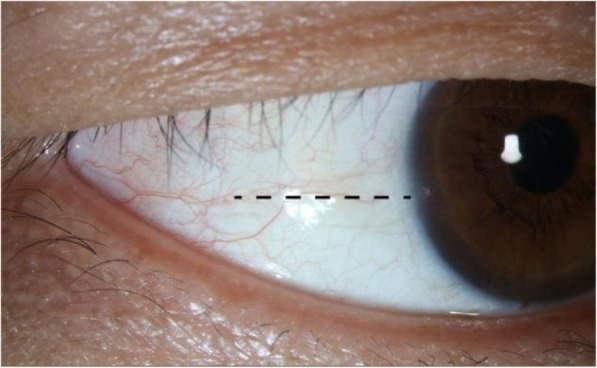
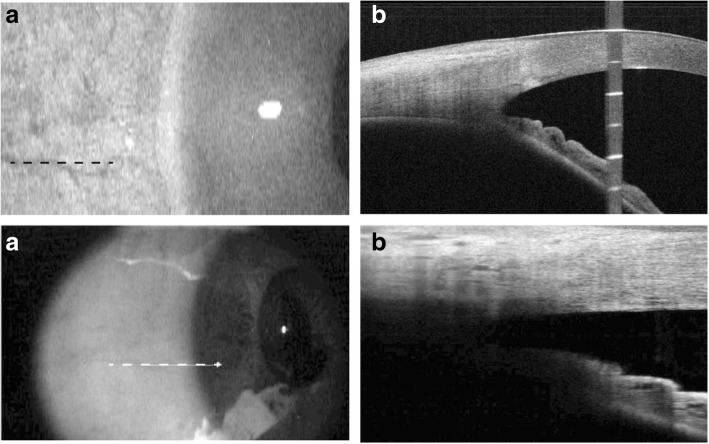
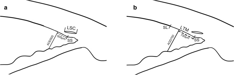
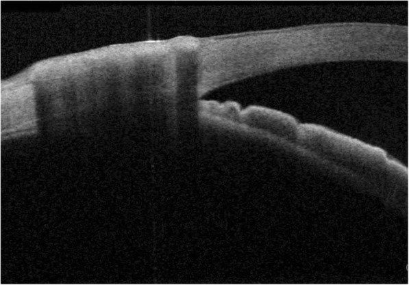
Sixty seven eyes from 67 healthy subjects underwent ACA imaging at the nasal and temporal sides using SS-OCT and SD-OCT with different wavelength (Tomey, 1310 nm and RTvue, 840 nm). Images were evaluated for the ability to distinguish angle structures including the Schwalbe's line (SL), the Schlemm's canal (SC) and the scleral spur (SS). The length of trabecular meshwork (LTM), the angle-opening distance (AOD500 and AOD750) and the length of Schlemm's canal (LSC) were also measured.

RESULTS

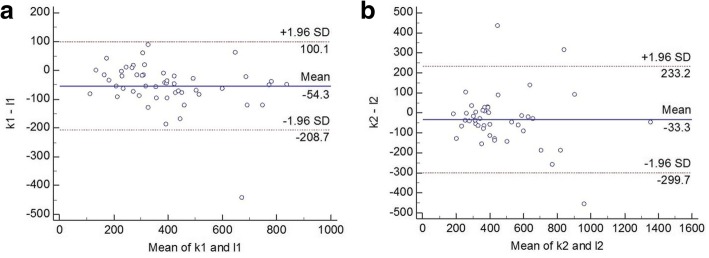
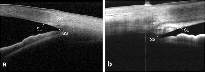
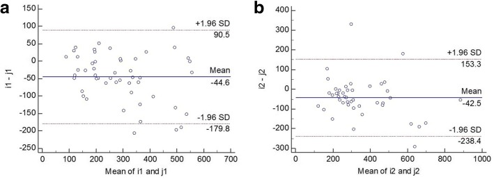
The nasal identification rate for SL, SC and SS were 91.04%/89.55%, 50.75%/40.30% and 100.0%/74.63% (SS-OCT/SD-OCT), respectively. The temporal identification rate for SL, SC and SS were 86.57%/91.04%, 68.66%/70.15% and 100.0%/65.67% (SS-OCT/SD-OCT), respectively. Differences between SS-OCT and SD-OCT were found in terms of the visualization of the SS. With respect to the measurements of angle, the evaluation of LTM at the nasal side, LSC at the temporal side and AOD500/750 at both sides showed significant difference between the two devices. However, there existed good correlation between the AOD500/750 measured by SS-OCT and SD-OCT (Spearman's rank correlation coefficient > 0.8, p < 0.000).

CONCLUSIONS

SS-OCT displayed a better performance in detecting deeper structures of the angle such as the SS. However, for discriminating structures lying in transparent or semi-transparent tissue such as the SL and the SC, the two devices showed good consistency. Although SS-OCT and SD-OCT demonstrated high correlation for angle measurement (AOD500/750), their agreement was poor.

摘要

背景

本对比研究旨在证明扫频光学相干断层扫描(SS-OCT)(1310 nm)和谱域光学相干断层扫描(SD-OCT)(840 nm)在眼前房角(ACA)结构识别和测量方面的差异。

方法

对67名健康受试者的67只眼睛使用不同波长的SS-OCT和SD-OCT(Tomey,1310 nm和RTvue,840 nm)在鼻侧和颞侧进行ACA成像。评估图像区分包括施瓦贝线(SL)、巩膜静脉窦(SC)和巩膜突(SS)在内的房角结构的能力。还测量了小梁网长度(LTM)、房角开放距离(AOD500和AOD750)以及巩膜静脉窦长度(LSC)。

结果

SL、SC和SS的鼻侧识别率分别为91.04%/89.55%、50.75%/40.30%和100.0%/74.63%(SS-OCT/SD-OCT)。SL、SC和SS的颞侧识别率分别为86.57%/91.04%、68.66%/70.15%和100.0%/65.67%(SS-OCT/SD-OCT)。在SS的可视化方面发现了SS-OCT和SD-OCT之间的差异。关于房角测量,在鼻侧LTM、颞侧LSC以及两侧的AOD500/750评估中,两种设备之间存在显著差异。然而,SS-OCT和SD-OCT测量的AOD500/750之间存在良好的相关性(斯皮尔曼等级相关系数>0.8,p<0.000)。

结论

SS-OCT在检测房角较深结构如SS方面表现更好。然而,对于区分位于透明或半透明组织中的结构如SL和SC,两种设备显示出良好的一致性。尽管SS-OCT和SD-OCT在房角测量(AOD500/750)方面显示出高度相关性,但它们的一致性较差。

https://cdn.ncbi.nlm.nih.gov/pmc/blobs/e4b1/6615428/22605027aee8/12886_2019_1145_Fig7_HTML.jpg
https://cdn.ncbi.nlm.nih.gov/pmc/blobs/e4b1/6615428/05aaf7bd0ae8/12886_2019_1145_Fig1_HTML.jpg
https://cdn.ncbi.nlm.nih.gov/pmc/blobs/e4b1/6615428/5675acb02e53/12886_2019_1145_Fig2_HTML.jpg
https://cdn.ncbi.nlm.nih.gov/pmc/blobs/e4b1/6615428/aebb9948d055/12886_2019_1145_Fig3_HTML.jpg
https://cdn.ncbi.nlm.nih.gov/pmc/blobs/e4b1/6615428/248f189adc30/12886_2019_1145_Fig4_HTML.jpg
https://cdn.ncbi.nlm.nih.gov/pmc/blobs/e4b1/6615428/b0b1c241542c/12886_2019_1145_Fig5_HTML.jpg
https://cdn.ncbi.nlm.nih.gov/pmc/blobs/e4b1/6615428/65041173a583/12886_2019_1145_Fig6_HTML.jpg
https://cdn.ncbi.nlm.nih.gov/pmc/blobs/e4b1/6615428/22605027aee8/12886_2019_1145_Fig7_HTML.jpg

相似文献

1
Comparison of spectral domain and swept source optical coherence tomography for angle assessment of Chinese elderly subjects.光谱域光学相干断层扫描与扫频源光学相干断层扫描用于中国老年受试者房角评估的比较
BMC Ophthalmol. 2019 Jul 8;19(1):142. doi: 10.1186/s12886-019-1145-7.
2
Comparison of two spectral domain optical coherence tomography devices for angle-closure assessment.两种用于评估闭角型青光眼的谱域光学相干断层扫描仪的比较。
Invest Ophthalmol Vis Sci. 2012 Aug 3;53(9):5131-6. doi: 10.1167/iovs.12-10132.
3
Anterior chamber angle imaging with swept-source optical coherence tomography: detecting the scleral spur, Schwalbe's Line, and Schlemm's Canal.前房角扫频源光学相干断层扫描成像:检测巩膜突、Schwalbe 线和 Schlemm 管。
J Glaucoma. 2013 Aug;22(6):468-72. doi: 10.1097/IJG.0b013e31824485fa.
4
Assessment of trabecular meshwork width using swept source optical coherence tomography.应用扫频源光学相干断层扫描评估小梁网宽度。
Graefes Arch Clin Exp Ophthalmol. 2013 Jun;251(6):1587-92. doi: 10.1007/s00417-013-2285-8. Epub 2013 Feb 23.
5
Spectral domain optical coherence tomography imaging of the aqueous outflow structures in normal participants of the EPIC-Norfolk Eye Study.EPIC-Norfolk 眼部研究中正常参与者房水流出结构的光谱域光相干断层扫描成像。
Br J Ophthalmol. 2013 Feb;97(2):189-95. doi: 10.1136/bjophthalmol-2012-302147. Epub 2012 Nov 30.
6
Anterior chamber angle measurements using Schwalbe's line with high-resolution fourier-domain optical coherence tomography.应用高分辨率傅里叶域光学相干断层扫描技术的 Schwalbe 线前房角测量。
J Glaucoma. 2013 Dec;22(9):684-8. doi: 10.1097/IJG.0b013e318264b921.
7
Understanding diagnostic disagreement in angle closure assessment between anterior segment optical coherence tomography and gonioscopy.了解前节光学相干断层扫描和房角镜检查在闭角型青光眼评估中的诊断分歧。
Br J Ophthalmol. 2020 Jun;104(6):795-799. doi: 10.1136/bjophthalmol-2019-314672. Epub 2019 Sep 6.
8
COMPARISON OF ANTERIOR SEGMENT OPTICAL COHERENCE TOMOGRAPHY PARAMETERS BETWEEN CENTRAL RETINAL VEIN OCCLUSION AND NORMAL EYES: Is Primary Angle Closure a Risk Factor for Central Retinal Vein Occlusion?视网膜中央静脉阻塞与正常眼的眼前节光学相干断层扫描参数比较:原发性闭角型青光眼是视网膜中央静脉阻塞的危险因素吗?
Retina. 2015 Sep;35(9):1795-9. doi: 10.1097/IAE.0000000000000549.
9
Assessment of Circumferential Angle Closure with Swept-Source Optical Coherence Tomography: a Community Based Study.应用扫频源光学相干断层扫描评估周边角关闭:基于社区的研究。
Am J Ophthalmol. 2019 Mar;199:133-139. doi: 10.1016/j.ajo.2018.11.015. Epub 2018 Nov 28.
10
Evaluation of the Anterior Segment Angle-to-Angle Scan of Cirrus High-Definition Optical Coherence Tomography and Comparison With Gonioscopy and With the Visante OCT.Cirrus高清光学相干断层扫描眼前节角对角度扫描的评估及其与前房角镜检查和Visante OCT的比较。
Invest Ophthalmol Vis Sci. 2017 Jan 1;58(1):59-64. doi: 10.1167/iovs.16-20886.

引用本文的文献

1
Utility of Swept-Source Anterior-Segment OCT as an In-Office Biomarker for Early Childhood Glaucoma.扫频源眼前节光学相干断层扫描作为儿童青光眼门诊生物标志物的效用
JAMA Ophthalmol. 2025 May 22. doi: 10.1001/jamaophthalmol.2025.1009.
2
In vivo analysis of iridocorneal angle parameters with spectral-domain optical coherence tomography in children with Neurofibromatosis type 1.利用光谱域光学相干断层扫描技术对1型神经纤维瘤病患儿的虹膜角膜角参数进行体内分析。
Eye (Lond). 2025 May 14. doi: 10.1038/s41433-025-03840-z.
3
Advances in swept-source optical coherence tomography and optical coherence tomography angiography.

本文引用的文献

1
Repeatability, reproducibility, agreement characteristics of 2 SD-OCT devices for anterior chamber angle measurements.两种扫频光学相干断层扫描(SD-OCT)设备用于前房角测量的重复性、再现性及一致性特征
Can J Ophthalmol. 2017 Apr;52(2):166-170. doi: 10.1016/j.jcjo.2016.08.019. Epub 2016 Nov 11.
2
Comparison Between Spectral-Domain and Swept-Source Optical Coherence Tomography Angiographic Imaging of Choroidal Neovascularization.光谱域与扫频源光学相干断层扫描血管造影成像对脉络膜新生血管的比较
Invest Ophthalmol Vis Sci. 2017 Mar 1;58(3):1499-1505. doi: 10.1167/iovs.16-20969.
3
Reproducibility of anterior segment angle metrics measurements derived from Cirrus spectral domain optical coherence tomography.
扫频源光学相干断层扫描及光学相干断层扫描血管造影术的进展。
Adv Ophthalmol Pract Res. 2022 Nov 25;3(2):67-79. doi: 10.1016/j.aopr.2022.10.005. eCollection 2023 May-Jun.
4
Comparison of a new implantable collamer lens formula to standards formulas using spectral domain optical coherence tomography.使用光谱域光学相干断层扫描技术将一种新型可植入式胶原晶状体配方与标准配方进行比较。
Int Ophthalmol. 2023 Dec;43(12):4613-4620. doi: 10.1007/s10792-023-02861-5. Epub 2023 Sep 4.
5
Visualization of Anterior Chamber Angle Structures With Scattering- and Polarization-Sensitive Anterior Segment Optical Coherence Tomography.散射和偏振敏感眼前节光学相干断层扫描对前房角结构的可视化。
Transl Vis Sci Technol. 2021 Dec 1;10(14):29. doi: 10.1167/tvst.10.14.29.
6
Lens-vault analysis and its correlation with other biometric parameters using swept-source OCT.应用扫频源光相干断层扫描的晶状体囊袋分析及其与其他生物测量参数的相关性。
J Optom. 2022 Jan-Mar;15(1):88-99. doi: 10.1016/j.optom.2021.04.001. Epub 2021 Nov 1.
7
Assessment of anterior chamber angle changes after phacoemulsification with swept-source OCT.使用扫频源光学相干断层扫描评估白内障超声乳化术后前房角的变化。
Int J Ophthalmol. 2021 Oct 18;14(10):1527-1532. doi: 10.18240/ijo.2021.10.08. eCollection 2021.
8
Imaging of iris vasculature: current limitations and future perspective.虹膜血管成像:当前的局限性和未来展望。
Eye (Lond). 2022 May;36(5):930-940. doi: 10.1038/s41433-021-01809-2. Epub 2021 Oct 14.
9
Detection of primary angleclosure suspect with different mechanisms of angle closure using multivariate prediction models.利用多元预测模型检测不同机制的原发性闭角型青光眼疑似患者。
Acta Ophthalmol. 2021 Jun;99(4):e576-e586. doi: 10.1111/aos.14634. Epub 2020 Sep 30.
基于Cirrus光谱域光学相干断层扫描技术的眼前节角度测量指标的可重复性
J Glaucoma. 2015 Jun-Jul;24(5):e47-51. doi: 10.1097/IJG.0000000000000136.
4
Analysis of Schwalbe's Line (Limbal Smooth Zone) by Scanning Electron Microscopy and Optical Coherence Tomography in Human Eye Bank Eyes.人眼库眼球中施瓦贝线(角膜缘平滑区)的扫描电子显微镜及光学相干断层扫描分析
J Ophthalmic Vis Res. 2013 Jan;8(1):9-16.
5
Methods of measuring the iridocorneal angle in tomographic images of the anterior segment of the eye.眼前节断层图像虹膜角测量方法。
Biomed Eng Online. 2013 May 10;12:40. doi: 10.1186/1475-925X-12-40.
6
Reproducibility of scleral spur identification and angle measurements using fourier domain anterior segment optical coherence tomography.使用傅里叶域眼前节光学相干断层扫描技术进行巩膜突识别和房角测量的可重复性
J Ophthalmol. 2012;2012:487309. doi: 10.1155/2012/487309. Epub 2012 Nov 1.
7
Comparison of two spectral domain optical coherence tomography devices for angle-closure assessment.两种用于评估闭角型青光眼的谱域光学相干断层扫描仪的比较。
Invest Ophthalmol Vis Sci. 2012 Aug 3;53(9):5131-6. doi: 10.1167/iovs.12-10132.
8
Identification of Schlemm's canal and its surrounding tissues by anterior segment fourier domain optical coherence tomography.眼前节傅立叶域光学相干断层扫描对施莱姆氏管及其周围组织的识别。
Invest Ophthalmol Vis Sci. 2011 Sep 1;52(9):6934-9. doi: 10.1167/iovs.10-7009.
9
Novel anterior-chamber angle measurements by high-definition optical coherence tomography using the Schwalbe line as the landmark.利用 Schwalbe 线作为标志,通过高清光学相干断层扫描进行新型前房角测量。
Br J Ophthalmol. 2011 Jul;95(7):955-9. doi: 10.1136/bjo.2010.189217. Epub 2010 Dec 22.
10
Identification and assessment of Schlemm's canal by spectral-domain optical coherence tomography.应用谱域光学相干断层扫描技术对施莱姆氏管进行识别和评估。
Invest Ophthalmol Vis Sci. 2010 Aug;51(8):4054-9. doi: 10.1167/iovs.09-4559. Epub 2010 Mar 17.