• 文献检索
  • 文档翻译
  • 深度研究
  • 学术资讯
  • Suppr Zotero 插件Zotero 插件
  • 邀请有礼
  • 套餐&价格
  • 历史记录
应用&插件
Suppr Zotero 插件Zotero 插件浏览器插件Mac 客户端Windows 客户端微信小程序
定价
高级版会员购买积分包购买API积分包
服务
文献检索文档翻译深度研究API 文档MCP 服务
关于我们
关于 Suppr公司介绍联系我们用户协议隐私条款
关注我们

Suppr 超能文献

核心技术专利:CN118964589B侵权必究
粤ICP备2023148730 号-1Suppr @ 2026

文献检索

告别复杂PubMed语法,用中文像聊天一样搜索,搜遍4000万医学文献。AI智能推荐,让科研检索更轻松。

立即免费搜索

文件翻译

保留排版,准确专业,支持PDF/Word/PPT等文件格式,支持 12+语言互译。

免费翻译文档

深度研究

AI帮你快速写综述,25分钟生成高质量综述,智能提取关键信息,辅助科研写作。

立即免费体验

FAF1 通过 JNK1 介导的线粒体功能障碍介导细胞坏死,导致缺血性损伤后神经节细胞层发生视网膜变性。

FAF1 mediates necrosis through JNK1-mediated mitochondrial dysfunction leading to retinal degeneration in the ganglion cell layer upon ischemic insult.

机构信息

Department of Biological Sciences, Chungnam National University, 99 Daehak-ro, Yuseong-gu, Daejeon, 34134, South Korea.

MOGAM Institute for Biomedical Research, 93, 30beon-gil, Ihyeon-ro, Gilheung-gu, Yongin-si, Gyeonggi-do, 16924, South Korea.

出版信息

Cell Commun Signal. 2018 Sep 10;16(1):56. doi: 10.1186/s12964-018-0265-7.

DOI:10.1186/s12964-018-0265-7
PMID:30200976
原文链接:https://pmc.ncbi.nlm.nih.gov/articles/PMC6131785/
Abstract

BACKGROUND

Aberrant cell death induced by ischemic stress is implicated in the pathogenesis of ischemic diseases. Fas-associated factor 1 (FAF1) has been identified as a death-promoting protein. This study demonstrates that FAF1 functions in death signaling triggered by ischemic insult.

METHODS

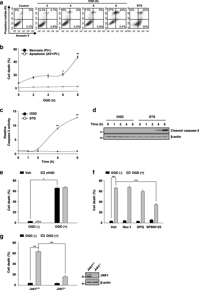
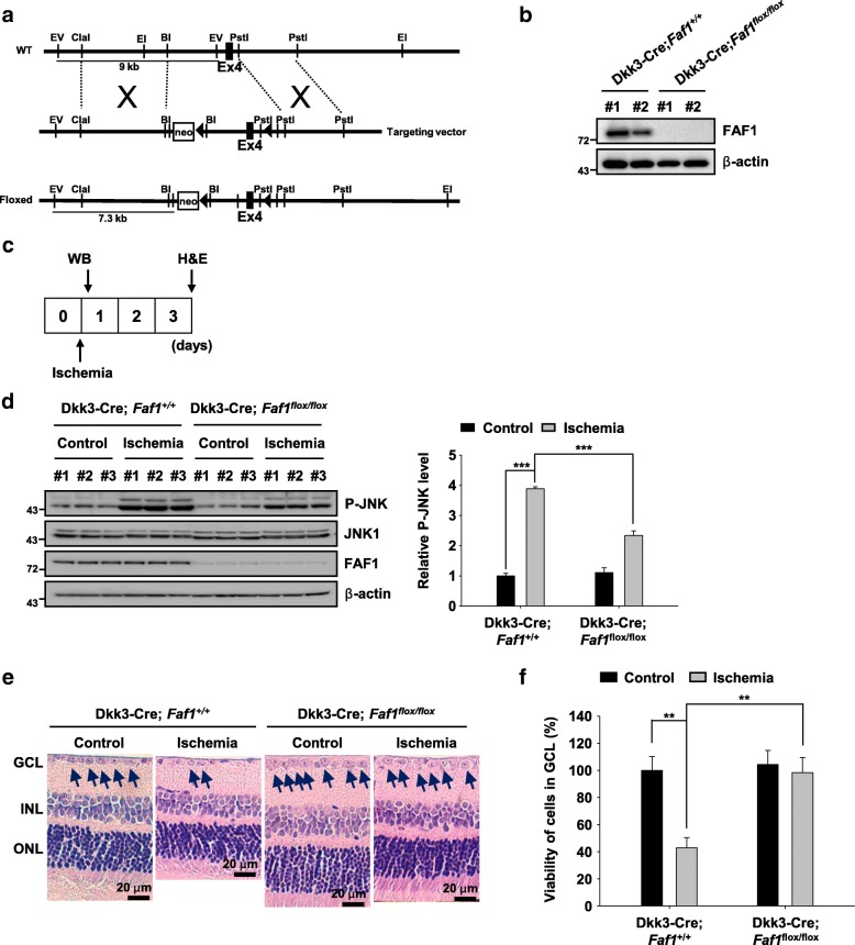
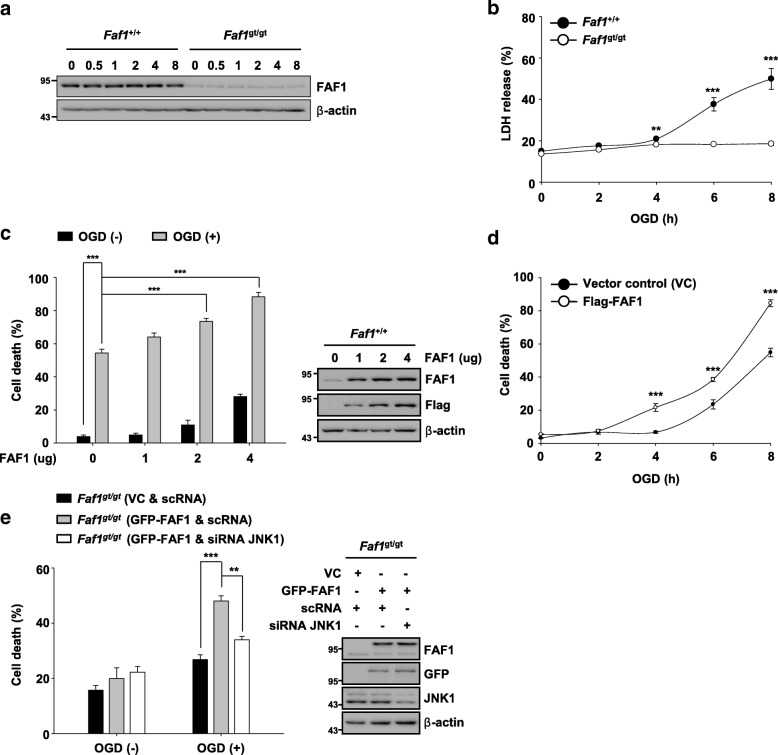
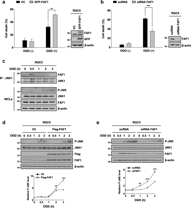
The expression changes of FAF1 and phophorylated JNK1 were detected by Western blotting. Immunoprecipitation was employed to investigate protein-protein interaction. We determined the cell death using flow cytometry and lactate dehydrogenase release measurement. To validate the death-promoting role of FAF1 in the retina, we generated conditional retinal FAF1 knockout mice. We used hematoxylin and eosin staining to detect retinal cell death in retinal ganglion cell layer.

RESULTS

FAF1 was found to function upstream of c-Jun N-terminal kinase 1 (JNK1), followed by mitochondrial dysregulation and necrotic cell death processes upon ischemic insult. We investigated whether FAF1 is involved in the pathogenesis of ischemic diseases using a retinal ischemia model. Indeed, FAF1 potentiated necrosis through JNK1 activation upon ischemic stress in retinal cells demonstrating retinal ganglion-like character. Conditional FAF1 depletion attenuated JNK1 activation in the retinas of Dkk3-Cre;Faf1 mice and ameliorated death of retinal cells due to elevated intraocular pressure (IOP).

CONCLUSIONS

Our results show that FAF1 plays a key role in ischemic retinal damage and may be implicated in the pathogenesis of retinal ischemic disease.

摘要

背景

缺血性应激诱导的异常细胞死亡与缺血性疾病的发病机制有关。Fas 相关因子 1(FAF1)已被确定为一种促进死亡的蛋白。本研究表明,FAF1 在缺血性损伤引发的死亡信号中起作用。

方法

通过 Western blot 检测 FAF1 和磷酸化 JNK1 的表达变化。采用免疫沉淀法研究蛋白质-蛋白质相互作用。通过流式细胞术和乳酸脱氢酶释放测定来确定细胞死亡。为了验证 FAF1 在视网膜中的促死亡作用,我们生成了条件性视网膜 FAF1 敲除小鼠。我们使用苏木精和伊红染色检测视网膜神经节细胞层中的视网膜细胞死亡。

结果

发现 FAF1 在 c-Jun N 端激酶 1(JNK1)上游起作用,随后在缺血性损伤后发生线粒体失调和坏死性细胞死亡过程。我们使用视网膜缺血模型研究 FAF1 是否参与缺血性疾病的发病机制。事实上,FAF1 通过 JNK1 激活增强了视网膜细胞中的坏死作用,表现出类似于视网膜神经节的特征。条件性 FAF1 缺失可减轻 Dkk3-Cre;Faf1 小鼠视网膜中 JNK1 的激活,并减轻因眼内压升高而导致的视网膜细胞死亡。

结论

我们的结果表明,FAF1 在缺血性视网膜损伤中起关键作用,可能与视网膜缺血性疾病的发病机制有关。

https://cdn.ncbi.nlm.nih.gov/pmc/blobs/6b2a/6131785/ad0ee164bc9e/12964_2018_265_Fig7_HTML.jpg
https://cdn.ncbi.nlm.nih.gov/pmc/blobs/6b2a/6131785/d4f2401584c4/12964_2018_265_Fig1_HTML.jpg
https://cdn.ncbi.nlm.nih.gov/pmc/blobs/6b2a/6131785/010453e5c2a5/12964_2018_265_Fig2_HTML.jpg
https://cdn.ncbi.nlm.nih.gov/pmc/blobs/6b2a/6131785/d28c83f15d4f/12964_2018_265_Fig3_HTML.jpg
https://cdn.ncbi.nlm.nih.gov/pmc/blobs/6b2a/6131785/70cabf796291/12964_2018_265_Fig4_HTML.jpg
https://cdn.ncbi.nlm.nih.gov/pmc/blobs/6b2a/6131785/a4b13ac4bf2d/12964_2018_265_Fig5_HTML.jpg
https://cdn.ncbi.nlm.nih.gov/pmc/blobs/6b2a/6131785/886a8cbccb22/12964_2018_265_Fig6_HTML.jpg
https://cdn.ncbi.nlm.nih.gov/pmc/blobs/6b2a/6131785/ad0ee164bc9e/12964_2018_265_Fig7_HTML.jpg

相似文献

1
FAF1 mediates necrosis through JNK1-mediated mitochondrial dysfunction leading to retinal degeneration in the ganglion cell layer upon ischemic insult.FAF1 通过 JNK1 介导的线粒体功能障碍介导细胞坏死,导致缺血性损伤后神经节细胞层发生视网膜变性。
Cell Commun Signal. 2018 Sep 10;16(1):56. doi: 10.1186/s12964-018-0265-7.
2
FAF1 mediates regulated necrosis through PARP1 activation upon oxidative stress leading to dopaminergic neurodegeneration.FAF1在氧化应激导致多巴胺能神经变性时通过PARP1激活介导程序性坏死。
Cell Death Differ. 2016 Nov 1;23(11):1873-1885. doi: 10.1038/cdd.2016.99. Epub 2016 Sep 23.
3
Retina-specific loss of causes mitochondrial dysfunction that leads to selective retinal ganglion cell degeneration in a mouse model of familial dysautonomia.家族性自主神经异常症小鼠模型中视锥细胞特异性缺失导致线粒体功能障碍,进而引发选择性视网膜神经节细胞变性。
Dis Model Mech. 2018 Jul 30;11(7):dmm033746. doi: 10.1242/dmm.033746.
4
Glutamate receptor activation triggers OPA1 release and induces apoptotic cell death in ischemic rat retina.谷氨酸受体激活触发视神经萎缩蛋白1(OPA1)释放,并诱导缺血大鼠视网膜发生凋亡性细胞死亡。
Mol Vis. 2008;14:2629-38. Epub 2008 Dec 31.
5
Inhibition of cyclophilin D by cyclosporin A promotes retinal ganglion cell survival by preventing mitochondrial alteration in ischemic injury.环孢素A抑制亲环蛋白D可通过防止缺血性损伤中的线粒体改变来促进视网膜神经节细胞存活。
Cell Death Dis. 2014 Mar 6;5(3):e1105. doi: 10.1038/cddis.2014.80.
6
Daxx is a key downstream component of receptor interacting protein kinase 3 mediating retinal ischemic cell death.Daxx 是受体相互作用蛋白激酶 3 的下游关键组成部分,介导视网膜缺血性细胞死亡。
FEBS Lett. 2013 Jan 31;587(3):266-71. doi: 10.1016/j.febslet.2012.12.004. Epub 2012 Dec 19.
7
Ubiquinol promotes retinal ganglion cell survival and blocks the apoptotic pathway in ischemic retinal degeneration.泛醇促进视网膜神经节细胞存活并阻断缺血性视网膜变性中的凋亡途径。
Biochem Biophys Res Commun. 2018 Sep 18;503(4):2639-2645. doi: 10.1016/j.bbrc.2018.08.016. Epub 2018 Aug 11.
8
Enhanced protein expressions of sortilin and p75NTR in retina of rat following elevated intraocular pressure-induced retinal ischemia.眼内压升高诱导视网膜缺血后大鼠视网膜中sortilin和p75NTR蛋白表达增强。
Neurosci Lett. 2007 Dec 18;429(2-3):169-74. doi: 10.1016/j.neulet.2007.10.012. Epub 2007 Oct 22.
9
Endoplasmic reticulum stress in retinal vascular degeneration: protective role of resveratrol.内质网应激在视网膜血管退变中的作用:白藜芦醇的保护作用。
Invest Ophthalmol Vis Sci. 2012 May 31;53(6):3241-9. doi: 10.1167/iovs.11-8406.
10
Inactivation of Endothelial ADAM17 Reduces Retinal Ischemia-Reperfusion Induced Neuronal and Vascular Damage.内皮细胞 ADAM17 的失活可减少视网膜缺血再灌注诱导的神经元和血管损伤。
Int J Mol Sci. 2020 Jul 29;21(15):5379. doi: 10.3390/ijms21155379.

引用本文的文献

1
Identification of Novel Biomarkers for Ischemic Stroke Through Integrated Bioinformatics Analysis and Machine Learning.通过综合生物信息学分析和机器学习识别缺血性中风的新型生物标志物
J Mol Neurosci. 2025 Jan 25;75(1):13. doi: 10.1007/s12031-025-02309-8.
2
Glaucomatous retinal ganglion cells: death and protection.青光眼性视网膜神经节细胞:死亡与保护
Int J Ophthalmol. 2025 Jan 18;18(1):160-167. doi: 10.18240/ijo.2025.01.20. eCollection 2025.
3
Trauma diagnostic-related target proteins and their detection techniques.创伤相关诊断靶蛋白及其检测技术。

本文引用的文献

1
661W is a retinal ganglion precursor-like cell line in which glaucoma-associated optineurin mutants induce cell death selectively.661W 是一种视网膜神经节前体细胞样细胞系,其中与青光眼相关的视神经丝蛋白突变体选择性诱导细胞死亡。
Sci Rep. 2017 Dec 4;7(1):16855. doi: 10.1038/s41598-017-17241-0.
2
Optic Nerve Degeneration after Retinal Ischemia/Reperfusion in a Rodent Model.啮齿动物模型中视网膜缺血/再灌注后的视神经变性
Front Cell Neurosci. 2017 Aug 22;11:254. doi: 10.3389/fncel.2017.00254. eCollection 2017.
3
Retinal metabolism: A comparative look at energetics in the retina.
Expert Rev Mol Med. 2024 Apr 11;26:e7. doi: 10.1017/erm.2024.3.
4
Expression and Localization of Fas-Associated Factor 1 in Testicular Tissues of Different Ages and Ovaries at Different Reproductive Cycle Phases of .Fas相关因子1在不同年龄睾丸组织及不同生殖周期阶段卵巢中的表达与定位
Animals (Basel). 2023 Jan 18;13(3):340. doi: 10.3390/ani13030340.
5
UBX Domain Protein 6 Positively Regulates JAK-STAT1/2 Signaling.UBX 结构域蛋白 6 正向调控 JAK-STAT1/2 信号通路。
J Immunol. 2021 Jun 1;206(11):2682-2691. doi: 10.4049/jimmunol.1901337. Epub 2021 May 21.
视网膜代谢:视网膜能量学的比较研究。
Brain Res. 2017 Oct 1;1672:50-57. doi: 10.1016/j.brainres.2017.07.025. Epub 2017 Jul 29.
4
FAS-associated factor-1 positively regulates type I interferon response to RNA virus infection by targeting NLRX1.FAS相关因子1通过靶向NLRX1正向调节I型干扰素对RNA病毒感染的反应。
PLoS Pathog. 2017 May 22;13(5):e1006398. doi: 10.1371/journal.ppat.1006398. eCollection 2017 May.
5
FAF1 mediates regulated necrosis through PARP1 activation upon oxidative stress leading to dopaminergic neurodegeneration.FAF1在氧化应激导致多巴胺能神经变性时通过PARP1激活介导程序性坏死。
Cell Death Differ. 2016 Nov 1;23(11):1873-1885. doi: 10.1038/cdd.2016.99. Epub 2016 Sep 23.
6
Regulation of mitochondrial morphology and function by stearoylation of TFR1.通过TFR1的硬脂酰化调节线粒体形态和功能
Nature. 2015 Sep 3;525(7567):124-8. doi: 10.1038/nature14601. Epub 2015 Jul 27.
7
Inhibition of JNK aggravates the recovery of rat hearts after global ischemia: the role of mitochondrial JNK.抑制JNK会加重大鼠心脏在全心缺血后的恢复:线粒体JNK的作用
PLoS One. 2014 Nov 25;9(11):e113526. doi: 10.1371/journal.pone.0113526. eCollection 2014.
8
Mitochondrial JNK activation triggers autophagy and apoptosis and aggravates myocardial injury following ischemia/reperfusion.线粒体JNK激活引发自噬和凋亡,并加重缺血/再灌注后的心肌损伤。
Biochim Biophys Acta. 2015 Feb;1852(2):262-70. doi: 10.1016/j.bbadis.2014.05.012. Epub 2014 May 21.
9
Retinal ganglion cell (RGC) programmed necrosis contributes to ischemia-reperfusion-induced retinal damage.视网膜神经节细胞(RGC)程序性坏死会导致缺血再灌注引起的视网膜损伤。
Exp Eye Res. 2014 Jun;123:1-7. doi: 10.1016/j.exer.2014.04.009. Epub 2014 Apr 19.
10
What is the nature of the RGC-5 cell line?RGC-5 细胞系的性质是什么?
Adv Exp Med Biol. 2014;801:145-54. doi: 10.1007/978-1-4614-3209-8_19.