• 文献检索
  • 文档翻译
  • 深度研究
  • 学术资讯
  • Suppr Zotero 插件Zotero 插件
  • 邀请有礼
  • 套餐&价格
  • 历史记录
应用&插件
Suppr Zotero 插件Zotero 插件浏览器插件Mac 客户端Windows 客户端微信小程序
定价
高级版会员购买积分包购买API积分包
服务
文献检索文档翻译深度研究API 文档MCP 服务
关于我们
关于 Suppr公司介绍联系我们用户协议隐私条款
关注我们

Suppr 超能文献

核心技术专利:CN118964589B侵权必究
粤ICP备2023148730 号-1Suppr @ 2026

文献检索

告别复杂PubMed语法,用中文像聊天一样搜索,搜遍4000万医学文献。AI智能推荐,让科研检索更轻松。

立即免费搜索

文件翻译

保留排版,准确专业,支持PDF/Word/PPT等文件格式,支持 12+语言互译。

免费翻译文档

深度研究

AI帮你快速写综述,25分钟生成高质量综述,智能提取关键信息,辅助科研写作。

立即免费体验

左心房壁分割和厚度算法 - 在开源 CT 和 MRI 图像数据库上的评估。

Algorithms for left atrial wall segmentation and thickness - Evaluation on an open-source CT and MRI image database.

机构信息

School of Biomedical Engineering & Imaging Sciences, King's College London, UK.

School of Biomedical Engineering & Imaging Sciences, King's College London, UK.

出版信息

Med Image Anal. 2018 Dec;50:36-53. doi: 10.1016/j.media.2018.08.004. Epub 2018 Aug 24.

DOI:10.1016/j.media.2018.08.004
PMID:30208355
原文链接:https://pmc.ncbi.nlm.nih.gov/articles/PMC6218662/
Abstract

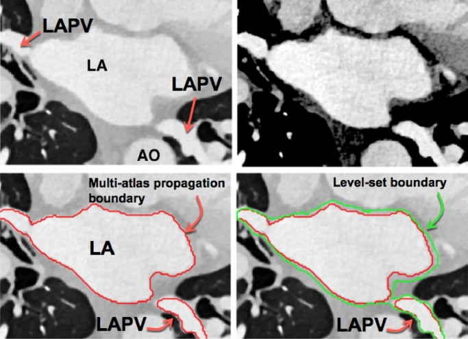
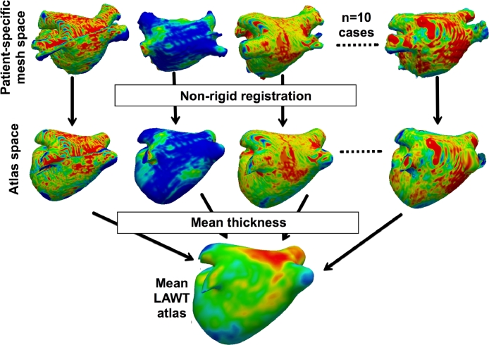
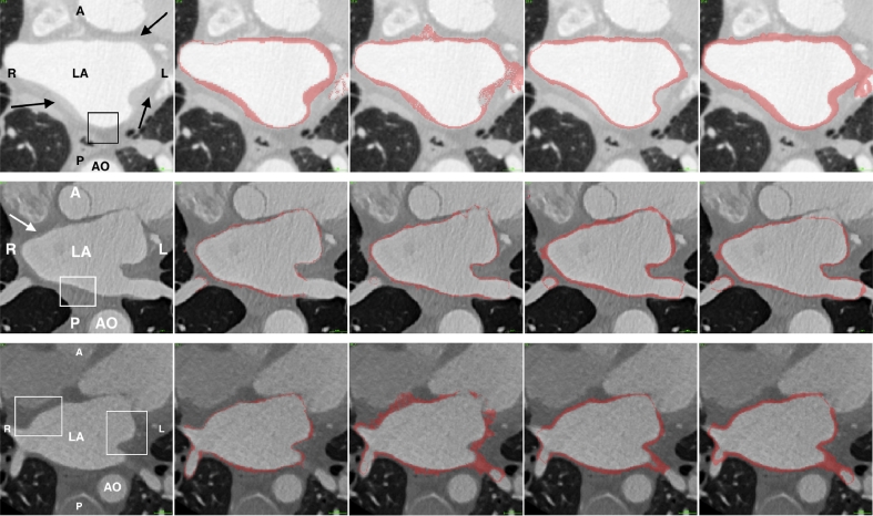
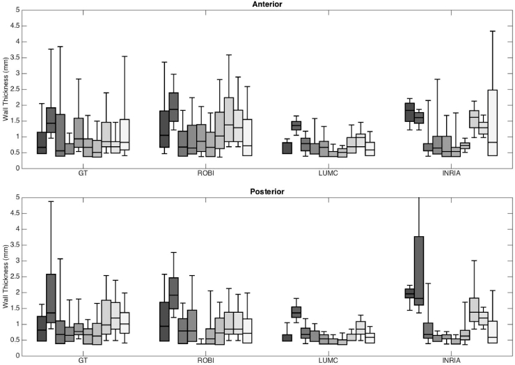
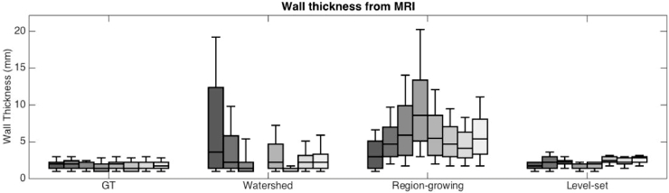
Structural changes to the wall of the left atrium are known to occur with conditions that predispose to Atrial fibrillation. Imaging studies have demonstrated that these changes may be detected non-invasively. An important indicator of this structural change is the wall's thickness. Present studies have commonly measured the wall thickness at few discrete locations. Dense measurements with computer algorithms may be possible on cardiac scans of Computed Tomography (CT) and Magnetic Resonance Imaging (MRI). The task is challenging as the atrial wall is a thin tissue and the imaging resolution is a limiting factor. It is unclear how accurate algorithms may get and how they compare in this new emerging area. We approached this problem of comparability with the Segmentation of Left Atrial Wall for Thickness (SLAWT) challenge organised in conjunction with MICCAI 2016 conference. This manuscript presents the algorithms that had participated and evaluation strategies for comparing them on the challenge image database that is now open-source. The image database consisted of cardiac CT (n=10) and MRI (n=10) of healthy and diseased subjects. A total of 6 algorithms were evaluated with different metrics, with 3 algorithms in each modality. Segmentation of the wall with algorithms was found to be feasible in both modalities. There was generally a lack of accuracy in the algorithms and inter-rater differences showed that algorithms could do better. Benchmarks were determined and algorithms were ranked to allow future algorithms to be ranked alongside the state-of-the-art techniques presented in this work. A mean atlas was also constructed from both modalities to illustrate the variation in thickness within this small cohort.

摘要

左心房壁的结构变化已知会发生在易患心房颤动的情况下。影像学研究表明,这些变化可以通过非侵入性方法检测到。这种结构变化的一个重要指标是壁的厚度。目前的研究通常在少数离散位置测量壁的厚度。计算机算法的密集测量可能在心脏 CT(计算机断层扫描)和磁共振成像(MRI)扫描上实现。由于心房壁是一种薄组织,成像分辨率是一个限制因素,因此这项任务具有挑战性。目前还不清楚算法的准确性如何,以及在这个新出现的领域中它们的比较情况如何。我们在 2016 年 MICCAI 会议上共同组织的左心房壁厚度分割挑战赛(SLAWT)中解决了这个可比性问题。本文介绍了参与挑战赛的算法,以及在现在开源的挑战图像数据库上比较它们的评估策略。该图像数据库包含了健康和患病受试者的心脏 CT(n=10)和 MRI(n=10)。共有 6 种算法使用不同的指标进行了评估,每种模态有 3 种算法。在两种模态中,使用算法分割壁是可行的。算法的准确性普遍不足,并且评分者之间的差异表明算法可以做得更好。确定了基准,并对算法进行了排名,以便未来的算法能够与本工作中提出的最新技术一起排名。还从两种模态构建了平均图谱,以说明在这个小队列中厚度的变化。

https://cdn.ncbi.nlm.nih.gov/pmc/blobs/10d0/6218662/be809973eab5/gr15.jpg
https://cdn.ncbi.nlm.nih.gov/pmc/blobs/10d0/6218662/15271654e507/fx1.jpg
https://cdn.ncbi.nlm.nih.gov/pmc/blobs/10d0/6218662/11627547bdcb/gr1.jpg
https://cdn.ncbi.nlm.nih.gov/pmc/blobs/10d0/6218662/f60dd0d16c7a/gr2.jpg
https://cdn.ncbi.nlm.nih.gov/pmc/blobs/10d0/6218662/a575b58e25b6/gr3.jpg
https://cdn.ncbi.nlm.nih.gov/pmc/blobs/10d0/6218662/025703d12e89/gr4.jpg
https://cdn.ncbi.nlm.nih.gov/pmc/blobs/10d0/6218662/f761c98bccc9/gr5.jpg
https://cdn.ncbi.nlm.nih.gov/pmc/blobs/10d0/6218662/14b30ab85c07/gr6.jpg
https://cdn.ncbi.nlm.nih.gov/pmc/blobs/10d0/6218662/798cca682c56/gr7.jpg
https://cdn.ncbi.nlm.nih.gov/pmc/blobs/10d0/6218662/b2cad1d7d57f/gr8.jpg
https://cdn.ncbi.nlm.nih.gov/pmc/blobs/10d0/6218662/3ad19cbc30b3/gr9.jpg
https://cdn.ncbi.nlm.nih.gov/pmc/blobs/10d0/6218662/12d9516733bb/gr10.jpg
https://cdn.ncbi.nlm.nih.gov/pmc/blobs/10d0/6218662/5fc18f4b0fad/gr11.jpg
https://cdn.ncbi.nlm.nih.gov/pmc/blobs/10d0/6218662/376ececc1896/gr12.jpg
https://cdn.ncbi.nlm.nih.gov/pmc/blobs/10d0/6218662/7d305d639416/gr13.jpg
https://cdn.ncbi.nlm.nih.gov/pmc/blobs/10d0/6218662/0b48a5349cf1/gr14.jpg
https://cdn.ncbi.nlm.nih.gov/pmc/blobs/10d0/6218662/be809973eab5/gr15.jpg

相似文献

1
Algorithms for left atrial wall segmentation and thickness - Evaluation on an open-source CT and MRI image database.左心房壁分割和厚度算法 - 在开源 CT 和 MRI 图像数据库上的评估。
Med Image Anal. 2018 Dec;50:36-53. doi: 10.1016/j.media.2018.08.004. Epub 2018 Aug 24.
2
Automated segmentation of the atrial region and fossa ovalis towards computer-aided planning of inter-atrial wall interventions.自动分割心房区域和卵圆窝,以辅助规划房间隔壁介入。
Comput Methods Programs Biomed. 2018 Jul;161:73-84. doi: 10.1016/j.cmpb.2018.04.014. Epub 2018 Apr 18.
3
Three-dimensional atrial wall thickness maps to inform catheter ablation procedures for atrial fibrillation.用于指导房颤导管消融手术的三维心房壁厚度图。
Europace. 2016 Mar;18(3):376-83. doi: 10.1093/europace/euv073. Epub 2015 Apr 4.
4
The role of myocardial wall thickness in atrial arrhythmogenesis.心肌壁厚度在房性心律失常发生机制中的作用。
Europace. 2016 Dec;18(12):1758-1772. doi: 10.1093/europace/euw014. Epub 2016 May 31.
5
Evaluation of current algorithms for segmentation of scar tissue from late gadolinium enhancement cardiovascular magnetic resonance of the left atrium: an open-access grand challenge.评估当前算法在左心房晚期钆增强心血管磁共振瘢痕组织分割中的应用:公开获取的大挑战。
J Cardiovasc Magn Reson. 2013 Dec 20;15(1):105. doi: 10.1186/1532-429X-15-105.
6
Measurements of the left atrium and pulmonary veins for analysis of reverse structural remodeling following cardiac ablation therapy.测量左心房和肺静脉以分析心脏消融治疗后的逆向结构重塑。
Comput Methods Programs Biomed. 2015 Feb;118(2):198-206. doi: 10.1016/j.cmpb.2014.11.001. Epub 2014 Nov 13.
7
GCW-UNet segmentation of cardiac magnetic resonance images for evaluation of left atrial enlargement.GCW-UNet 分割心脏磁共振图像用于评估左心房增大。
Comput Methods Programs Biomed. 2022 Jun;221:106915. doi: 10.1016/j.cmpb.2022.106915. Epub 2022 May 25.
8
Automated left atrial time-resolved segmentation in MRI long-axis cine images using active contours.基于主动轮廓的 MRI 左心房长轴电影图像的自动左心房时间分辨分割。
BMC Med Imaging. 2021 Jun 19;21(1):101. doi: 10.1186/s12880-021-00630-3.
9
Evaluation of algorithms for Multi-Modality Whole Heart Segmentation: An open-access grand challenge.多模态全心脏分割算法评估:一个开放获取的大型挑战赛。
Med Image Anal. 2019 Dec;58:101537. doi: 10.1016/j.media.2019.101537. Epub 2019 Aug 1.
10
A global benchmark of algorithms for segmenting the left atrium from late gadolinium-enhanced cardiac magnetic resonance imaging.一种用于从晚期钆增强心脏磁共振成像中分割左心房的算法的全球基准。
Med Image Anal. 2021 Jan;67:101832. doi: 10.1016/j.media.2020.101832. Epub 2020 Oct 16.

引用本文的文献

1
Utilizing multimodal artificial intelligence to advance cardiovascular diseases.利用多模态人工智能推进心血管疾病研究。
Precis Clin Med. 2025 Jul 17;8(3):pbaf016. doi: 10.1093/pcmedi/pbaf016. eCollection 2025 Sep.
2
On preserving anatomical detail in statistical shape analysis for clustering: focus on left atrial appendage morphology.关于在用于聚类的统计形状分析中保留解剖细节:聚焦于左心耳形态
Front Netw Physiol. 2024 Oct 10;4:1467180. doi: 10.3389/fnetp.2024.1467180. eCollection 2024.
3
Chamber-specific wall thickness features in human atrial fibrillation.

本文引用的文献

1
Real-Time X-MRI-Guided Left Ventricular Lead Implantation for Targeted Delivery of Cardiac Resynchronization Therapy.实时 X 线磁共振引导左心室导线植入术用于心脏再同步治疗的靶向递送。
JACC Clin Electrophysiol. 2017 Aug;3(8):803-814. doi: 10.1016/j.jacep.2017.01.018. Epub 2017 Apr 26.
2
Novel MRI Technique Enables Non-Invasive Measurement of Atrial Wall Thickness.新型磁共振成像技术可实现心房壁厚度的无创测量。
IEEE Trans Med Imaging. 2017 Aug;36(8):1607-1614. doi: 10.1109/TMI.2017.2671839. Epub 2017 Apr 13.
3
Effect of Left Atrial Wall Thickness on Radiofrequency Ablation Success.
人类心房颤动中特定腔室的壁厚度特征
Interface Focus. 2023 Dec 15;13(6):20230044. doi: 10.1098/rsfs.2023.0044. eCollection 2023 Dec 6.
4
Posterior wall thickness of the confluent inferior pulmonary veins measured by left atrial intracardiac echocardiography: implications for catheter ablation.左房心内超声测量融合下肺静脉后壁厚度:对导管消融的影响。
J Interv Card Electrophysiol. 2024 Jan;67(1):193-201. doi: 10.1007/s10840-023-01613-w. Epub 2023 Jul 25.
5
Beyond CFD: Emerging methodologies for predictive simulation in cardiovascular health and disease.超越计算流体动力学:心血管健康与疾病预测模拟的新兴方法
Biophys Rev (Melville). 2023 Mar;4(1):011301. doi: 10.1063/5.0109400. Epub 2023 Jan 13.
6
Extent of Left Atrial Fibrosis Correlates with Descending Aorta Proximity at 3D Late Gadolinium Enhancement Cardiac MRI in Patients with Atrial Fibrillation.心房颤动患者3D延迟钆增强心脏磁共振成像中左心房纤维化程度与降主动脉毗邻关系的相关性研究
Radiol Cardiothorac Imaging. 2022 Jan 13;4(1):e210192. doi: 10.1148/ryct.210192. eCollection 2022 Feb.
7
Medical image analysis on left atrial LGE MRI for atrial fibrillation studies: A review.用于房颤研究的左心房 LGE MRI 的医学图像分析:综述。
Med Image Anal. 2022 Apr;77:102360. doi: 10.1016/j.media.2022.102360. Epub 2022 Jan 29.
8
A deep-learning approach for direct whole-heart mesh reconstruction.深度学习方法实现直接整体心脏网格重建。
Med Image Anal. 2021 Dec;74:102222. doi: 10.1016/j.media.2021.102222. Epub 2021 Sep 8.
9
Simultaneous Multi-Structure Segmentation of the Heart and Peripheral Tissues in Contrast Enhanced Cardiac Computed Tomography Angiography.对比增强心脏计算机断层扫描血管造影中心脏和周围组织的同步多结构分割
IEEE Access. 2020;8:16187-16202. doi: 10.1109/access.2020.2966985. Epub 2020 Jan 15.
10
Machine Learning-Derived Fractal Features of Shape and Texture of the Left Atrium and Pulmonary Veins From Cardiac Computed Tomography Scans Are Associated With Risk of Recurrence of Atrial Fibrillation Postablation.机器学习衍生的左心房和肺静脉的形状和纹理的分形特征与消融后心房颤动复发的风险相关。
Circ Arrhythm Electrophysiol. 2021 Mar;14(3):e009265. doi: 10.1161/CIRCEP.120.009265. Epub 2021 Feb 12.
左心房壁厚度对射频消融术成功率的影响。
J Cardiovasc Electrophysiol. 2016 Nov;27(11):1298-1303. doi: 10.1111/jce.13065. Epub 2016 Sep 16.
4
ISLES 2015 - A public evaluation benchmark for ischemic stroke lesion segmentation from multispectral MRI.ISLES 2015 - 多光谱 MRI 缺血性脑卒中病灶分割的公共评估基准。
Med Image Anal. 2017 Jan;35:250-269. doi: 10.1016/j.media.2016.07.009. Epub 2016 Jul 21.
5
The role of myocardial wall thickness in atrial arrhythmogenesis.心肌壁厚度在房性心律失常发生机制中的作用。
Europace. 2016 Dec;18(12):1758-1772. doi: 10.1093/europace/euw014. Epub 2016 May 31.
6
A Method to Standardize Quantification of Left Atrial Scar From Delayed-Enhancement MR Images.一种从延迟增强磁共振图像标准化左心房瘢痕量化的方法。
IEEE J Transl Eng Health Med. 2014 Mar 18;2:1800615. doi: 10.1109/JTEHM.2014.2312191. eCollection 2014.
7
Evaluation of state-of-the-art segmentation algorithms for left ventricle infarct from late Gadolinium enhancement MR images.评估基于晚期钆增强磁共振图像的左心室梗死的最新分割算法。
Med Image Anal. 2016 May;30:95-107. doi: 10.1016/j.media.2016.01.004. Epub 2016 Jan 28.
8
Fully automatic segmentation of left atrium and pulmonary veins in late gadolinium-enhanced MRI: Towards objective atrial scar assessment.钆增强磁共振成像晚期左心房和肺静脉的全自动分割:迈向客观的心房瘢痕评估
J Magn Reson Imaging. 2016 Aug;44(2):346-54. doi: 10.1002/jmri.25148. Epub 2016 Jan 11.
9
Multi-atlas segmentation of biomedical images: A survey.生物医学图像的多图谱分割:一项综述。
Med Image Anal. 2015 Aug;24(1):205-219. doi: 10.1016/j.media.2015.06.012. Epub 2015 Jul 6.
10
Three-dimensional atrial wall thickness maps to inform catheter ablation procedures for atrial fibrillation.用于指导房颤导管消融手术的三维心房壁厚度图。
Europace. 2016 Mar;18(3):376-83. doi: 10.1093/europace/euv073. Epub 2015 Apr 4.