Department of Imaging Sciences & Biomedical Engineering, King's College London, London, UK.
J Cardiovasc Magn Reson. 2013 Dec 20;15(1):105. doi: 10.1186/1532-429X-15-105.
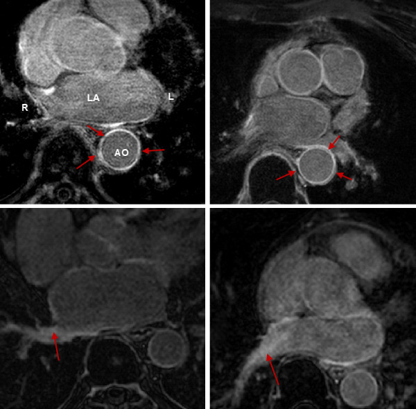
Late Gadolinium enhancement (LGE) cardiovascular magnetic resonance (CMR) imaging can be used to visualise regions of fibrosis and scarring in the left atrium (LA) myocardium. This can be important for treatment stratification of patients with atrial fibrillation (AF) and for assessment of treatment after radio frequency catheter ablation (RFCA). In this paper we present a standardised evaluation benchmarking framework for algorithms segmenting fibrosis and scar from LGE CMR images. The algorithms reported are the response to an open challenge that was put to the medical imaging community through an ISBI (IEEE International Symposium on Biomedical Imaging) workshop.
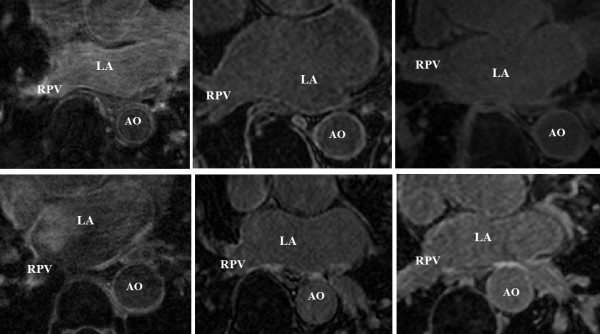
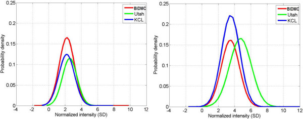
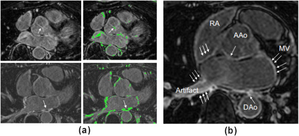
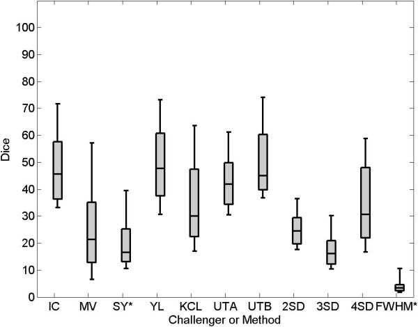
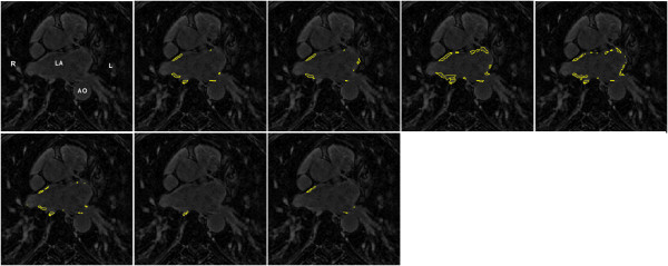
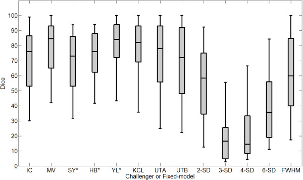
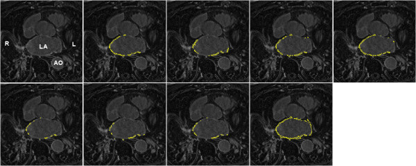
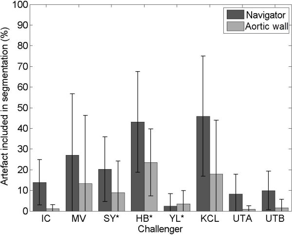
The image database consisted of 60 multicenter, multivendor LGE CMR image datasets from patients with AF, with 30 images taken before and 30 after RFCA for the treatment of AF. A reference standard for scar and fibrosis was established by merging manual segmentations from three observers. Furthermore, scar was also quantified using 2, 3 and 4 standard deviations (SD) and full-width-at-half-maximum (FWHM) methods. Seven institutions responded to the challenge: Imperial College (IC), Mevis Fraunhofer (MV), Sunnybrook Health Sciences (SY), Harvard/Boston University (HB), Yale School of Medicine (YL), King's College London (KCL) and Utah CARMA (UTA, UTB). There were 8 different algorithms evaluated in this study.
Some algorithms were able to perform significantly better than SD and FWHM methods in both pre- and post-ablation imaging. Segmentation in pre-ablation images was challenging and good correlation with the reference standard was found in post-ablation images. Overlap scores (out of 100) with the reference standard were as follows: Pre: IC = 37, MV = 22, SY = 17, YL = 48, KCL = 30, UTA = 42, UTB = 45; Post: IC = 76, MV = 85, SY = 73, HB = 76, YL = 84, KCL = 78, UTA = 78, UTB = 72.
The study concludes that currently no algorithm is deemed clearly better than others. There is scope for further algorithmic developments in LA fibrosis and scar quantification from LGE CMR images. Benchmarking of future scar segmentation algorithms is thus important. The proposed benchmarking framework is made available as open-source and new participants can evaluate their algorithms via a web-based interface.
迟发钆增强(LGE)心血管磁共振(CMR)成像可用于可视化左心房(LA)心肌中的纤维化和瘢痕区域。这对于房颤(AF)患者的治疗分层以及射频导管消融(RFCA)后的治疗评估非常重要。在本文中,我们提出了一个用于从 LGE CMR 图像分割纤维化和瘢痕的算法的标准化评估基准。报告的算法是对通过 ISBI(IEEE 生物医学成像国际研讨会)研讨会向医学成像社区提出的公开挑战的回应。
图像数据库由 60 个多中心、多供应商的 AF 患者 LGE CMR 图像数据集组成,其中 30 个图像在 RFCA 治疗 AF 之前采集,30 个图像在之后采集。通过合并三位观察者的手动分割,建立了瘢痕和纤维化的参考标准。此外,还使用 2、3 和 4 个标准差(SD)和半峰全宽(FWHM)方法量化了瘢痕。有 7 个机构对该挑战做出了回应:帝国理工学院(IC)、Mevis Fraunhofer(MV)、森尼布鲁克健康科学(SY)、哈佛/波士顿大学(HB)、耶鲁大学医学院(YL)、伦敦国王学院(KCL)和犹他州 CARMA(UTA、UTB)。在这项研究中评估了 8 种不同的算法。
在术前和术后成像中,一些算法的表现明显优于 SD 和 FWHM 方法。术前图像的分割具有挑战性,与参考标准的相关性在术后图像中得到很好的验证。与参考标准的重叠评分(满分 100)如下:术前:IC=37,MV=22,SY=17,YL=48,KCL=30,UTA=42,UTB=45;术后:IC=76,MV=85,SY=73,HB=76,YL=84,KCL=78,UTA=78,UTB=72。
研究得出的结论是,目前还没有一种算法被认为明显优于其他算法。从 LGE CMR 图像量化 LA 纤维化和瘢痕方面,仍有进一步开发算法的空间。因此,未来瘢痕分割算法的基准测试非常重要。所提出的基准测试框架是开源的,新的参与者可以通过基于网络的界面评估他们的算法。