• 文献检索
  • 文档翻译
  • 深度研究
  • 学术资讯
  • Suppr Zotero 插件Zotero 插件
  • 邀请有礼
  • 套餐&价格
  • 历史记录
应用&插件
Suppr Zotero 插件Zotero 插件浏览器插件Mac 客户端Windows 客户端微信小程序
定价
高级版会员购买积分包购买API积分包
服务
文献检索文档翻译深度研究API 文档MCP 服务
关于我们
关于 Suppr公司介绍联系我们用户协议隐私条款
关注我们

Suppr 超能文献

核心技术专利:CN118964589B侵权必究
粤ICP备2023148730 号-1Suppr @ 2026

文献检索

告别复杂PubMed语法,用中文像聊天一样搜索,搜遍4000万医学文献。AI智能推荐,让科研检索更轻松。

立即免费搜索

文件翻译

保留排版,准确专业,支持PDF/Word/PPT等文件格式,支持 12+语言互译。

免费翻译文档

深度研究

AI帮你快速写综述,25分钟生成高质量综述,智能提取关键信息,辅助科研写作。

立即免费体验

无标记法预测透射光显微镜的三维荧光图像。

Label-free prediction of three-dimensional fluorescence images from transmitted-light microscopy.

机构信息

Allen Institute for Cell Science, Seattle, WA, USA.

Allen Institute for Brain Science, Seattle, WA, USA.

出版信息

Nat Methods. 2018 Nov;15(11):917-920. doi: 10.1038/s41592-018-0111-2. Epub 2018 Sep 17.

DOI:10.1038/s41592-018-0111-2
PMID:30224672
原文链接:https://pmc.ncbi.nlm.nih.gov/articles/PMC6212323/
Abstract

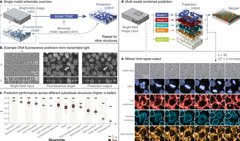
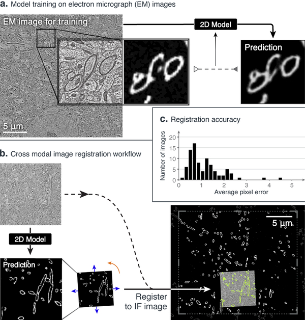
Understanding cells as integrated systems is central to modern biology. Although fluorescence microscopy can resolve subcellular structure in living cells, it is expensive, is slow, and can damage cells. We present a label-free method for predicting three-dimensional fluorescence directly from transmitted-light images and demonstrate that it can be used to generate multi-structure, integrated images. The method can also predict immunofluorescence (IF) from electron micrograph (EM) inputs, extending the potential applications.

摘要

将细胞视为一个集成系统是现代生物学的核心。尽管荧光显微镜可以解析活细胞中的亚细胞结构,但它昂贵、缓慢,并且可能会对细胞造成损伤。我们提出了一种无标记的方法,可以直接从透射光图像预测三维荧光,并证明它可以用于生成多结构、集成的图像。该方法还可以从电子显微镜输入预测免疫荧光(IF),从而扩展了潜在的应用。

https://cdn.ncbi.nlm.nih.gov/pmc/blobs/f134/6212323/16eac305ed5c/nihms-1501928-f0002.jpg
https://cdn.ncbi.nlm.nih.gov/pmc/blobs/f134/6212323/d9608e44e419/nihms-1501928-f0001.jpg
https://cdn.ncbi.nlm.nih.gov/pmc/blobs/f134/6212323/16eac305ed5c/nihms-1501928-f0002.jpg
https://cdn.ncbi.nlm.nih.gov/pmc/blobs/f134/6212323/d9608e44e419/nihms-1501928-f0001.jpg
https://cdn.ncbi.nlm.nih.gov/pmc/blobs/f134/6212323/16eac305ed5c/nihms-1501928-f0002.jpg

相似文献

1
Label-free prediction of three-dimensional fluorescence images from transmitted-light microscopy.无标记法预测透射光显微镜的三维荧光图像。
Nat Methods. 2018 Nov;15(11):917-920. doi: 10.1038/s41592-018-0111-2. Epub 2018 Sep 17.
2
Cell biology of the future: Nanometer-scale cellular cartography.未来的细胞生物学:纳米级细胞图谱
J Cell Biol. 2015 Oct 26;211(2):211-4. doi: 10.1083/jcb.201508021. Epub 2015 Oct 19.
3
Advances in high-resolution imaging--techniques for three-dimensional imaging of cellular structures.高分辨率成像技术——细胞结构三维成像技术。
J Cell Sci. 2012 Jun 1;125(Pt 11):2571-80. doi: 10.1242/jcs.090027. Epub 2012 Jun 8.
4
Super-resolution for a 3D world.面向三维世界的超分辨率技术。
Nat Methods. 2008 Jun;5(6):471-2. doi: 10.1038/nmeth0608-471.
5
Overcoming obstacles in localization microscopy.克服定位显微镜技术中的障碍。
Nat Methods. 2016 Apr;13(4):301-2. doi: 10.1038/nmeth.3813.
6
Development in the STORM.STORM 的发展。
Dev Cell. 2012 Dec 11;23(6):1103-10. doi: 10.1016/j.devcel.2012.10.003.
7
Live CLEM imaging to analyze nuclear structures at high resolution.通过实时细胞成像与电子显微镜关联技术在高分辨率下分析核结构。
Methods Mol Biol. 2015;1262:89-103. doi: 10.1007/978-1-4939-2253-6_6.
8
Nanometer-resolution fluorescence electron microscopy (nano-EM) in cultured cells.培养细胞中的纳米分辨率荧光电子显微镜技术(纳米电镜)
Methods Mol Biol. 2014;1117:503-26. doi: 10.1007/978-1-62703-776-1_22.
9
3D reconstruction of high-resolution STED microscope images.高分辨率受激发射损耗(STED)显微镜图像的三维重建
Microsc Res Tech. 2008 Sep;71(9):644-50. doi: 10.1002/jemt.20602.
10
Correlative light and electron microscopy for complex cellular structures on PDMS substrates with coded micro-patterns.用编码微图案的 PDMS 基底进行复杂细胞结构的相关光镜和电子显微镜检查。
Lab Chip. 2018 Dec 4;18(24):3840-3848. doi: 10.1039/c8lc00703a.

引用本文的文献

1
Inverse-scattering in biological samples via beam-propagation.通过光束传播对生物样本进行逆散射。
bioRxiv. 2025 Aug 22:2025.08.17.670744. doi: 10.1101/2025.08.17.670744.
2
On the Utility of Virtual Staining for Downstream Applications as it relates to Task Network Capacity.虚拟染色在下游应用中的效用及其与任务网络容量的关系
bioRxiv. 2025 Aug 6:2025.08.04.668552. doi: 10.1101/2025.08.04.668552.
3
On the Utility of Virtual Staining for Downstream Applications as it relates to Task Network Capacity.虚拟染色在与任务网络容量相关的下游应用中的效用

本文引用的文献

1
Content-aware image restoration: pushing the limits of fluorescence microscopy.内容感知图像恢复:推动荧光显微镜的极限。
Nat Methods. 2018 Dec;15(12):1090-1097. doi: 10.1038/s41592-018-0216-7. Epub 2018 Nov 26.
2
In Silico Labeling: Predicting Fluorescent Labels in Unlabeled Images.计算机标记:在未标记的图像中预测荧光标记。
Cell. 2018 Apr 19;173(3):792-803.e19. doi: 10.1016/j.cell.2018.03.040. Epub 2018 Apr 12.
3
Systematic gene tagging using CRISPR/Cas9 in human stem cells to illuminate cell organization.利用CRISPR/Cas9对人类干细胞进行系统基因标记以阐明细胞组织。
ArXiv. 2025 Jul 31:arXiv:2508.00164v1.
4
BlurryScope enables compact, cost-effective scanning microscopy for HER2 scoring using deep learning on blurry images.BlurryScope可通过对模糊图像进行深度学习,实现用于HER2评分的紧凑、经济高效的扫描显微镜技术。
NPJ Digit Med. 2025 Aug 6;8(1):506. doi: 10.1038/s41746-025-01882-x.
5
Virtual staining of label-free tissue in imaging mass spectrometry.成像质谱中无标记组织的虚拟染色
Sci Adv. 2025 Aug;11(31):eadv0741. doi: 10.1126/sciadv.adv0741. Epub 2025 Aug 1.
6
Foreground-aware Virtual Staining for Accurate 3D Cell Morphological Profiling.用于精确3D细胞形态分析的前景感知虚拟染色
ArXiv. 2025 Jul 7:arXiv:2507.05383v1.
7
SwinCell: a 3D transformer and flow-based framework for improved cell segmentation.SwinCell:一种用于改进细胞分割的3D变压器和基于流的框架。
Commun Biol. 2025 Jul 1;8(1):962. doi: 10.1038/s42003-025-08397-x.
8
Doubling multiplexed imaging capability via spatial expression pattern-guided protein pairing and computational unmixing.通过空间表达模式引导的蛋白质配对和计算解混实现多重成像能力翻倍。
Commun Biol. 2025 Jun 14;8(1):928. doi: 10.1038/s42003-025-08357-5.
9
Preserving spatial and quantitative information in unpaired biomedical image-to-image translation.在非配对生物医学图像到图像转换中保留空间和定量信息。
Cell Rep Methods. 2025 Jun 16;5(6):101074. doi: 10.1016/j.crmeth.2025.101074. Epub 2025 Jun 9.
10
Extensible Immunofluorescence (ExIF) accessibly generates high-plexity datasets by integrating standard 4-plex imaging data.可扩展免疫荧光技术(ExIF)通过整合标准的四重成像数据,可轻松生成高复杂性数据集。
Nat Commun. 2025 May 17;16(1):4606. doi: 10.1038/s41467-025-59592-7.
Mol Biol Cell. 2017 Oct 15;28(21):2854-2874. doi: 10.1091/mbc.E17-03-0209. Epub 2017 Aug 16.
4
A survey on deep learning in medical image analysis.深度学习在医学图像分析中的应用研究综述。
Med Image Anal. 2017 Dec;42:60-88. doi: 10.1016/j.media.2017.07.005. Epub 2017 Jul 26.
5
Challenges in long-term imaging and quantification of single-cell dynamics.单细胞动力学的长期成像和定量的挑战。
Nat Biotechnol. 2016 Nov 8;34(11):1137-1144. doi: 10.1038/nbt.3713.
6
3D correlative light and electron microscopy of cultured cells using serial blockface scanning electron microscopy.使用连续块面扫描电子显微镜对培养细胞进行三维相关光电子显微镜观察。
J Cell Sci. 2017 Jan 1;130(1):278-291. doi: 10.1242/jcs.188433. Epub 2016 Jul 21.
7
Mapping synapses by conjugate light-electron array tomography.通过共轭光电子阵列断层扫描绘制突触图谱。
J Neurosci. 2015 Apr 8;35(14):5792-807. doi: 10.1523/JNEUROSCI.4274-14.2015.
8
Lattice light-sheet microscopy: imaging molecules to embryos at high spatiotemporal resolution.晶格层光片显微镜:以高时空分辨率对分子和胚胎进行成像。
Science. 2014 Oct 24;346(6208):1257998. doi: 10.1126/science.1257998. Epub 2014 Oct 23.
9
Correlative photoactivated localization and scanning electron microscopy.相关的光激活定位和扫描电子显微镜。
PLoS One. 2013 Oct 25;8(10):e77209. doi: 10.1371/journal.pone.0077209. eCollection 2013.
10
TrakEM2 software for neural circuit reconstruction.TrakEM2 软件用于神经回路重建。
PLoS One. 2012;7(6):e38011. doi: 10.1371/journal.pone.0038011. Epub 2012 Jun 19.