Synopsis of Golden Chamber, Chinese Medicine College, Beijing University of Chinese Medicine, Chaoyang, Beijing 100029, P.R. China.
Pharmacology Departments, Chinese Medicine College, Beijing University of Chinese Medicine, Chaoyang, Beijing 100029, P.R. China.
Int J Mol Med. 2018 Nov;42(5):2515-2526. doi: 10.3892/ijmm.2018.3859. Epub 2018 Sep 5.
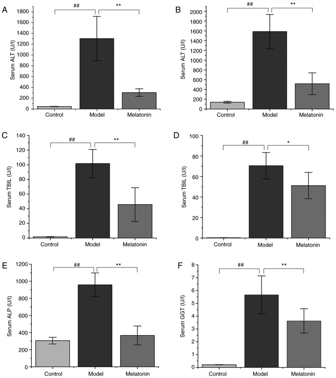
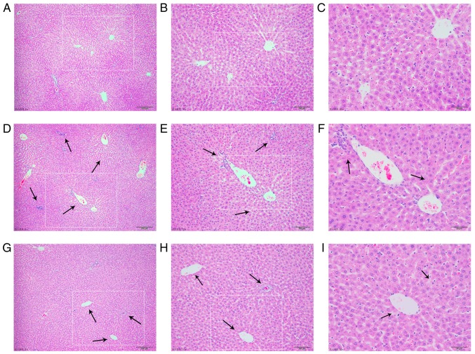
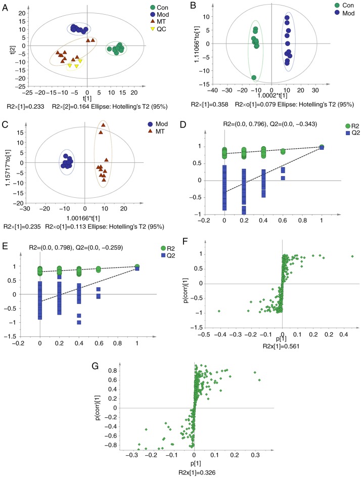
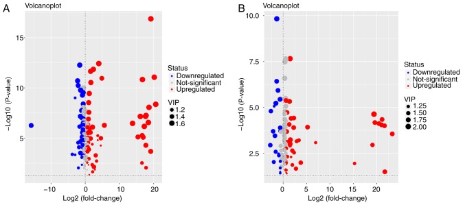
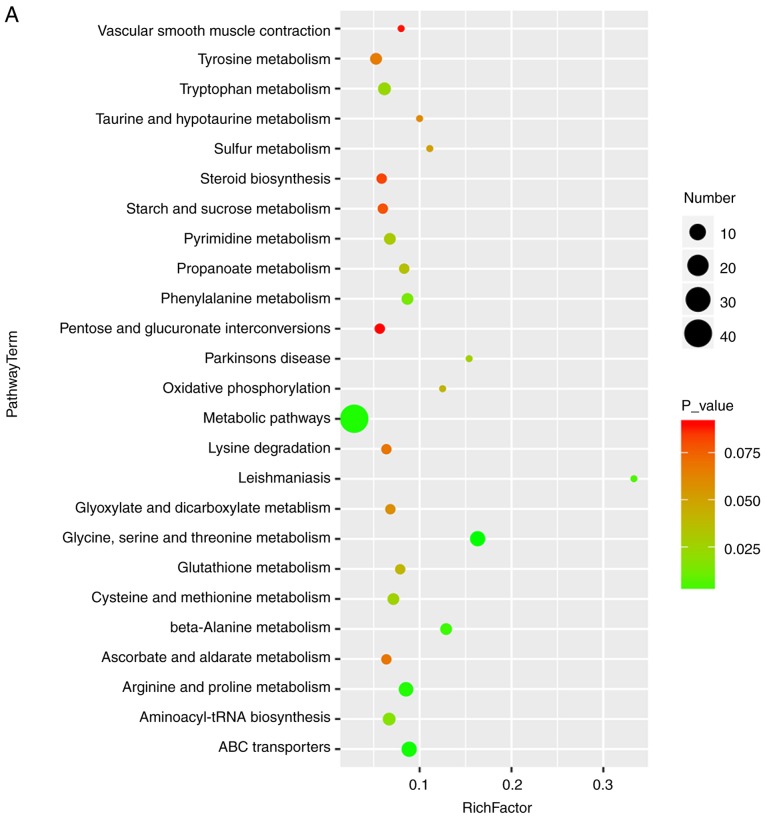
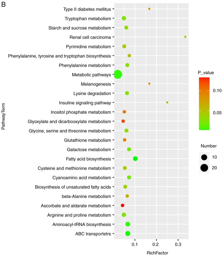
The present study investigated the anti‑cholestatic effect of melatonin (MT) against α‑naphthyl isothiocyanate (ANIT)‑induced liver injury in rats and screened for potential biomarkers of cholestasis. Rats were administered ANIT by intraperitoneal injection and then sacrificed 36 h later. Serum biochemical parameters were measured and liver tissue samples were subjected to histological analysis. Active components in the serum were identified by gas chromatography‑mass spectrometry, while biomarkers and biochemical pathways were identified by multivariate data analysis. The results revealed that the serum levels of alanine aminotransferase, aspartate aminotransferase, total bilirubin, direct bilirubin, γ‑glutamyl transpeptidase, and alkaline phosphatase were reduced in rats with ANIT‑induced cholestasis that were treated with MT. The histological observations indicated that MT had a protective effect against ANIT‑induced hepatic tissue damage. Metabolomics analysis revealed that this effect was likely to be associated with the regulation of compounds related to MT synthesis and catabolism, and amino acid metabolism, including 5‑aminopentanoate, 5‑methoxytryptamine, L‑tryptophan, threonine, glutathione, L‑methionine, and indolelactate. In addition, principal component analysis demonstrated that the levels of these metabolites differed significantly between the MT and control groups, providing further evidence that they may be responsible for the effects induced by MT. These results provide an insight into the mechanisms underlying cholestasis development and highlight potential biomarkers for disease diagnosis.
本研究探讨了褪黑素(MT)对α-萘异硫氰酸酯(ANIT)诱导的大鼠肝损伤的抗胆汁淤积作用,并筛选了潜在的胆汁淤积生物标志物。大鼠通过腹腔注射给予 ANIT,然后在 36 小时后处死。测量血清生化参数,并对肝组织样本进行组织学分析。通过气相色谱-质谱联用仪鉴定血清中的活性成分,通过多元数据分析鉴定生物标志物和生化途径。结果表明,MT 治疗 ANIT 诱导的胆汁淤积大鼠的血清丙氨酸氨基转移酶、天冬氨酸氨基转移酶、总胆红素、直接胆红素、γ-谷氨酰转肽酶和碱性磷酸酶水平降低。组织学观察表明,MT 对 ANIT 诱导的肝组织损伤具有保护作用。代谢组学分析表明,这种作用可能与 MT 合成和分解代谢以及氨基酸代谢相关化合物的调节有关,包括 5-氨基戊酸、5-甲氧基色胺、L-色氨酸、苏氨酸、谷胱甘肽、L-蛋氨酸和吲哚乳酸。此外,主成分分析表明,这些代谢物在 MT 和对照组之间的水平有显著差异,进一步证明它们可能是 MT 诱导作用的原因。这些结果深入了解了胆汁淤积发展的机制,并强调了疾病诊断的潜在生物标志物。