Liver Cancer Institute, Zhongshan Hospital, Fudan University and Key Laboratory of Carcinogenesis and Cancer Invasion, Ministry of Education, Shanghai, China.
BMC Cancer. 2018 Sep 18;18(1):901. doi: 10.1186/s12885-018-4820-9.
Accelerated malignant behaviors induced by insufficient thermal ablation have been increasingly reported, however, the exact mechanisms are still unclear. Here, we investigated the importance of the extracellular matrix (ECM) in modulating the progression of residual hepatocellular carcinoma (HCC) after heat treatment.
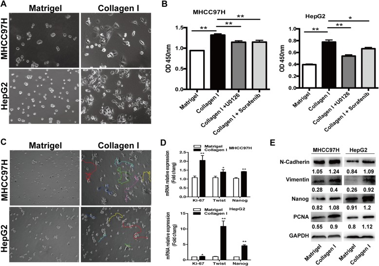
Heat-exposed residual HCC cells were cultured in different ECM gels. We used basement membrane gel (Matrigel) to simulate the normal microenvironment and collagen I to model the pathological stromal ECM. The alterations of morphology and parameters of proliferation, epithelial-mesenchymal transition (EMT) and stemness were analyzed in vitro and in vivo.
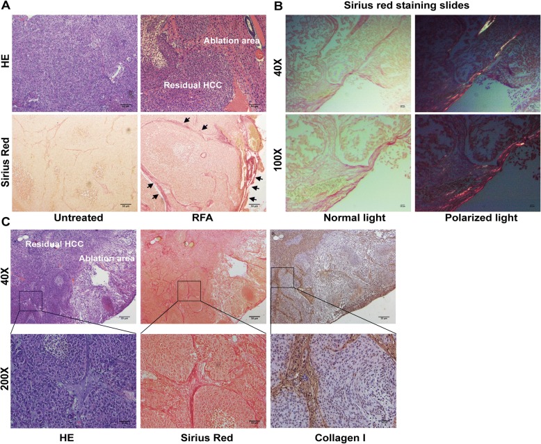
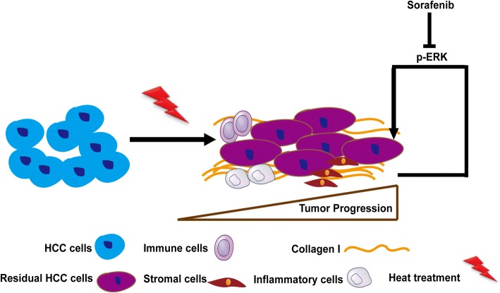
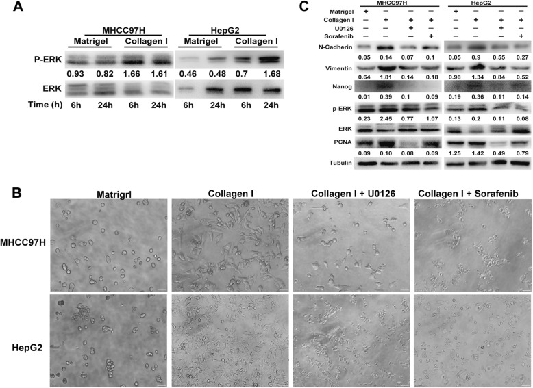
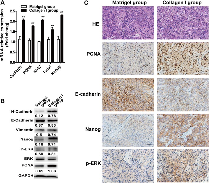
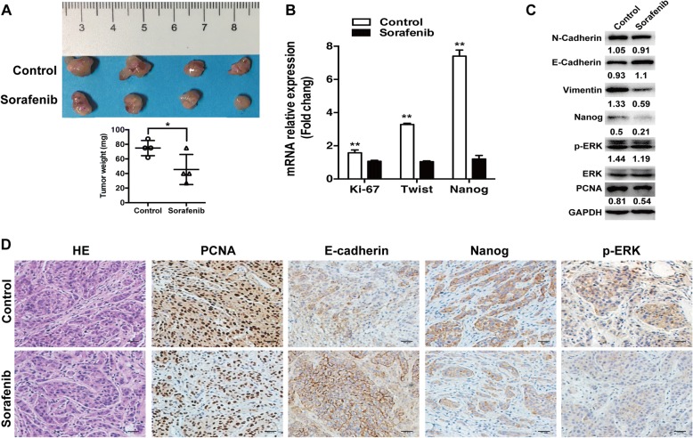
Increased collagen I deposition was observed at the periablational zone after incomplete RFA of HCC in a xenograft model. The markers of cell proliferation, EMT, motility and progenitor-like traits of heat-exposed residual HCC cells were significantly induced by collagen I as compared to Matrigel (p values all < 0.05). Importantly, collagen I induced the activation of ERK phosphorylation in heat-exposed residual HCC cells. ERK1/2 inhibitor reversed the collagen I-promoted ERK phosphorylation, cell proliferative, protrusive and spindle-like appearance of heat-treated residual HCC cells in vitro. Moreover, collagen I promoted the in vivo tumor progression of heat-exposed residual HCC cells, and sorafenib markedly reversed the collagen I-mediated protumor effects.
Our findings demonstrate that collagen I could enhance the aggressive progression of residual HCC cells after suboptimal heat treatment and sorafenib may be a treatment approach to thwart this process.
热消融治疗后,加速的恶性行为已被越来越多地报道,但确切的机制仍不清楚。在这里,我们研究了细胞外基质(ECM)在调节热治疗后残留肝细胞癌(HCC)进展中的重要性。
将热暴露的残留 HCC 细胞在不同的 ECM 凝胶中培养。我们使用基底膜凝胶(Matrigel)模拟正常微环境,并用胶原 I 模拟病理性基质 ECM。在体外和体内分析了形态和增殖、上皮-间充质转化(EMT)和干性参数的变化。
在 HCC 的不完全 RFA 后,异种移植模型中在围消融区观察到胶原 I 沉积增加。与 Matrigel 相比,热暴露的残留 HCC 细胞的细胞增殖、EMT、迁移和祖细胞样特征的标志物明显被胶原 I 诱导(p 值均<0.05)。重要的是,胶原 I 诱导了热暴露的残留 HCC 细胞中 ERK 磷酸化的激活。ERK1/2 抑制剂逆转了胶原 I 促进的 ERK 磷酸化、热处理残留 HCC 细胞的体外增殖、突起和纺锤形外观。此外,胶原 I 促进了热暴露的残留 HCC 细胞在体内的肿瘤进展,而索拉非尼显著逆转了胶原 I 介导的促肿瘤作用。
我们的研究结果表明,胶原 I 可以增强亚最佳热治疗后残留 HCC 细胞的侵袭性进展,而索拉非尼可能是阻止这一过程的治疗方法。