Stulpinas Rokas, Morkunas Mindaugas, Rasmusson Allan, Drachneris Julius, Augulis Renaldas, Gulla Aiste, Strupas Kestutis, Laurinavicius Arvydas
Faculty of Medicine, Institute of Biomedical Sciences, Department of Pathology and Forensic Medicine, Vilnius University, 03101 Vilnius, Lithuania.
National Center of Pathology, Affiliate of Vilnius University Hospital Santaros Klinikos, 08406 Vilnius, Lithuania.
Cancers (Basel). 2023 Dec 24;16(1):106. doi: 10.3390/cancers16010106.
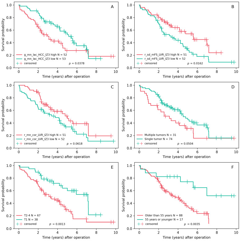
Despite advances in diagnostic and treatment technologies, predicting outcomes of patients with hepatocellular carcinoma (HCC) remains a challenge. Prognostic models are further obscured by the variable impact of the tumor properties and the remaining liver parenchyma, often affected by cirrhosis or non-alcoholic fatty liver disease that tend to precede HCC. This study investigated the prognostic value of reticulin and collagen microarchitecture in liver resection samples. We analyzed 105 scanned tissue sections that were stained using a Gordon and Sweet's silver impregnation protocol combined with Picric Acid-Sirius Red. A convolutional neural network was utilized to segment the red-staining collagen and black linear reticulin strands, generating a detailed map of the fiber structure within the HCC and adjacent liver tissue. Subsequent hexagonal grid subsampling coupled with automated epithelial edge detection and computational fiber morphometry provided the foundation for region-specific tissue analysis. Two penalized Cox regression models using LASSO achieved a concordance index (C-index) greater than 0.7. These models incorporated variables such as patient age, tumor multifocality, and fiber-derived features from the epithelial edge in both the tumor and liver compartments. The prognostic value at the tumor edge was derived from the reticulin structure, while collagen characteristics were significant at the epithelial edge of peritumoral liver. The prognostic performance of these models was superior to models solely reliant on conventional clinicopathologic parameters, highlighting the utility of AI-extracted microarchitectural features for the management of HCC.
尽管诊断和治疗技术取得了进展,但预测肝细胞癌(HCC)患者的预后仍然是一项挑战。肿瘤特性和剩余肝实质的可变影响进一步模糊了预后模型,这些肝实质通常受肝硬化或非酒精性脂肪性肝病影响,而这些疾病往往先于HCC出现。本研究调查了肝切除样本中网硬蛋白和胶原微结构的预后价值。我们分析了105个扫描组织切片,这些切片采用戈登-斯威特银浸染法结合苦味酸-天狼星红染色。利用卷积神经网络对红色染色的胶原和黑色线性网硬蛋白链进行分割,生成HCC及相邻肝组织内纤维结构的详细图谱。随后的六边形网格二次抽样,结合自动上皮边缘检测和计算纤维形态学,为区域特异性组织分析奠定了基础。使用LASSO的两个惩罚Cox回归模型的一致性指数(C指数)大于0.7。这些模型纳入了患者年龄、肿瘤多灶性以及肿瘤和肝区上皮边缘的纤维衍生特征等变量。肿瘤边缘的预后价值源于网硬蛋白结构,而胶原特征在肿瘤周围肝组织的上皮边缘具有显著性。这些模型的预后性能优于仅依赖传统临床病理参数的模型,突出了人工智能提取的微结构特征在HCC管理中的实用性。