Department of Cell and Molecular Biology, Faculty of Biotechnology and Biomolecular Sciences, Universiti Putra Malaysia, Serdang, Selangor, Malaysia,
Material Synthesis and Characterization Laboratory, Institute of Advanced Technology, Universiti Putra Malaysia, Serdang, Selangor, Malaysia,
Int J Nanomedicine. 2018 Sep 5;13:5075-5095. doi: 10.2147/IJN.S164843. eCollection 2018.
Inefficient cellular delivery and poor intracellular accumulation are major drawbacks towards achieving favorable therapeutic responses from many therapeutic drugs and biomolecules. To tackle this issue, nanoparticle-mediated delivery vectors have been aptly explored as a promising delivery strategy capable of enhancing the cellular localization of biomolecules and improve their therapeutic efficacies. However, the dynamics of intracellular biomolecule release and accumulation from such nanoparticle systems has currently remained scarcely studied.
The objective of this study was to utilize a chitosan-based nanoparticle system as the delivery carrier for glutamic acid, a model for encapsulated biomolecules to visualize the in vitro release and accumulation of the encapsulated glutamic acid from chitosan nanoparticle (CNP) systems.
CNP was synthesized via ionic gelation routes utilizing tripolyphosphate (TPP) as a cross-linker. In order to track glutamic acid release, the glutamic acid was fluorescently-labeled with fluorescein isothiocyanate prior encapsulation into CNP.
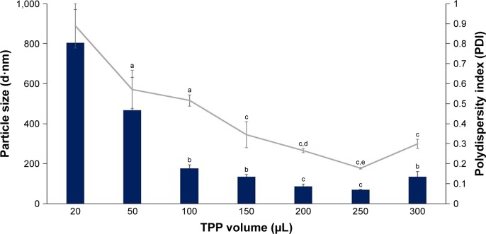
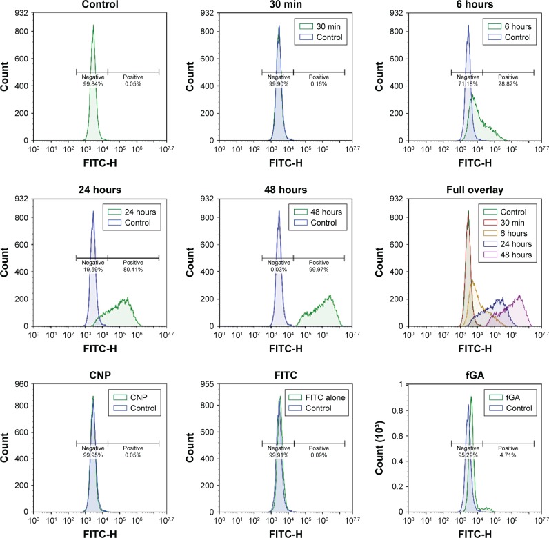
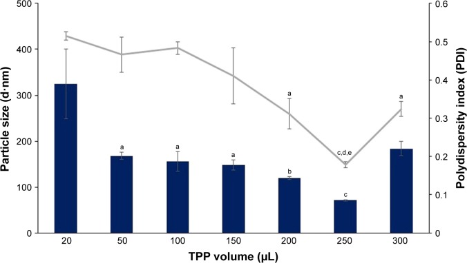
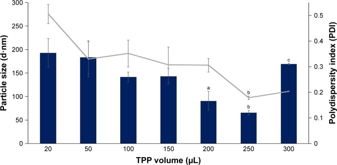
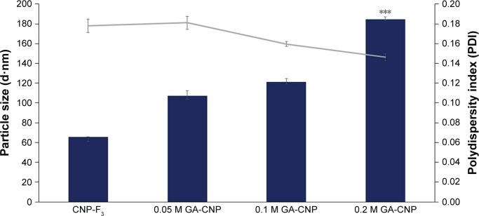
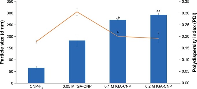
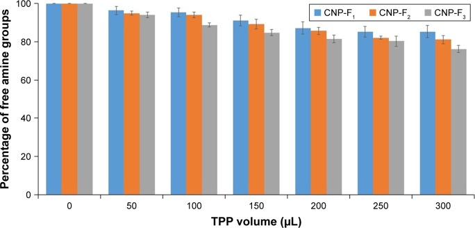
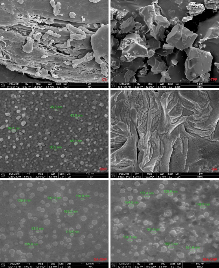
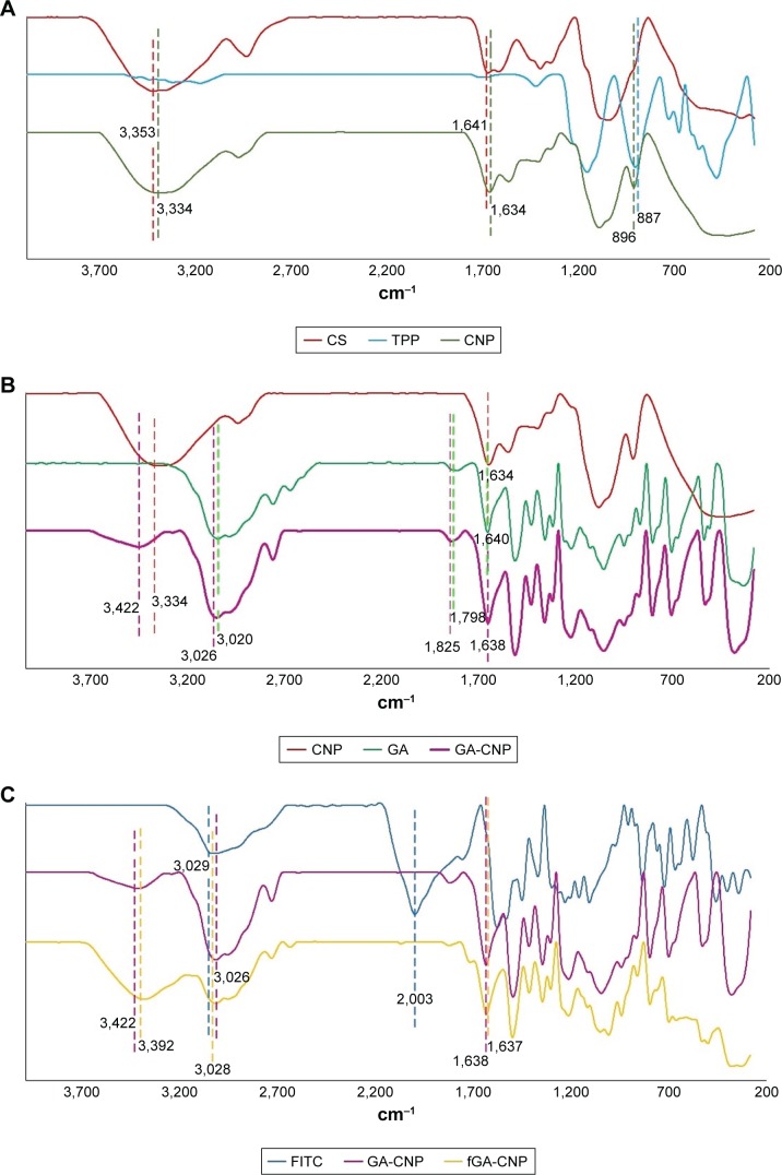
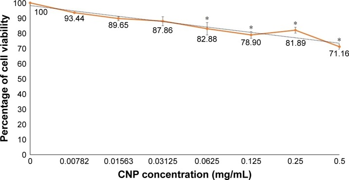
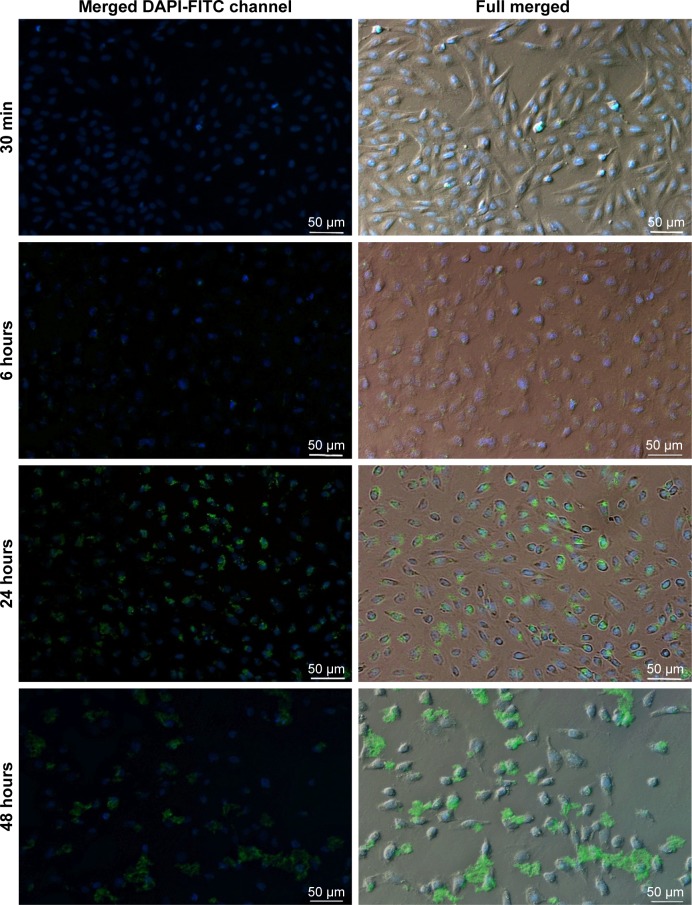
Light Scattering data concluded the successful formation of small-sized and mono-dispersed CNP at a specific volume ratio of chitosan to TPP. Encapsulation of glutamic acid as a model cargo into CNP led to an increase in particle size to >100 nm. The synthesized CNP exhibited spherical shape under Electron Microscopy. The formation of CNP was reflected by the reduction in free amine groups of chitosan following ionic crosslinking reactions. The encapsulation of glutamic acid was further confirmed by Fourier Transform Infrared (FTIR) analysis. Cell viability assay showed 70% cell viability at the maximum concentration of 0.5 mg/mL CS and 0.7 mg/mL TPP used, indicating the low inherent toxicity property of this system. In vitro release study using fluorescently-tagged glutamic acids demonstrated the release and accumulation of the encapsulated glutamic acids at 6 hours post treatment. A significant accumulation was observed at 24 hours and 48 hours later. Flow cytometry data demonstrated a gradual increase in intracellular fluorescence signal from 30 minutes to 48 hours post treatment with fluorescently-labeled glutamic acids encapsulated CNP.
These results therefore suggested the potential of CNP system towards enhancing the intracellular delivery and release of the encapsulated glutamic acids. This CNP system thus may serves as a potential candidate vector capable to improve the therapeutic efficacy for drugs and biomolecules in medical as well as pharmaceutical applications through the enhanced intracellular release and accumulation of the encapsulated cargo.
许多治疗药物和生物分子的细胞递送效率低和细胞内积累差是实现良好治疗反应的主要障碍。为了解决这个问题,纳米颗粒介导的递药载体作为一种有前途的递药策略得到了恰当的探索,该策略能够增强生物分子的细胞定位并提高其治疗效果。然而,目前对于这种纳米颗粒系统中细胞内生物分子释放和积累的动力学仍知之甚少。
本研究旨在利用壳聚糖纳米颗粒作为载体,对谷氨酸进行包裹,作为模型包裹生物分子,以可视化壳聚糖纳米颗粒(CNP)系统中包裹谷氨酸的体外释放和积累。
通过使用三聚磷酸钠(TPP)作为交联剂的离子凝胶化途径合成 CNP。为了跟踪谷氨酸的释放,在将谷氨酸包裹到 CNP 之前,用异硫氰酸荧光素对谷氨酸进行荧光标记。
光散射数据表明,在壳聚糖与 TPP 的特定体积比下,成功形成了小尺寸和单分散的 CNP。将谷氨酸作为模型货物包裹到 CNP 中会导致粒径增加到> 100nm。电子显微镜下合成的 CNP 呈球形。壳聚糖的游离胺基减少反映了离子交联反应后 CNP 的形成。傅里叶变换红外(FTIR)分析进一步证实了谷氨酸的包封。细胞活力测定显示,在使用的最大浓度 0.5mg/mLCS 和 0.7mg/mLTPP 下,细胞存活率为 70%,表明该系统的固有毒性低。使用荧光标记的谷氨酸进行体外释放研究表明,在处理后 6 小时内释放和积累了包裹的谷氨酸。24 小时和 48 小时后观察到明显的积累。流式细胞术数据表明,用荧光标记的谷氨酸包裹的 CNP 处理后 30 分钟至 48 小时,细胞内荧光信号逐渐增加。
这些结果表明 CNP 系统具有增强包裹谷氨酸的细胞内递送和释放的潜力。因此,该 CNP 系统可能是一种有前途的载体,通过增强包裹货物的细胞内释放和积累,能够提高药物和生物分子在医学和制药领域的治疗效果。