Liubaviciute Ausra, Kaseta Vytautas, Vaitkuviene Aida, Mackiewicz Zygmunt, Biziuleviciene Gene
State Research Institute Centre for Innovative Medicine, Department of Stem Cell Biology, Santariskiu str. 5, LT-08406 Vilnius, Lithuania.
State Research Institute Centre for Innovative Medicine, Department of Regenerative Medicine, Santariskiu str. 5, LT-08406 Vilnius, Lithuania.
EXCLI J. 2018 Aug 31;17:871-888. doi: 10.17179/excli2018-1504. eCollection 2018.
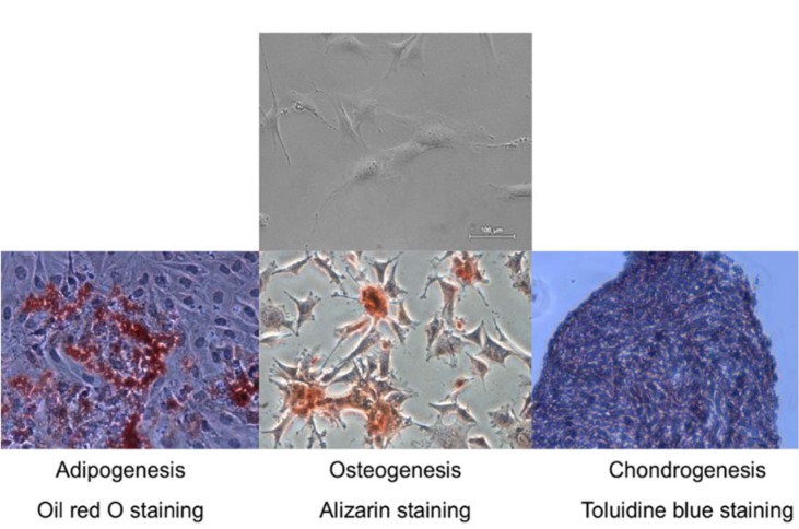
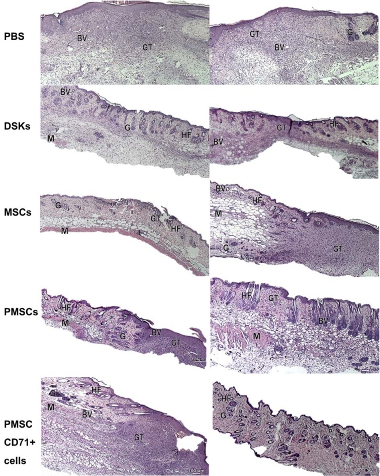
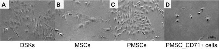
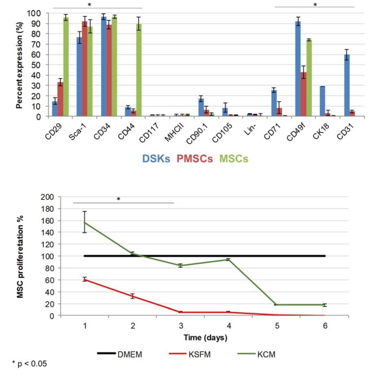
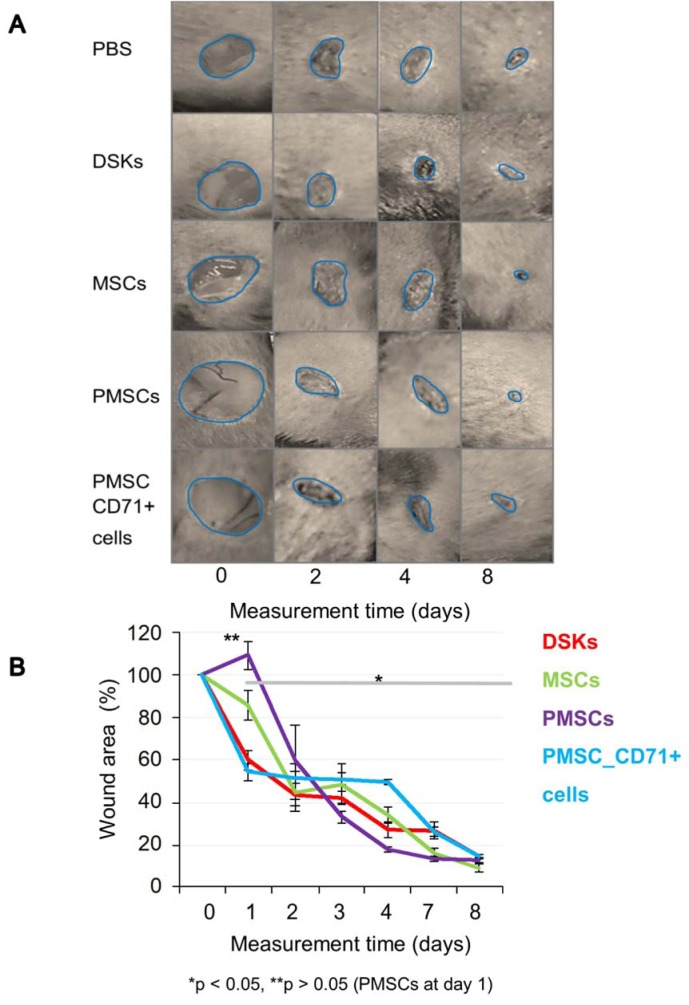
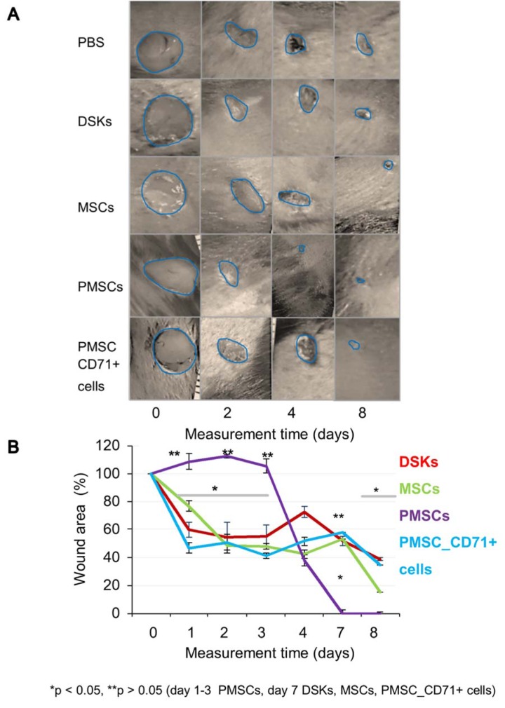
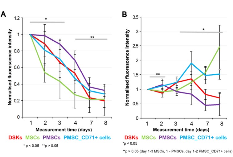
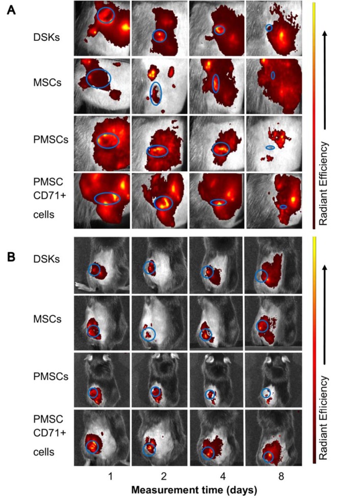
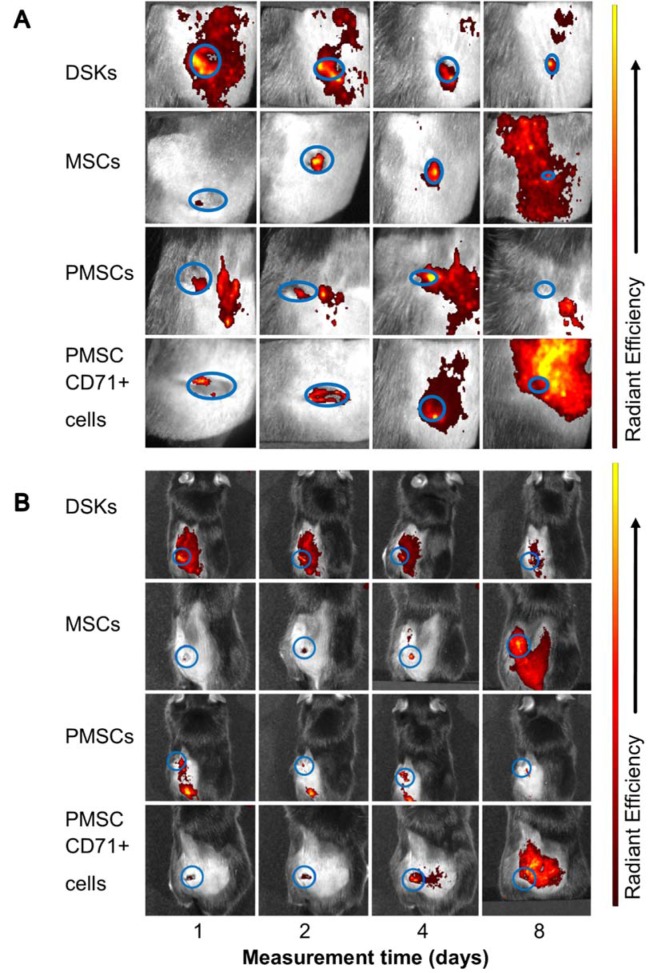
Mesenchymal stromal cells (MSCs, known as mesenchymal stem cells) are considered to be a promising therapeutic tool for many diseases. But it is still unclear which cells are more efficient and safe for wound healing and tissue regeneration for clinical applications: undifferentiated, partially differentiated stem cells or differentiated cells. In this study, we modified MSCs with keratinocyte-conditioned medium (KCM) and examined MSCs, partially differentiated MSCs (PMSCs) and differentiated cell migration, accumulation in the wounded area as well as cell regenerative efficiency in a full-thickness skin wound model. In addition to that, the impact of intradermal and intravenous cell delivery methods of wound healing was evaluated. C57BL/6J mouse compact bone MSCs were treated with a KCM for 14 days. Flow cytometry analysis showed the appearance of keratinocyte surface markers which were absent in MSCs, whereas the specific markers for MSCs were lost. Cells were injected either intravenously or intradermally in C57BL/6J mice. Wound closure, cell migration and accumulation in the wounded area were further analysed. Wound healing was assessed by the rate of wound closure and by histological evaluation. Cells were monitored using optical imaging. We demonstrated that PMSCs showed morphology similar to keratinocyte cells, had enhanced migration and increased survival at the site of injury. PMSCs had a beneficial effect on wound healing and tissue regeneration. This effect was reinforced when these cells were injected intravenously. Due to their partial differentiation status, we assume that PMSCs can differentiate more rapidly into epidermal cell lineages thus causing faster and qualitatively improved wound healing.
间充质基质细胞(MSCs,即间充质干细胞)被认为是治疗多种疾病的一种很有前景的治疗工具。但对于临床应用中,哪种细胞在伤口愈合和组织再生方面更有效且更安全仍不清楚:未分化的、部分分化的干细胞还是已分化的细胞。在本研究中,我们用角质形成细胞条件培养基(KCM)对MSCs进行修饰,并在全层皮肤伤口模型中检测了MSCs、部分分化的MSCs(PMSCs)和已分化细胞的迁移、在伤口区域的聚集以及细胞再生效率。此外,还评估了皮内和静脉内细胞递送方法对伤口愈合的影响。用KCM处理C57BL/6J小鼠致密骨MSCs 14天。流式细胞术分析显示MSCs中不存在的角质形成细胞表面标志物出现,而MSCs的特异性标志物丢失。将细胞静脉内或皮内注射到C57BL/6J小鼠体内。进一步分析伤口闭合情况、细胞在伤口区域的迁移和聚集。通过伤口闭合率和组织学评估来评估伤口愈合情况。使用光学成像监测细胞。我们证明PMSCs表现出与角质形成细胞相似的形态,在损伤部位具有增强的迁移能力和更高的存活率。PMSCs对伤口愈合和组织再生有有益作用。当这些细胞静脉内注射时,这种作用会增强。由于它们的部分分化状态,我们推测PMSCs可以更快地分化为表皮细胞谱系,从而导致更快且质量上更好的伤口愈合。