Department of Neurology, Seoul National University Hospital, 101, Daehak-ro, Jongno-gu, Seoul, 03080, South Korea.
Department of Neurology, National Center for Mental Health, Seoul, South Korea.
J Neuroinflammation. 2018 Sep 25;15(1):279. doi: 10.1186/s12974-018-1314-2.
While brain asymmetry has been a fascinating issue in neuroscience, the critical mechanism remains to be elucidated. Based on some index cases with asymmetric 18F-fluoro-2-deoxy-D-glucose positron emission tomography (FDG-PET) uptake in leucine-rich glioma-inactivated 1 (LGI1)-antibody encephalitis, we hypothesized LGI1 expression could be asymmetrically distributed in the human brain.
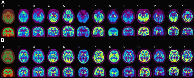
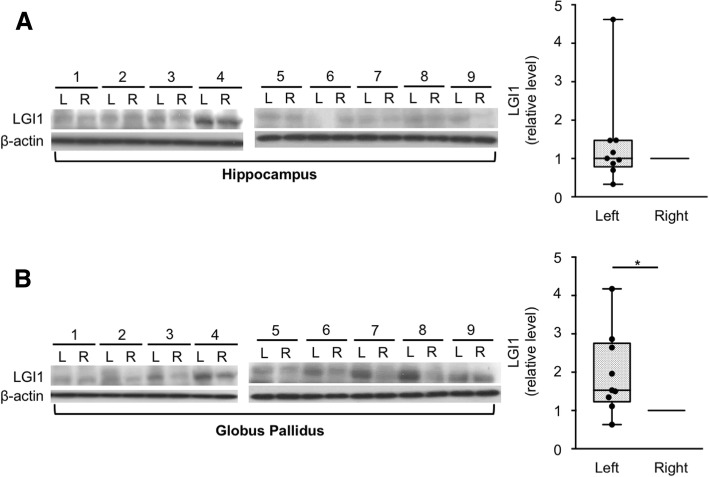
We enrolled 13 patients who were diagnosed with LGI1-antibody encephalitis between June 2012 and January 2018 at Seoul National University Hospital. Their pretreatment 18F-FDG-PET images were analyzed to find asymmetry between the left and right hemispheres. Guided by these observations, expression of LGI1 in the human hippocampus and the globus pallidus of both cerebral hemispheres was studied in nine post-mortem human brains.
Eleven of the 13 LGI1-antibody encephalitis patients (84.6%) showed asymmetrical FDG high uptake in the hippocampus: nine (81.8%) on the left hippocampus and two (18.2%) on the right. In the basal ganglia, seven patients (53.8%) showed asymmetry: four (57.1%) on the left and three (42.9%) on the right. The asymmetry was not evident in the laterality of faciobrachial dystonic seizures, brain MRI, and EEG. When the expression of LGI1 protein was analyzed in nine post-mortem human brains by western blotting, LGI1 expression was higher on eight left globus pallidus samples (88.89%, P = 0.019) and on four left hippocampal samples (44.44%, P = 0.652), compared to their right hemisphere samples.
Imaging parameters from patients with LGI1-antibody encephalitis and studies of LGI1 protein expression suggest that LGI1 is asymmetrically distributed in the human brain. These observations have implications for our understanding of human brain development.
大脑不对称性一直是神经科学中的一个迷人问题,但关键机制仍有待阐明。基于一些具有不对称性 18F-氟-2-脱氧-D-葡萄糖正电子发射断层扫描(FDG-PET)摄取的亮氨酸丰富胶质瘤失活 1(LGI1)-抗体脑炎的指数病例,我们假设 LGI1 表达可能在人脑中有不对称分布。
我们招募了 13 名于 2012 年 6 月至 2018 年 1 月在首尔国立大学医院被诊断为 LGI1-抗体脑炎的患者。分析了他们的预处理 18F-FDG-PET 图像,以发现左右半球之间的不对称性。根据这些观察结果,在九个人死后的大脑中研究了左右半球 LGI1 在海马体和苍白球中的表达。
13 名 LGI1-抗体脑炎患者中有 11 名(84.6%)表现出海马体 FDG 摄取的不对称性高:左海马体 9 名(81.8%),右海马体 2 名(18.2%)。在基底节中,7 名患者(53.8%)表现出不对称性:左 4 名(57.1%),右 3 名(42.9%)。偏侧性面肩肱型肌阵挛发作、脑 MRI 和 EEG 中没有明显的不对称性。通过 Western blot 分析九个人死后大脑中的 LGI1 蛋白表达,在 8 个左苍白球样本中(88.89%,P=0.019)和 4 个左海马体样本中(44.44%,P=0.652),LGI1 表达高于右半球样本。
LGI1-抗体脑炎患者的影像学参数和 LGI1 蛋白表达研究表明,LGI1 在人脑中有不对称分布。这些观察结果对我们理解人类大脑发育具有重要意义。