Department of Radiology, Zhongshan Hospital of Xiamen University, School of Medicine, Xiamen University, Xiamen, Fujian, China.
Department of Pathology, Zhongshan Hospital of Xiamen University, School of Medicine, Xiamen University, Xiamen, Fujian, China.
Eur J Med Res. 2024 Aug 22;29(1):431. doi: 10.1186/s40001-024-02017-w.
Accurate assessment of the depth of tumor invasion in gastric cancer (GC) is vital for the selection of suitable patients for neoadjuvant chemotherapy (NAC). Current problem is that preoperative differentiation between T1-2 and T3-4 stage cases in GC is always highly challenging for radiologists.
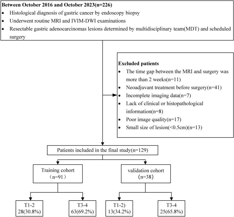
A total of 129 GC patients were divided into training (91 cases) and validation (38 cases) cohorts. Pathology from surgical specimens categorized patients into T1-2 and T3-4 stages. IVIM-DWI and MRI morphological characteristics were evaluated, and a multimodal nomogram was developed. The MRI morphological model, IVIM-DWI model, and combined model were constructed using logistic regression. Their effectiveness was assessed using receiver operating characteristic (ROC) curves, calibration curves, decision curve analysis (DCA), and clinical impact curves (CIC).
The combined nomogram, integrating preoperative IVIM-DWI parameters (D value) and MRI morphological characteristics (maximum tumor thickness, extra-serosal invasion), achieved the highest area under the curve (AUC) values of 0.901 and 0.883 in the training and validation cohorts, respectively. No significant difference was observed between the AUCs of the IVIM-DWI and MRI morphological models in either cohort (training: 0.796 vs. 0.835, p = 0.593; validation: 0.794 vs. 0.766, p = 0.79).
The multimodal nomogram, combining IVIM-DWI parameters and MRI morphological characteristics, emerges as a promising tool for assessing tumor invasion depth in GC, potentially guiding the selection of suitable candidates for neoadjuvant chemotherapy (NAC) treatment.
准确评估胃癌(GC)的肿瘤侵袭深度对于选择适合新辅助化疗(NAC)的患者至关重要。目前的问题是,对于放射科医生来说,术前区分 GC 的 T1-2 期和 T3-4 期病例始终极具挑战性。
共纳入 129 例 GC 患者,分为训练(91 例)和验证(38 例)队列。手术标本的病理学将患者分为 T1-2 和 T3-4 期。评估 IVIM-DWI 和 MRI 形态特征,并开发多模态列线图。使用逻辑回归构建 MRI 形态模型、IVIM-DWI 模型和联合模型。使用受试者工作特征(ROC)曲线、校准曲线、决策曲线分析(DCA)和临床影响曲线(CIC)评估其有效性。
结合术前 IVIM-DWI 参数(D 值)和 MRI 形态特征(最大肿瘤厚度、浆膜外侵犯)的联合列线图在训练和验证队列中的曲线下面积(AUC)值最高,分别为 0.901 和 0.883。在两个队列中,IVIM-DWI 模型和 MRI 形态模型的 AUC 值之间没有显著差异(训练:0.796 与 0.835,p=0.593;验证:0.794 与 0.766,p=0.79)。
结合 IVIM-DWI 参数和 MRI 形态特征的多模态列线图,是评估 GC 肿瘤侵袭深度的一种有前途的工具,可能有助于指导新辅助化疗(NAC)治疗的合适患者选择。