Sir Peter Mansfield Imaging Centre, School of Physics and Astronomy, University of Nottingham, University Park, Nottingham, NG7 2RD, UK.
Clinical Neurology, Division of Clinical Neuroscience, School of Medicine, University of Nottingham, Queen's Medical Centre, Nottingham, NG7 2UH, UK.
Eur Radiol. 2019 Apr;29(4):2027-2033. doi: 10.1007/s00330-018-5707-5. Epub 2018 Oct 2.
To assess the feasibility of using an optimised ultra-high-field high-spatial-resolution low-distortion arterial spin labelling (ASL) MRI acquisition to measure focal haemodynamic pathology in cortical lesions (CLs) in multiple sclerosis (MS).
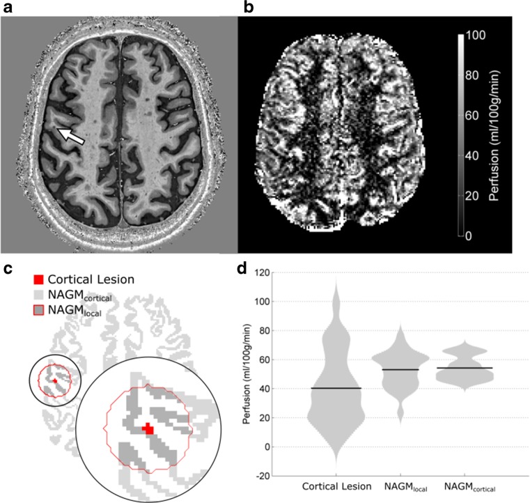
Twelve MS patients (eight female, mean age 50 years; range 35-64 years) gave informed consent and were scanned on a 7 Tesla Philips Achieva scanner. Perfusion data were collected at multiple post-labelling delay times using a single-slice flow-sensitive alternating inversion recovery ASL protocol with a balanced steady-state free precession readout scheme. CLs were identified using a high-resolution Phase-Sensitive Inversion Recovery (PSIR) scan. Significant differences in perfusion within CLs compared to immediately surrounding normal appearing grey matter (NAGM) and total cortical normal appearing grey matter (NAGM) were assessed using paired t-tests.
Forty CLs were identified in PSIR scans that overlapped with the ASL acquisition coverage. After excluding lesions due to small size or intravascular contamination, 27 lesions were eligible for analysis. Mean perfusion was 40 ± 25 ml/100 g/min in CLs, 53 ± 12 ml/100 g/min in NAGM, and 53 ± 8 ml/100 g/min in NAGM. CL perfusion was significantly reduced by 23 ± 9% (mean ± SE, p = 0.013) and 26 ± 9% (p = 0.006) relative to NAGM and NAGM perfusion, respectively.
This is the first ASL MRI study quantifying CL perfusion in MS at 7 Tesla, demonstrating that an optimised ASL acquisition is sensitive to focal haemodynamic pathology previously observed using dynamic susceptibility contrast MRI. ASL requires no exogenous contrast agent, making it a more appropriate tool to monitor longitudinal perfusion changes in MS, providing a new window to study lesion development.
• Perfusion can be quantified within cortical lesions in multiple sclerosis using an optimised high spatial resolution arterial spin Labelling MRI acquisition at ultra-high-field. • The majority of cortical lesions assessed using arterial spin labelling are hypo-perfused compared to normal appearing grey matter, in agreement with dynamic susceptibility contrast MRI literature. • Arterial spin labelling MRI, which does not involve the injection of a contrast agent, is a safe and appropriate technique for repeat scanning of an individual patient.
评估使用优化的超高场高空间分辨率低失真动脉自旋标记(ASL)MRI 采集来测量多发性硬化(MS)皮质病变(CL)中局灶性血液动力学病理的可行性。
12 名 MS 患者(8 名女性,平均年龄 50 岁;范围 35-64 岁)知情同意并在 7T 飞利浦 Achieva 扫描仪上进行扫描。使用单层面流动敏感交替反转恢复 ASL 协议和平衡稳态自由进动读出方案,在多个标记后延迟时间采集灌注数据。使用高分辨率相位敏感反转恢复(PSIR)扫描识别 CL。使用配对 t 检验评估 CL 内与紧邻正常表现灰质(NAGM)和总皮质正常表现灰质(NAGM)之间的灌注差异。
PSIR 扫描中识别出 40 个与 ASL 采集覆盖范围重叠的 CL。排除因体积小或血管内污染而导致的病变后,27 个病变可用于分析。CL 中的平均灌注为 40±25ml/100g/min,NAGM 中的灌注为 53±12ml/100g/min,NAGM 中的灌注为 53±8ml/100g/min。CL 灌注分别比 NAGM 和 NAGM 灌注降低 23±9%(平均值±SE,p=0.013)和 26±9%(p=0.006)。
这是首次在 7T 磁共振成像上定量研究多发性硬化 CL 灌注的 ASL MRI 研究,表明优化的 ASL 采集对以前使用动态对比磁共振成像观察到的局灶性血液动力学病理敏感。ASL 不需要外源性造影剂,因此更适合监测多发性硬化的纵向灌注变化,为研究病变发展提供了新的窗口。
使用优化的超高场高空间分辨率动脉自旋标记 MRI 采集,可以在多发性硬化的皮质病变内定量灌注。
使用动脉自旋标记 MRI 评估的大多数皮质病变与正常表现灰质相比灌注不足,与动态对比磁共振成像文献一致。
动脉自旋标记 MRI 不涉及造影剂的注射,是一种安全且适合个体患者重复扫描的技术。