Division of Veterinary Anatomy, Vetsuisse Faculty, University of Bern, Bern, Switzerland.
Geistlich Pharma AG, Wolhusen, Switzerland.
PLoS One. 2018 Oct 3;13(10):e0205027. doi: 10.1371/journal.pone.0205027. eCollection 2018.
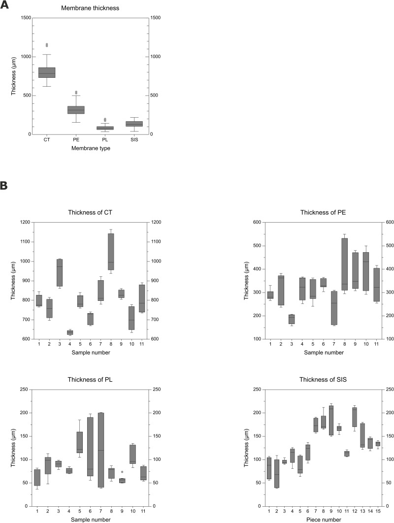
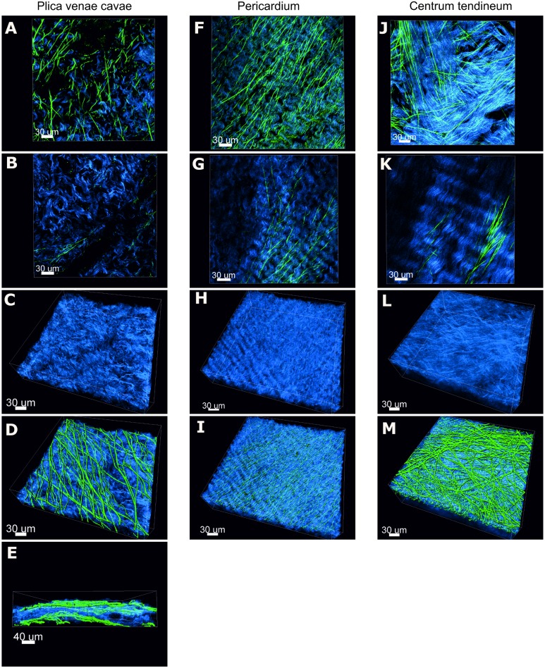
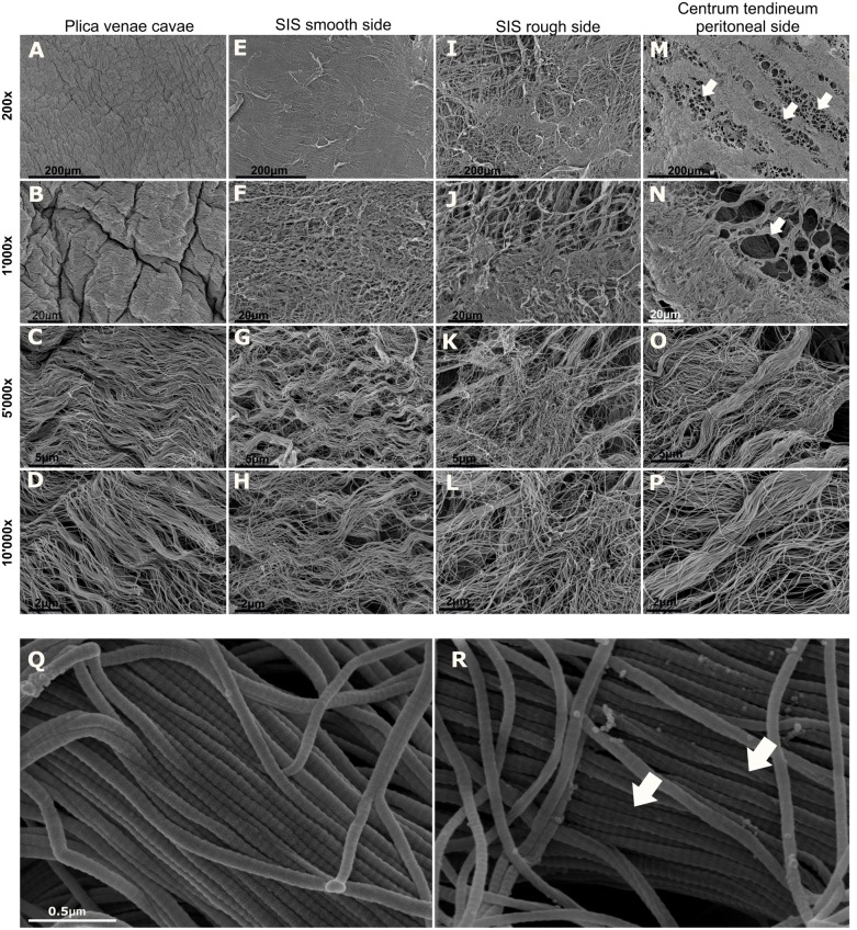
Collagen is the main structural element of connective tissues, and its favorable properties make it an ideal biomaterial for regenerative medicine. In dental medicine, collagen barrier membranes fabricated from naturally occurring tissues are used for guided bone regeneration. Since the morphological characteristics of collagen membranes play a crucial role in their mechanical properties and affect the cellular behavior at the defect site, in-depth knowledge of the structure is key. As a base for the development of novel collagen membranes, an extensive morphological analysis of four porcine membranes, including centrum tendineum, pericardium, plica venae cavae and small intestinal submucosa, was performed. Native membranes were analyzed in terms of their thickness. Second harmonic generation and two-photon excitation microscopy of the native membranes showed the 3D architecture of the collagen and elastic fibers, as well as a volumetric index of these two membrane components. The surface morphology, fiber arrangement, collagen fibril diameter and D-periodicity of decellularized membranes were investigated by scanning electron microscopy. All the membrane types showed significant differences in thickness. In general, undulating collagen fibers were arranged in stacked layers, which were parallel to the membrane surface. Multiphoton microscopy revealed a conspicuous superficial elastic fiber network, while the elastin content in deeper layers varied. The elastin/collagen volumetric index was very similar in the investigated membranes and indicated that the collagen content was clearly higher than the elastin content. The surface of both the pericardium and plica venae cavae and the cranial surface of the centrum tendineum revealed a smooth, tightly arranged and crumpled morphology. On the caudal face of the centrum tendineum, a compact collagen arrangement was interrupted by clusters of circular discontinuities. In contrast, both surfaces of the small intestinal submucosa were fibrous, fuzzy and irregular. All the membranes consisted of largely uniform fibrils displaying the characteristic D-banding. This study reveals similarities and relevant differences among the investigated porcine membranes, suggesting that each membrane represents a unique biomaterial suitable for specific applications.
胶原蛋白是结缔组织的主要结构元素,其优良的性能使其成为再生医学理想的生物材料。在牙科医学中,从天然组织制备的胶原屏障膜用于引导骨再生。由于胶原膜的形态特征对其力学性能起着至关重要的作用,并影响缺陷部位的细胞行为,因此深入了解其结构至关重要。作为新型胶原膜开发的基础,对包括中心腱、心包、腔静脉褶和小肠黏膜下层在内的四种猪源膜进行了广泛的形态分析。对天然膜的厚度进行了分析。天然膜的二次谐波产生和双光子激发显微镜显示了胶原和弹性纤维的 3D 结构,以及这两种膜成分的体积指数。通过扫描电子显微镜研究了脱细胞化膜的表面形态、纤维排列、胶原原纤维直径和 D 周期。所有膜类型的厚度均存在显著差异。一般来说,起伏的胶原纤维呈层叠排列,与膜表面平行。多光子显微镜显示出明显的表面弹性纤维网络,而深层的弹性蛋白含量则有所不同。所研究的膜中弹性蛋白/胶原体积指数非常相似,表明胶原含量明显高于弹性蛋白含量。心包和腔静脉褶的表面以及中心腱的颅面均呈现出光滑、紧密排列和起皱的形态。在中心腱的尾侧表面,致密的胶原排列被圆形不连续性的簇所中断。相比之下,小肠黏膜下层的两个表面均呈纤维状、模糊和不规则。所有的膜都由基本上均匀的原纤维组成,显示出特征性的 D 带。本研究揭示了所研究的猪源膜之间的相似性和相关差异,表明每种膜都是一种独特的生物材料,适用于特定的应用。