Clark Michael G, Smallwood Shoukry Rachel, Huang Caleb J, Danielian Laura E, Bageac Devin, Floeter Mary Kay
a National Institute of Neurological Disorders and Stroke , National Institutes of Health , Bethesda , MD , USA.
Amyotroph Lateral Scler Frontotemporal Degener. 2018 Nov;19(7-8):562-569. doi: 10.1080/21678421.2018.1517180. Epub 2018 Oct 9.
The clinical diagnosis of primary lateral sclerosis can only be made after upper motor neuron symptoms have progressed for several years without developing lower motor neuron signs. The goal of the study was to identify neuroimaging changes that occur early in primary lateral sclerosis, prior to clinical diagnosis.
MRI scans were obtained on 13 patients with adult-onset progressive spasticity for five years or less who were followed longitudinally to confirm a clinical diagnosis of primary lateral sclerosis. Resting state functional MRI, diffusion tensor imaging, and anatomical images were obtained. These "pre-PLS" patients were compared to 18 patients with longstanding, established primary lateral sclerosis and 28 controls.
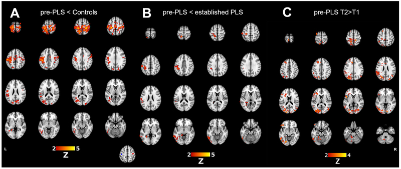
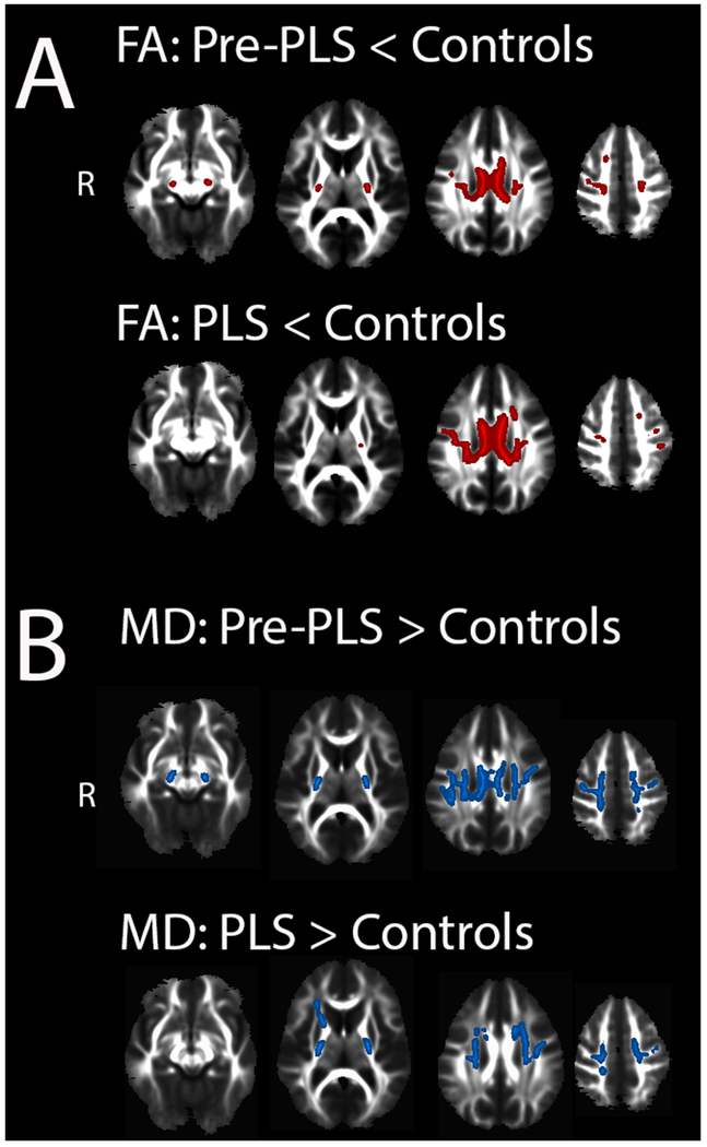
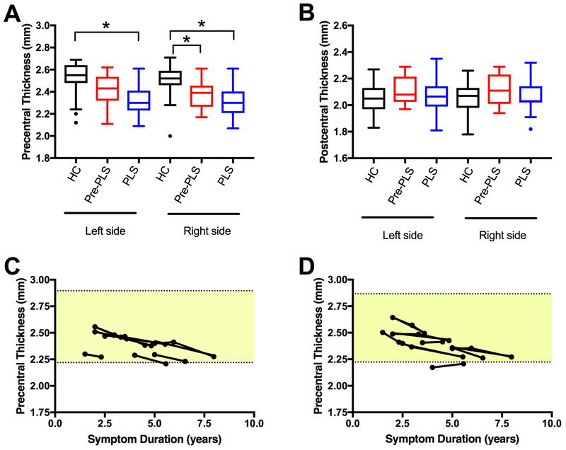
Pre-PLS patients had a marked reduction in seed-based resting-state motor network connectivity compared to the controls and patients with longstanding disease. White matter regions with reduced fractional anisotropy were similar in the two patient groups compared to the controls. Patients with longstanding disease had cortical thinning of the precentral gyrus. A slight thinning of the right precentral gyrus was detected in initial pre-PLS patients' scans. Follow-up scans in eight pre-PLS patients 1-2 years later showed increasing motor connectivity, thinning of the precentral gyrus, and no change in diffusion measures of the corticospinal tract or callosal motor region.
Loss of motor functional connectivity is an early imaging marker in primary lateral sclerosis. This differs from literature descriptions of amyotrophic lateral sclerosis, warranting further studies to test whether resting-state functional MRI can differentiate between amyotrophic lateral sclerosis and primary lateral sclerosis at early disease stages.
原发性侧索硬化症的临床诊断只能在上运动神经元症状进展数年且未出现下运动神经元体征后才能做出。本研究的目的是确定在临床诊断之前原发性侧索硬化症早期出现的神经影像学变化。
对13例成人起病、进行性痉挛5年或更短时间的患者进行了MRI扫描,并对其进行纵向随访以确诊原发性侧索硬化症。获取了静息态功能MRI、弥散张量成像和解剖图像。将这些“原发性侧索硬化症前期”患者与18例长期确诊的原发性侧索硬化症患者和28例对照组进行比较。
与对照组和长期患病患者相比,原发性侧索硬化症前期患者基于种子点的静息态运动网络连接性显著降低。与对照组相比,两组患者中各向异性分数降低的白质区域相似。长期患病患者中央前回皮质变薄。在原发性侧索硬化症前期患者的初始扫描中检测到右侧中央前回略有变薄。1 - 2年后对8例原发性侧索硬化症前期患者的随访扫描显示运动连接性增加、中央前回变薄,皮质脊髓束或胼胝体运动区的弥散测量无变化。
运动功能连接性丧失是原发性侧索硬化症的早期影像学标志物。这与肌萎缩侧索硬化症的文献描述不同,需要进一步研究以测试静息态功能MRI在疾病早期能否区分肌萎缩侧索硬化症和原发性侧索硬化症。