• 文献检索
  • 文档翻译
  • 深度研究
  • 学术资讯
  • Suppr Zotero 插件Zotero 插件
  • 邀请有礼
  • 套餐&价格
  • 历史记录
应用&插件
Suppr Zotero 插件Zotero 插件浏览器插件Mac 客户端Windows 客户端微信小程序
定价
高级版会员购买积分包购买API积分包
服务
文献检索文档翻译深度研究API 文档MCP 服务
关于我们
关于 Suppr公司介绍联系我们用户协议隐私条款
关注我们

Suppr 超能文献

核心技术专利:CN118964589B侵权必究
粤ICP备2023148730 号-1Suppr @ 2026

文献检索

告别复杂PubMed语法,用中文像聊天一样搜索,搜遍4000万医学文献。AI智能推荐,让科研检索更轻松。

立即免费搜索

文件翻译

保留排版,准确专业,支持PDF/Word/PPT等文件格式,支持 12+语言互译。

免费翻译文档

深度研究

AI帮你快速写综述,25分钟生成高质量综述,智能提取关键信息,辅助科研写作。

立即免费体验

骨质疏松性椎体终板和皮质骨折:图文综述

Osteoporotic vertebral endplate and cortex fractures: A pictorial review.

作者信息

Wáng Yì Xiáng J, Deng Min, He Lai-Chang, Che-Nordin Nazmi, Santiago Fernando Ruiz

机构信息

Department of Imaging and Interventional Radiology, The Chinese University of Hong Kong, Prince of Wales Hospital, Shatin, New Territories, Hong Kong Special Administrative Region.

Department of Radiology, The First Affiliated Hospital of Nanchang University, Nanchang, Jiangxi Province, China.

出版信息

J Orthop Translat. 2018 Sep 6;15:35-49. doi: 10.1016/j.jot.2018.08.004. eCollection 2018 Oct.

DOI:10.1016/j.jot.2018.08.004
PMID:30306044
原文链接:https://pmc.ncbi.nlm.nih.gov/articles/PMC6169255/
Abstract

UNLABELLED

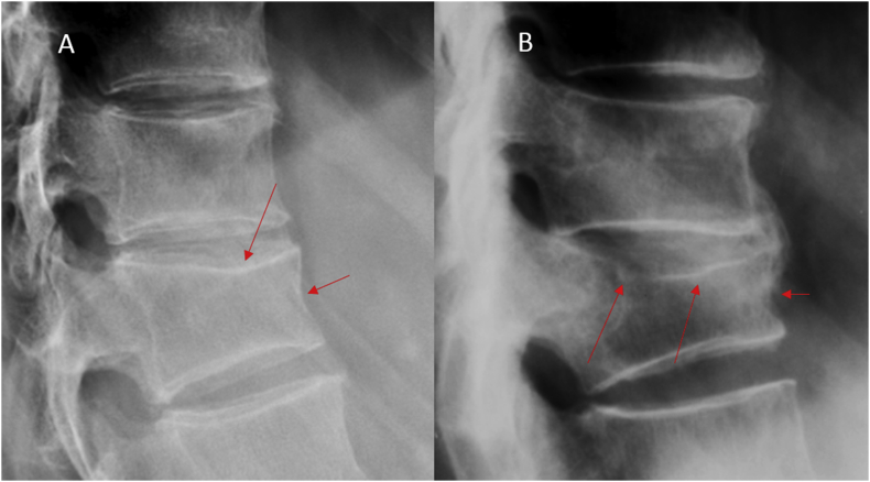
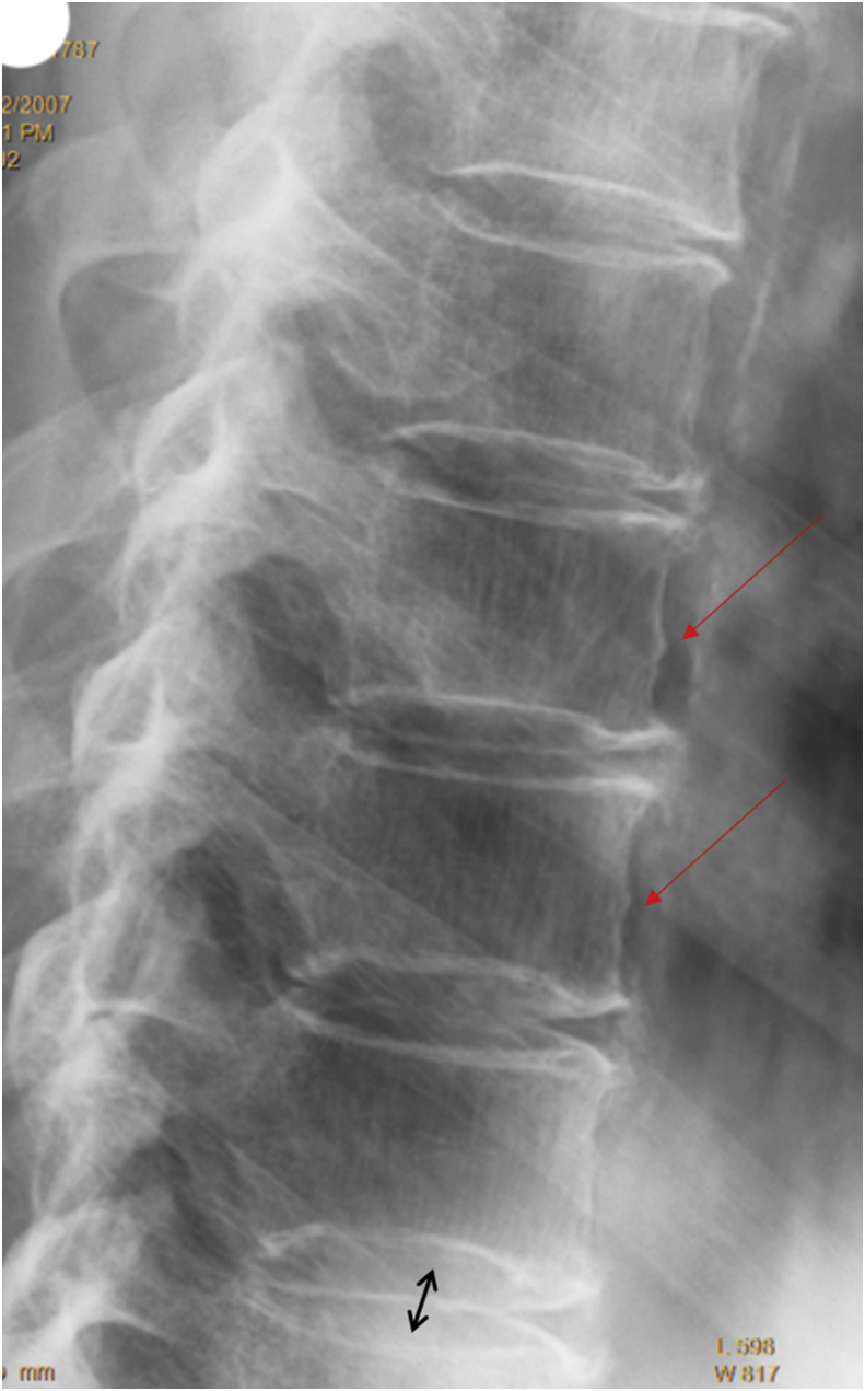
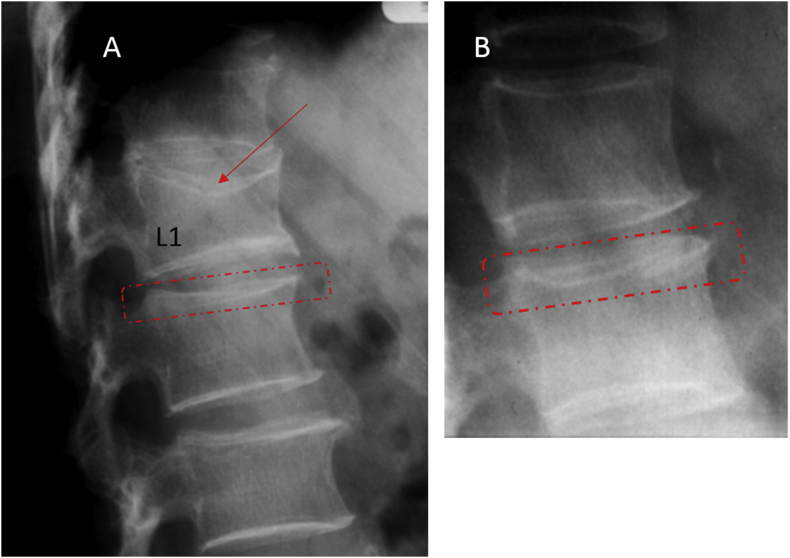
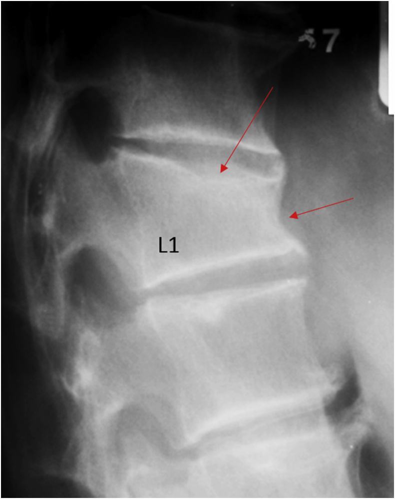
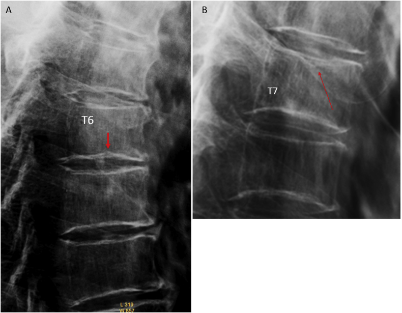
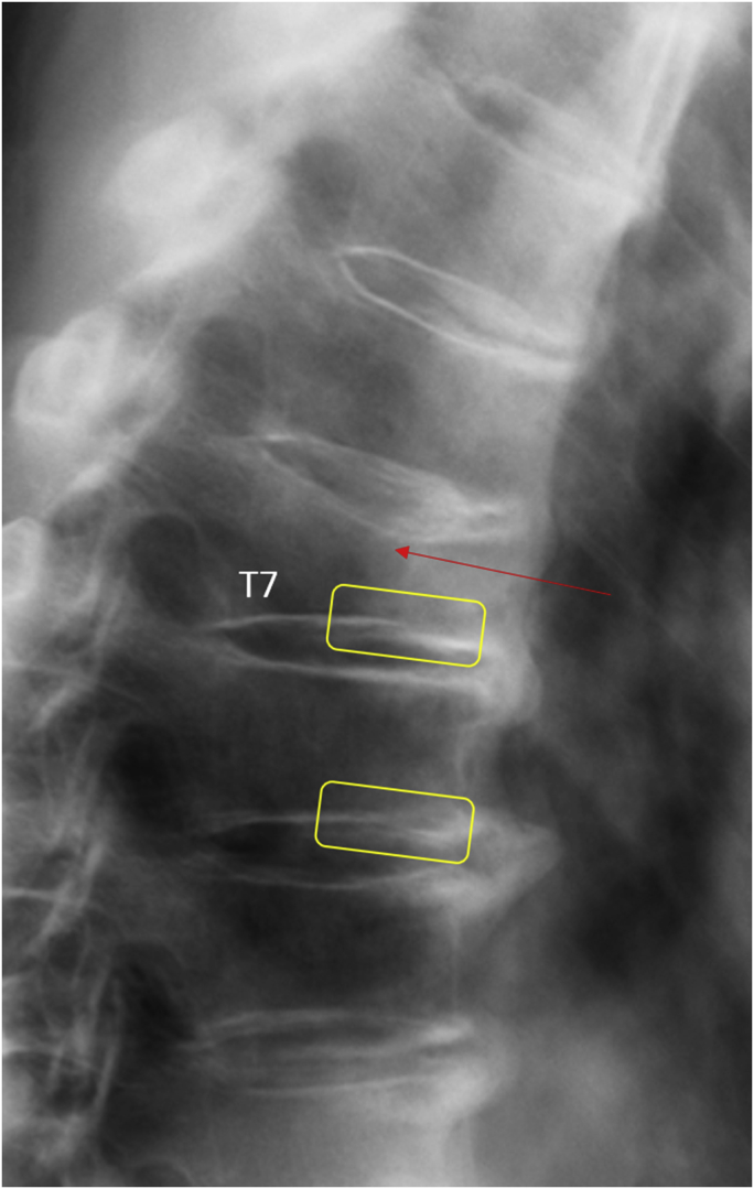
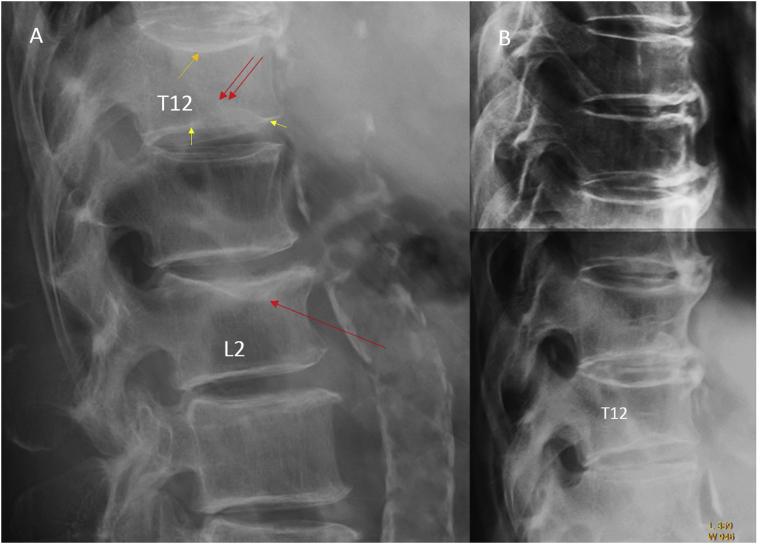
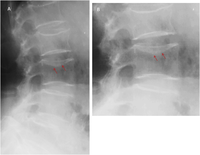
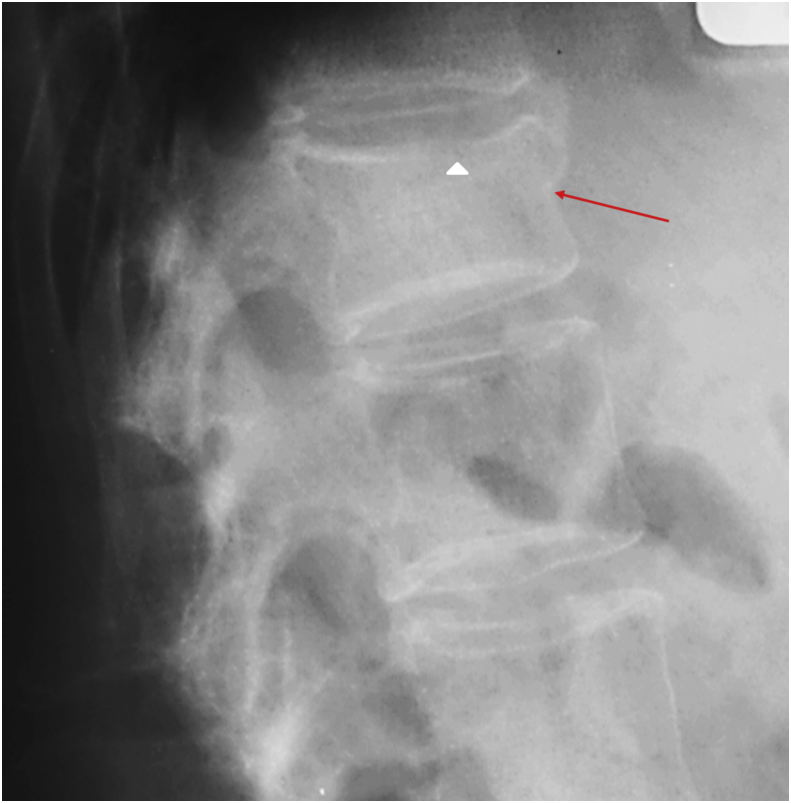
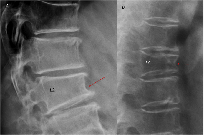
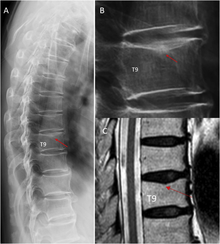
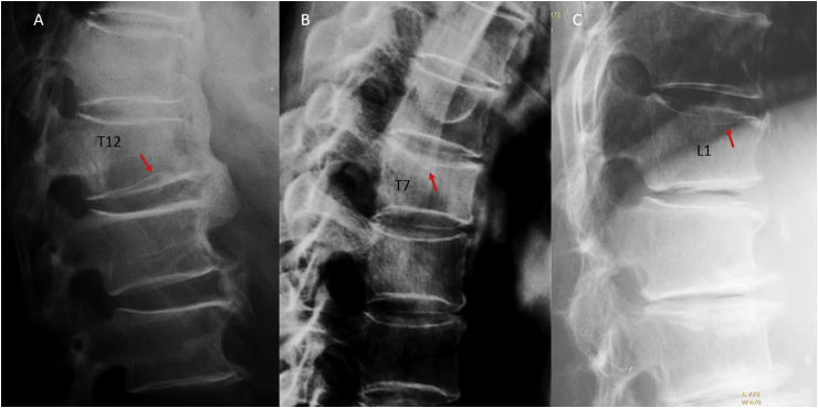
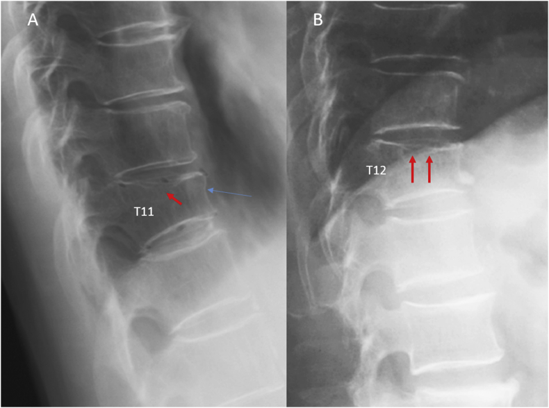
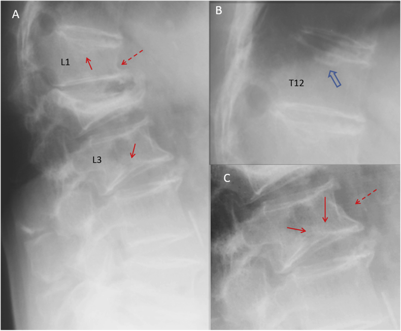
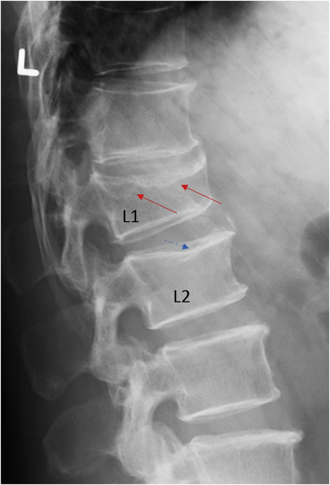
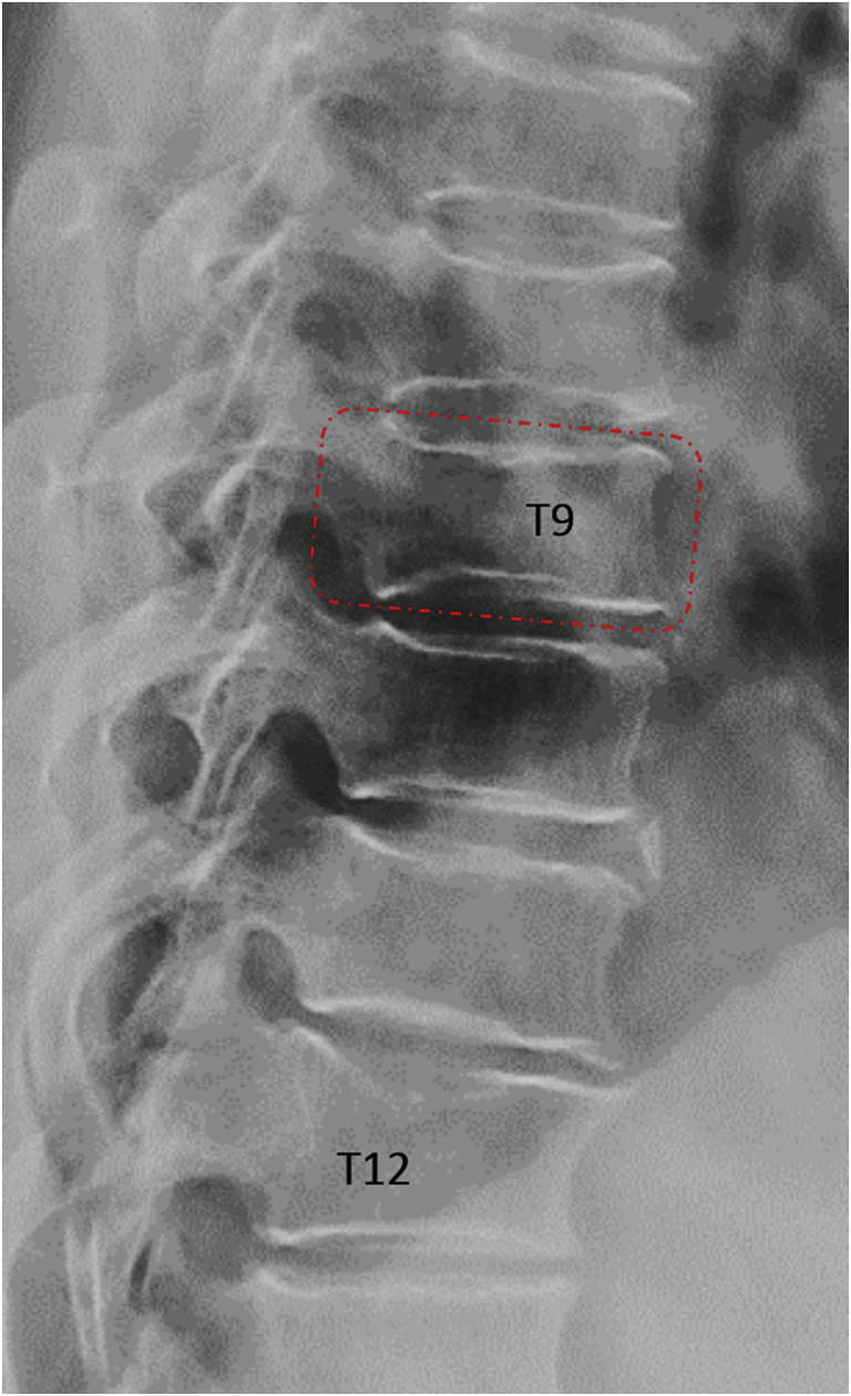
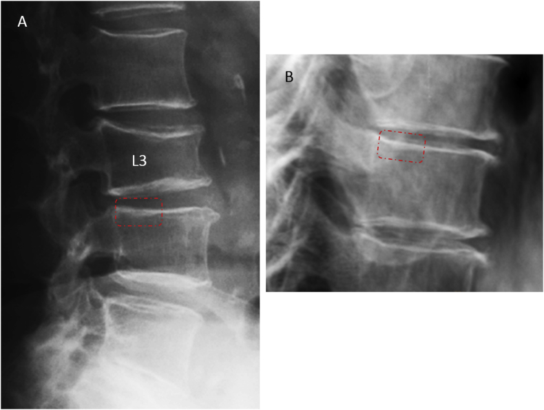
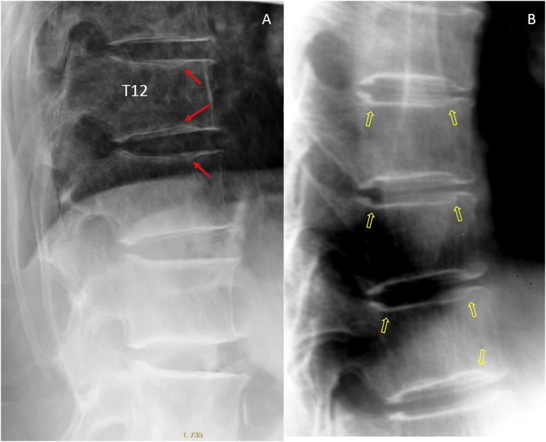
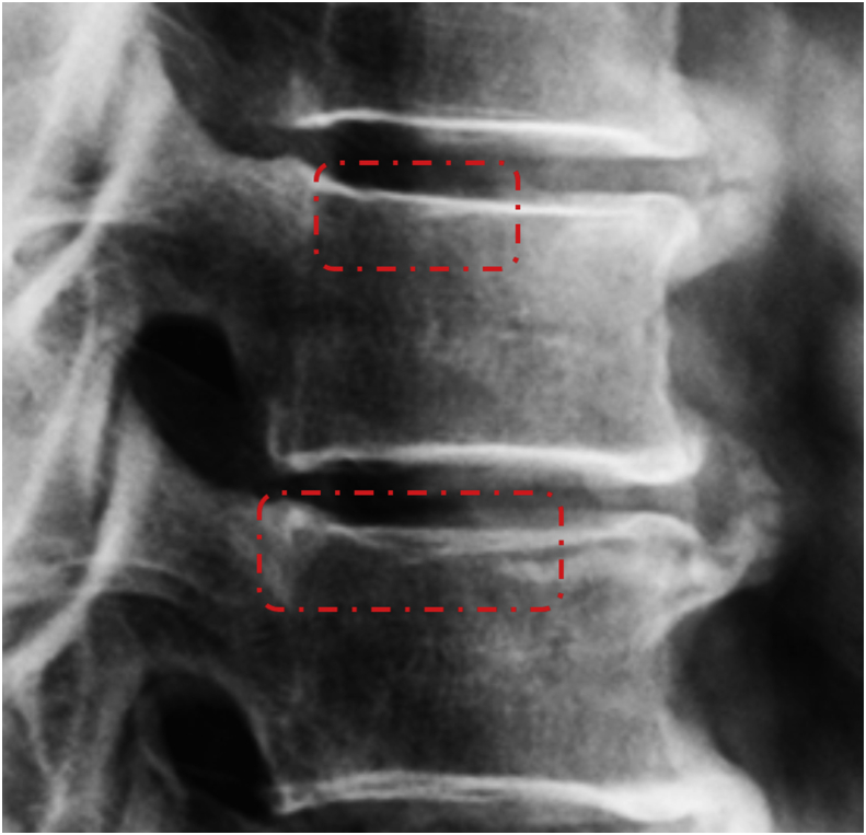
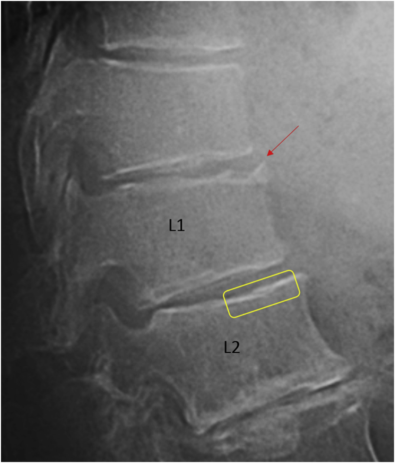
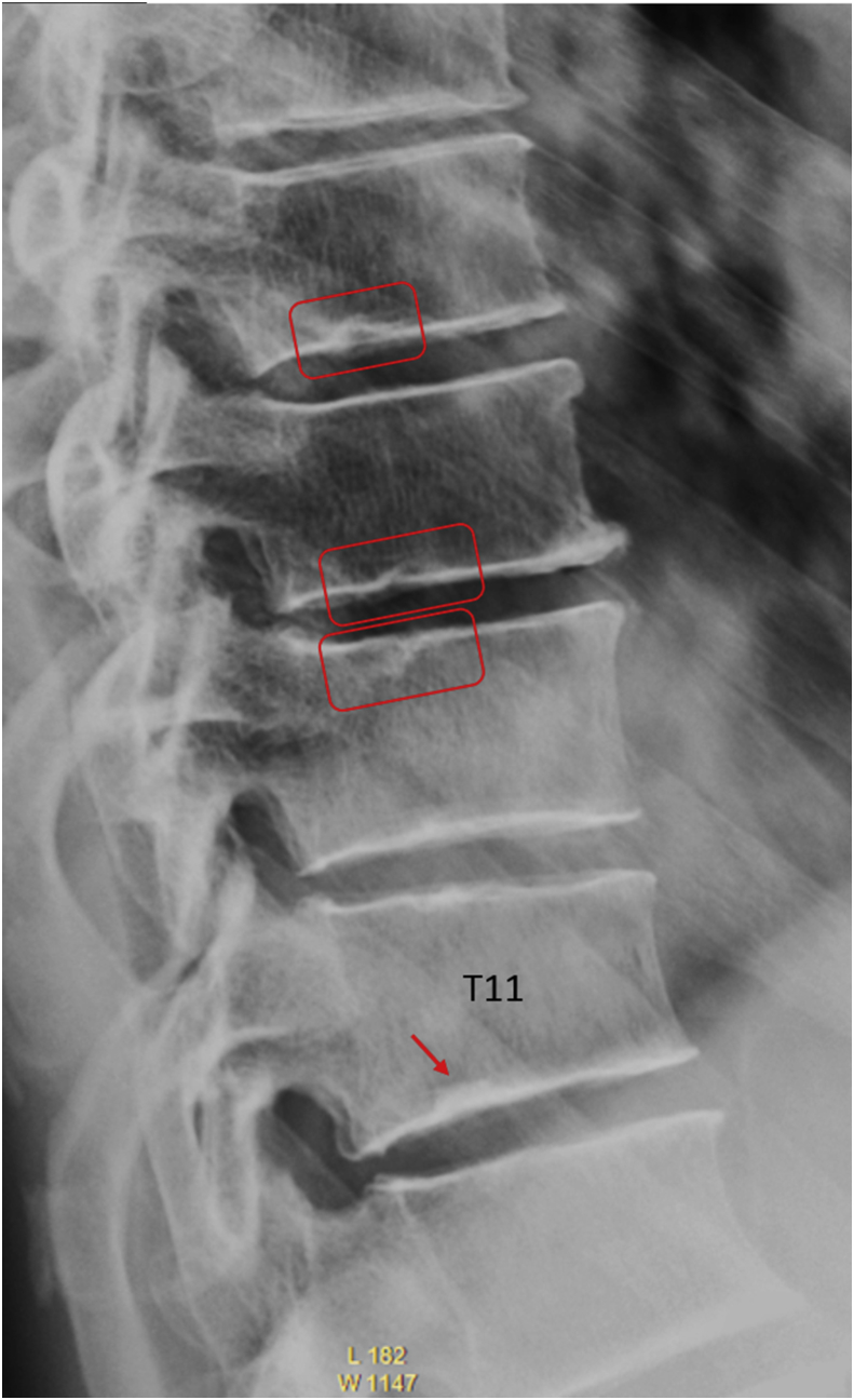
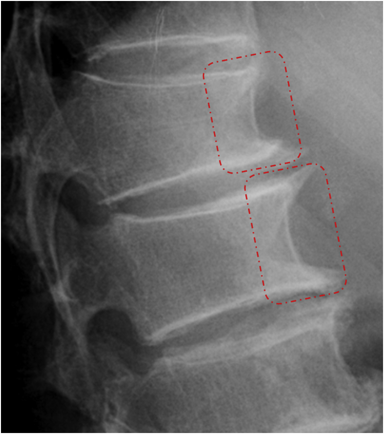
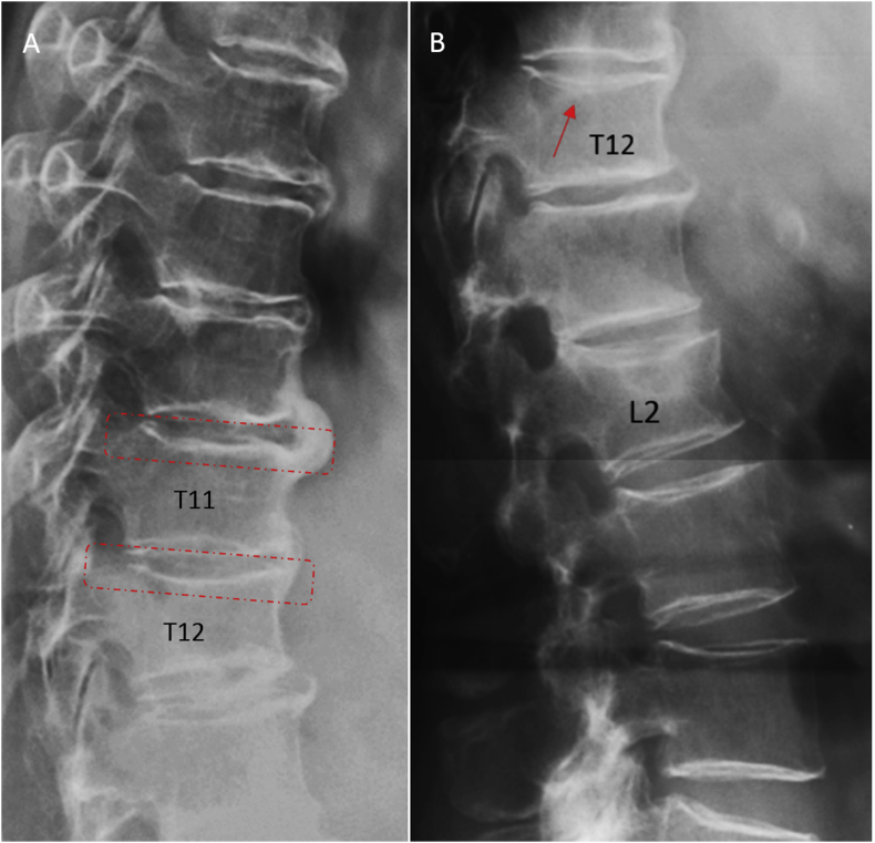
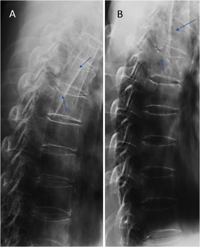
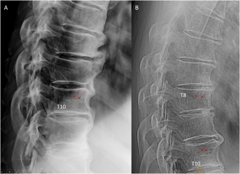
Despite years' research, the radiographic criteria for osteoporotic vertebral fracture and its grading remain debated. The importance of identifying vertebral endplate/cortex fracture (ECF) is being recognised; however, evaluation of osteoporotic ECF requires training and experience. This article aims to serve as a teaching material for radiologists/physicians or researchers to evaluate osteoporotic ECF. Emphasis is particularly dedicated to identifying ECF that may not be associated with apparent vertebral body collapse. We suggest a combined approach based on standardised radiologic evaluation by experts and morphometry measurement is the most appropriate approach to detect and classify osteoporotic vertebral fractures.

THE TRANSLATIONAL POTENTIAL

A good understanding of radiologic anatomy of vertebrae and fracture signs of endplate/cortex are essential for spine fragility fracture assessment.

摘要

未标注

尽管经过多年研究,骨质疏松性椎体骨折的影像学标准及其分级仍存在争议。识别椎体终板/皮质骨折(ECF)的重要性正得到认可;然而,评估骨质疏松性ECF需要培训和经验。本文旨在作为放射科医生/内科医生或研究人员评估骨质疏松性ECF的教材。重点特别在于识别可能与明显椎体塌陷无关的ECF。我们建议,基于专家的标准化放射学评估和形态测量的联合方法是检测和分类骨质疏松性椎体骨折的最合适方法。

转化潜力

对椎体的放射解剖学和终板/皮质骨折征象有良好的理解对于脊柱脆性骨折评估至关重要。

https://cdn.ncbi.nlm.nih.gov/pmc/blobs/e095/6169255/f3d7c0ae2e35/gr25.jpg
https://cdn.ncbi.nlm.nih.gov/pmc/blobs/e095/6169255/713c10f83089/gr1.jpg
https://cdn.ncbi.nlm.nih.gov/pmc/blobs/e095/6169255/2c42eb9aa0c3/gr2.jpg
https://cdn.ncbi.nlm.nih.gov/pmc/blobs/e095/6169255/15eff815947e/gr3.jpg
https://cdn.ncbi.nlm.nih.gov/pmc/blobs/e095/6169255/6a1638e804cb/gr4.jpg
https://cdn.ncbi.nlm.nih.gov/pmc/blobs/e095/6169255/d2026efbba45/gr5.jpg
https://cdn.ncbi.nlm.nih.gov/pmc/blobs/e095/6169255/3bd70a3fddf6/gr6.jpg
https://cdn.ncbi.nlm.nih.gov/pmc/blobs/e095/6169255/49bd9b69d604/gr7.jpg
https://cdn.ncbi.nlm.nih.gov/pmc/blobs/e095/6169255/8795839a29d9/gr8.jpg
https://cdn.ncbi.nlm.nih.gov/pmc/blobs/e095/6169255/4e93c8176594/gr9.jpg
https://cdn.ncbi.nlm.nih.gov/pmc/blobs/e095/6169255/3cc0ddcb5d3b/gr10.jpg
https://cdn.ncbi.nlm.nih.gov/pmc/blobs/e095/6169255/bf844051ff41/gr11.jpg
https://cdn.ncbi.nlm.nih.gov/pmc/blobs/e095/6169255/6090ca824758/gr12.jpg
https://cdn.ncbi.nlm.nih.gov/pmc/blobs/e095/6169255/cfc152d7f2de/gr13.jpg
https://cdn.ncbi.nlm.nih.gov/pmc/blobs/e095/6169255/8f7033dd310e/gr14.jpg
https://cdn.ncbi.nlm.nih.gov/pmc/blobs/e095/6169255/f92cf593841f/gr15.jpg
https://cdn.ncbi.nlm.nih.gov/pmc/blobs/e095/6169255/4011c5f8d75d/gr16.jpg
https://cdn.ncbi.nlm.nih.gov/pmc/blobs/e095/6169255/b07268850406/gr17.jpg
https://cdn.ncbi.nlm.nih.gov/pmc/blobs/e095/6169255/367f2e0c5eab/gr18.jpg
https://cdn.ncbi.nlm.nih.gov/pmc/blobs/e095/6169255/337e53fbdd17/gr19.jpg
https://cdn.ncbi.nlm.nih.gov/pmc/blobs/e095/6169255/91541f43c4c8/gr20.jpg
https://cdn.ncbi.nlm.nih.gov/pmc/blobs/e095/6169255/8cb5c9634d58/gr21.jpg
https://cdn.ncbi.nlm.nih.gov/pmc/blobs/e095/6169255/d365ea255923/gr22.jpg
https://cdn.ncbi.nlm.nih.gov/pmc/blobs/e095/6169255/3706d580f3ea/gr23.jpg
https://cdn.ncbi.nlm.nih.gov/pmc/blobs/e095/6169255/4c753f44dab5/gr24.jpg
https://cdn.ncbi.nlm.nih.gov/pmc/blobs/e095/6169255/f3d7c0ae2e35/gr25.jpg

相似文献

1
Osteoporotic vertebral endplate and cortex fractures: A pictorial review.骨质疏松性椎体终板和皮质骨折:图文综述
J Orthop Translat. 2018 Sep 6;15:35-49. doi: 10.1016/j.jot.2018.08.004. eCollection 2018 Oct.
2
Identifying osteoporotic vertebral endplate and cortex fractures.识别骨质疏松性椎体终板和皮质骨折。
Quant Imaging Med Surg. 2017 Oct;7(5):555-591. doi: 10.21037/qims.2017.10.05.
3
Osteoporotic vertebral deformity with endplate/cortex fracture is associated with higher further vertebral fracture risk: the Ms. OS (Hong Kong) study results.伴有终板/皮质骨折的骨质疏松性椎体变形与更高的进一步椎体骨折风险相关:香港 Ms. OS 研究结果。
Osteoporos Int. 2019 Apr;30(4):897-905. doi: 10.1007/s00198-019-04856-4. Epub 2019 Jan 22.
4
Underreporting characteristics of osteoporotic vertebral fracture in back pain clinic patients of a tertiary hospital in China.中国一家三级医院背痛门诊患者骨质疏松性椎体骨折的漏报特征
J Orthop Translat. 2019 Nov 9;23:152-158. doi: 10.1016/j.jot.2019.10.007. eCollection 2020 Jul.
5
Recognizing and reporting vertebral fractures: reducing the risk of future osteoporotic fractures.识别和报告椎体骨折:降低未来骨质疏松性骨折的风险。
Can Assoc Radiol J. 2007 Feb;58(1):27-36.
6
Osteoporotic Vertebral Fracture Prevalence in Elderly Chinese Men and Women: A Comparison of Endplate/Cortex Fracture-Based and Morphometrical Deformity-Based Methods.老年男女骨质疏松性椎体骨折患病率:基于终板/皮质骨折的方法与基于形态计量学畸形的方法比较。
J Clin Densitom. 2019 Jul-Sep;22(3):409-419. doi: 10.1016/j.jocd.2017.11.004. Epub 2017 Dec 2.
7
Radiological features of traumatic vertebral endplate fracture: an analysis of 194 cases with 263 vertebral fractures.创伤性椎体终板骨折的放射学特征:263 个椎体骨折的 194 例分析。
Chin Med J (Engl). 2020 Nov 20;133(22):2696-2702. doi: 10.1097/CM9.0000000000000919.
8
How to define an osteoporotic vertebral fracture?如何定义骨质疏松性椎体骨折?
Quant Imaging Med Surg. 2019 Sep;9(9):1485-1494. doi: 10.21037/qims.2019.09.10.
9
Prevalent osteoporotic vertebral fractures more likely involve the upper endplate than the lower endplate and even more so in males.与下端椎相比,骨质疏松性椎体骨折更常累及上端椎,男性更是如此。
Ann Transl Med. 2018 Nov;6(22):442. doi: 10.21037/atm.2018.10.61.
10
Elderly males with or without existing osteoporotic vertebral fracture have much lower future vertebral fracture risk than elderly females: the MrOS (Hong Kong) year-4 follow-up spine radiograph study.老年男性无论是否存在骨质疏松性椎体骨折,其未来椎体骨折风险均明显低于老年女性:MrOS(香港)研究 4 年随访脊柱 X 线片研究。
Osteoporos Int. 2019 Dec;30(12):2505-2514. doi: 10.1007/s00198-019-05136-x. Epub 2019 Sep 2.

引用本文的文献

1
The prevention of osteoporotic vertebral fractures in eastern and in western countries.东西方国家骨质疏松性椎体骨折的预防
Bone Rep. 2025 May 15;25:101851. doi: 10.1016/j.bonr.2025.101851. eCollection 2025 Jun.
2
Lower osteoporotic-like vertebral fractural deformity (OLVF) prevalence and severity among older Thais and Indonesians than among older Chinese.泰国和印度尼西亚老年人中骨质疏松样椎体骨折畸形(OLVF)的患病率和严重程度低于中国老年人。
Quant Imaging Med Surg. 2025 Jan 2;15(1):786-800. doi: 10.21037/qims-24-430. Epub 2024 Dec 2.
3
Radiological diagnosis of prevalent osteoporotic vertebral fracture on radiographs: an interim consensus from a group of experts of the ESSR osteoporosis and metabolism subcommittee.

本文引用的文献

1
All osteoporotically deformed vertebrae with >34% height loss have radiographically identifiable endplate/cortex fracture.所有椎体高度丢失超过34%的骨质疏松性变形椎体在X线片上都有可识别的终板/皮质骨折。
J Orthop Translat. 2018 Jul 6;14:63-66. doi: 10.1016/j.jot.2018.06.004. eCollection 2018 Jul.
2
Vertebral Fractures and Morphometric Deformities.椎体骨折与形态学畸形
J Bone Miner Res. 2018 Aug;33(8):1544-1545. doi: 10.1002/jbmr.3470. Epub 2018 Jun 21.
3
Vertebral Fracture: Diagnostic Difficulties of a Major Medical Problem.
放射学诊断常见的骨质疏松性椎体骨折:ESSR 骨质疏松和代谢小组委员会专家组的临时共识。
Skeletal Radiol. 2024 Dec;53(12):2563-2574. doi: 10.1007/s00256-024-04678-4. Epub 2024 Apr 25.
4
Artificial Intelligence-Assisted Detection of Osteoporotic Vertebral Fractures on Lateral Chest Radiographs in Post-Menopausal Women.人工智能辅助检测绝经后女性胸部侧位X线片上的骨质疏松性椎体骨折
J Clin Med. 2023 Dec 16;12(24):7730. doi: 10.3390/jcm12247730.
5
Conversion of osteoporotic-like vertebral fracture severity score to osteoporosis T-score equivalent status: A framework study for older Chinese men.将骨质疏松样椎体骨折严重程度评分转换为等效的骨质疏松T评分状态:一项针对中国老年男性的框架性研究。
Osteoporos Sarcopenia. 2023 Mar;9(1):14-21. doi: 10.1016/j.afos.2023.03.004. Epub 2023 Mar 24.
6
Osteoporotic-like vertebral fracture with less than 20% height loss is associated with increased further vertebral fracture risk in older women: the MrOS and MsOS (Hong Kong) year-18 follow-up radiograph results.椎体高度丢失小于20%的骨质疏松样椎体骨折与老年女性进一步发生椎体骨折的风险增加相关:骨质疏松性骨折男性纵向研究(MrOS)和骨质疏松性骨折女性纵向研究(MsOS,香港)第18年随访X线片结果
Quant Imaging Med Surg. 2023 Feb 1;13(2):1115-1125. doi: 10.21037/qims-2022-06. Epub 2022 Dec 1.
7
Chest radiograph prevalence of vertebral deformity among young and middle-aged population of mixed city dwellers and rural residents.城市居民和农村居民混合的中青年人群中胸部X线片显示的椎体畸形患病率。
J Thorac Dis. 2022 Dec;14(12):4685-4698. doi: 10.21037/jtd-22-1386.
8
Conversion of osteoporotic vertebral fracture severity score to osteoporosis T-score equivalent status: a framework and a comparative study of Hong Kong Chinese and Rome Caucasian older women.将骨质疏松性椎体骨折严重程度评分转换为骨质疏松 T 评分等效状态:一个框架及香港华裔和罗马白人老年女性的比较研究。
Arch Osteoporos. 2022 Dec 3;18(1):1. doi: 10.1007/s11657-022-01178-7.
9
A software program for automated compressive vertebral fracture detection on elderly women's lateral chest radiograph: Ofeye 1.0.一款用于老年女性胸部侧位X光片自动检测椎体压缩性骨折的软件程序:Ofeye 1.0。
Quant Imaging Med Surg. 2022 Aug;12(8):4259-4271. doi: 10.21037/qims-22-433.
10
An update of our understanding of radiographic diagnostics for prevalent osteoporotic vertebral fracture in elderly women.老年女性常见骨质疏松性椎体骨折影像学诊断的认识更新
Quant Imaging Med Surg. 2022 Jul;12(7):3495-3514. doi: 10.21037/qims-22-360.
椎体骨折:一个重大医学问题的诊断难点
J Bone Miner Res. 2018 Apr;33(4):553-559. doi: 10.1002/jbmr.3404. Epub 2018 Mar 5.
4
Osteoporotic Vertebral Fracture Prevalence in Elderly Chinese Men and Women: A Comparison of Endplate/Cortex Fracture-Based and Morphometrical Deformity-Based Methods.老年男女骨质疏松性椎体骨折患病率:基于终板/皮质骨折的方法与基于形态计量学畸形的方法比较。
J Clin Densitom. 2019 Jul-Sep;22(3):409-419. doi: 10.1016/j.jocd.2017.11.004. Epub 2017 Dec 2.
5
Identifying osteoporotic vertebral endplate and cortex fractures.识别骨质疏松性椎体终板和皮质骨折。
Quant Imaging Med Surg. 2017 Oct;7(5):555-591. doi: 10.21037/qims.2017.10.05.
6
Thoracolumbar Intervertebral Disc Area Morphometry in Elderly Chinese Men and Women: Radiographic Quantifications at Baseline and Changes at Year-4 Follow-up.老年中国男性和女性胸腰椎间盘面积形态计量学:基线时的放射学定量和 4 年随访时的变化。
Spine (Phila Pa 1976). 2018 May 15;43(10):E607-E614. doi: 10.1097/BRS.0000000000002482.
7
Association of Incident, Clinically Undiagnosed Radiographic Vertebral Fractures With Follow-Up Back Pain Symptoms in Older Men: the Osteoporotic Fractures in Men (MrOS) Study.老年男性新发、临床未诊断的放射学椎体骨折与随访后背痛症状的相关性:男性骨质疏松性骨折研究(MrOS)。
J Bone Miner Res. 2017 Nov;32(11):2263-2268. doi: 10.1002/jbmr.3215. Epub 2017 Sep 7.
8
Comparative Analysis of the Radiology of Osteoporotic Vertebral Fractures in Women and Men: Cross-Sectional and Longitudinal Observations from the Canadian Multicentre Osteoporosis Study (CaMos).女性和男性骨质疏松性椎体骨折的放射学比较分析:来自加拿大多中心骨质疏松研究(CaMos)的横断面和纵向观察。
J Bone Miner Res. 2018 Apr;33(4):569-579. doi: 10.1002/jbmr.3222. Epub 2017 Aug 7.
9
Osteoporotic Vertebral Fracture Prevalence Varies Widely Between Qualitative and Quantitative Radiological Assessment Methods: The Rotterdam Study.骨质疏松性椎体骨折的患病率在定性和定量放射学评估方法之间存在很大差异:鹿特丹研究。
J Bone Miner Res. 2018 Apr;33(4):560-568. doi: 10.1002/jbmr.3220. Epub 2017 Sep 6.
10
Misdiagnosis of vertebral fractures on local radiographic readings of the multicentre POINT (Prevalence of Osteoporosis in INTernal medicine) study.多中心POINT(内科骨质疏松症患病率)研究中局部影像学读数对椎体骨折的误诊情况。
Bone. 2017 Aug;101:230-235. doi: 10.1016/j.bone.2017.05.008. Epub 2017 May 13.