Department of Radiology, Brigham and Women's Hospital, Harvard Medical School, Boston, Massachusetts.
Department of Imaging Systems, Philips Healthcare, Taipei, Taiwan.
Magn Reson Med. 2019 Mar;81(3):1699-1713. doi: 10.1002/mrm.27525. Epub 2018 Oct 15.
Quantitative parameter maps, as opposed to qualitative grayscale images, may represent the future of diagnostic MRI. A new quantitative MRI method is introduced here that requires a single 3D acquisition, allowing good spatial coverage to be achieved in relatively short scan times.
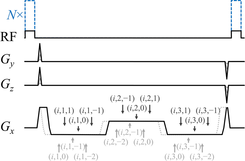
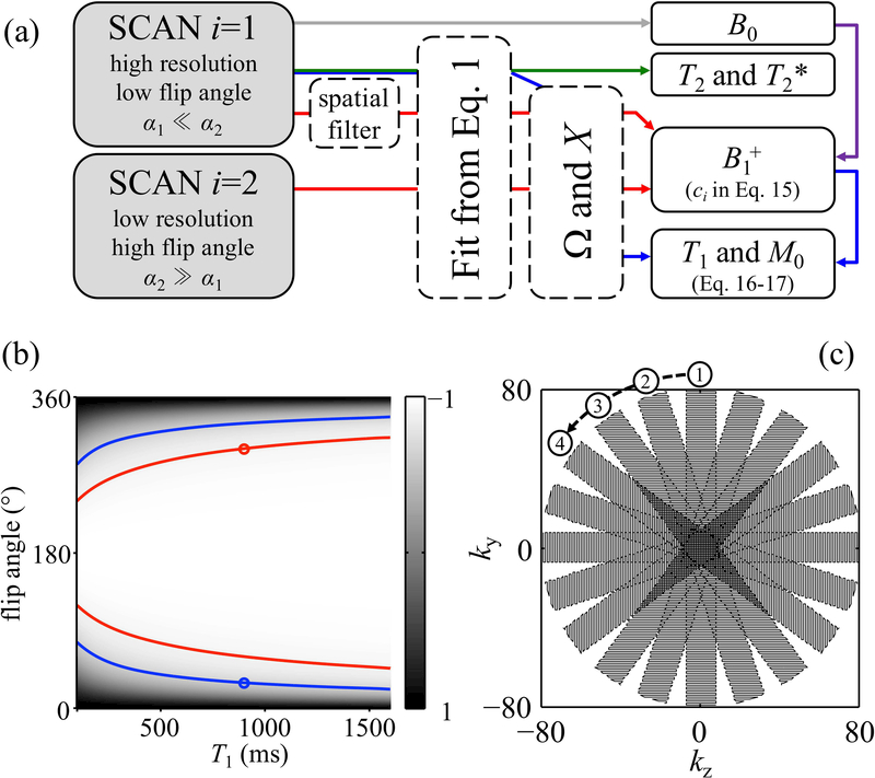
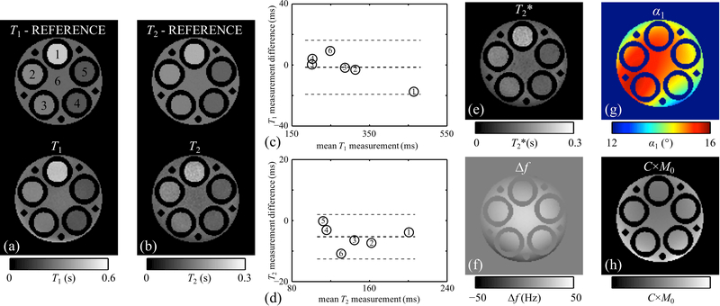
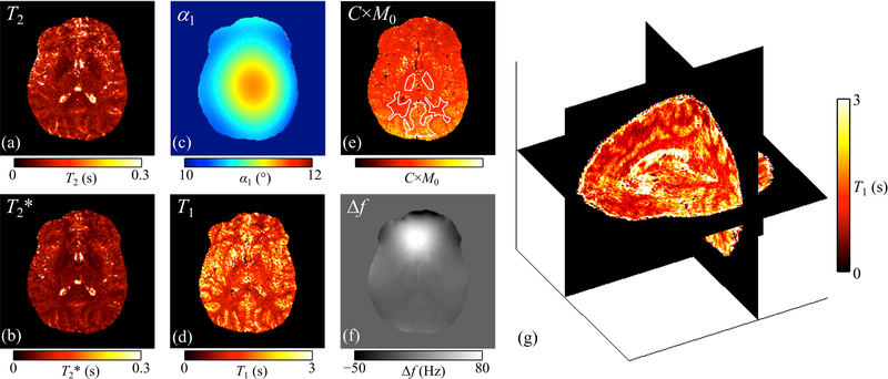
A multipathway multi-echo sequence was developed, and at least 3 pathways with 2 TEs were needed to generate T , T , T , B , and B maps. The method required the central k-space region to be sampled twice, with the same sequence but with 2 very different nominal flip angle settings. Consequently, scan time was only slightly longer than that of a single scan. The multipathway multi-echo data were reconstructed into parameter maps, for phantom as well as brain acquisitions, in 5 healthy volunteers at 3 T. Spatial resolution, matrix size, and FOV were 1.2 × 1.0 × 1.2 mm , 160 × 192 × 160, and 19.2 × 19.2 × 19.2 cm (whole brain), acquired in 11.5 minutes with minimal acceleration. Validation was performed against T , T , and T maps calculated from gradient-echo and spin-echo data.
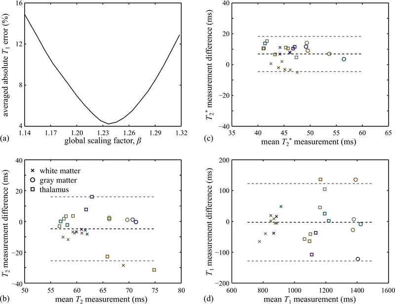
In Bland-Altman plots, bias and limits of agreement for T and T results in vivo and in phantom were -2.9/±125.5 ms (T in vivo), -4.8/±20.8 ms (T in vivo), -1.5/±18.1 ms (T in phantom), and -5.3/±7.4 ms (T in phantom), for regions of interest including given brain structures or phantom compartments. Due to relatively high noise levels, the current implementation of the approach may prove more useful for region of interest-based as opposed to pixel-based interpretation.
We proposed a novel approach to quantitatively map MR parameters based on a multipathway multi-echo acquisition.
与定性灰度图像相比,定量参数图可能代表了诊断 MRI 的未来。本文介绍了一种新的定量 MRI 方法,该方法仅需单次 3D 采集,即可在相对较短的扫描时间内实现良好的空间覆盖。
开发了一种多路径多回波序列,需要至少 3 条路径和 2 个 TE 来生成 T1、T2、T2*、B0 和 B图。该方法需要对中心 k 空间区域进行两次采样,使用相同的序列,但具有两个非常不同的标称翻转角设置。因此,扫描时间仅比单次扫描略长。多路径多回波数据被重建为参数图,用于在 3T 下对 5 名健康志愿者的体模和大脑采集进行重建。空间分辨率、矩阵大小和 FOV 分别为 1.2×1.0×1.2mm、160×192×160 和 19.2×19.2×19.2cm(全脑),在最小加速的情况下,采集时间为 11.5 分钟。通过与梯度回波和自旋回波数据计算得出的 T1、T2 和 T2图进行验证。
在 Bland-Altman 图中,体内和体模中 T1 和 T2 结果的偏倚和一致性界限为-2.9/±125.5ms(体内 T1)、-4.8/±20.8ms(体内 T2)、-1.5/±18.1ms(体模 T1)和-5.3/±7.4ms(体模 T2),包括给定的脑结构或体模腔室的感兴趣区域。由于相对较高的噪声水平,当前方法的实现可能更适合基于感兴趣区域的解释,而不是基于像素的解释。
我们提出了一种基于多路径多回波采集的定量映射 MR 参数的新方法。