Institute of Medical Sciences, School of Medicine, Medical Sciences and Nutrition, University of Aberdeen, Foresterhill, Aberdeen, AB25 2ZD, UK.
Department of Human Anatomy and Cell Science, Rady College of Medicine, Faculty of Health Sciences, University of Manitoba, Winnipeg, MB, Canada.
BMC Med. 2018 Oct 23;16(1):194. doi: 10.1186/s12916-018-1183-7.
Maternal lifestyle factors, including smoking and increased body weight, increase risks of adult diseases such as metabolic syndrome and infertility. The fetal thyroid gland is essential for the control of fetal metabolic rate, cardiac output, and brain development. Altered fetal thyroid function may contribute to increased disease onset later in life. Here, we investigated the impact of maternal smoking and high maternal weight on human fetal thyroid function during the second trimester.
Thyroid glands and plasma were collected from fetuses electively terminated in the second trimester (normally progressing pregnancies). Plasma total triiodothyronine (T3) and total thyroxine (T4) were measured by solid-phase extraction-liquid chromatography-tandem mass spectrometry. Fetal plasma thyroid-stimulating hormone (TSH) levels were measured using a multiplex assay for human pituitary hormones. Histology and immunolocalization of thyroid developmental markers were examined in thyroid sections. Transcript levels of developmental, functional, apoptotic, and detoxification markers were measured by real-time PCR. Statistical analyses were performed using multivariate linear regression models with fetal age, sex, and maternal smoking or maternal body mass index (BMI) as covariates.
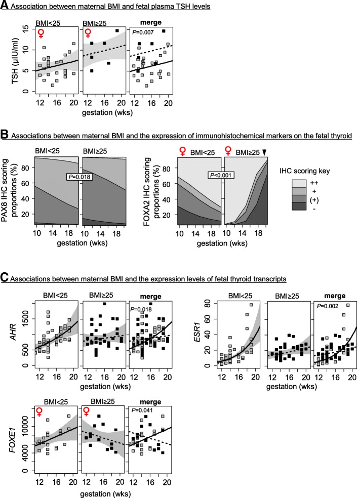
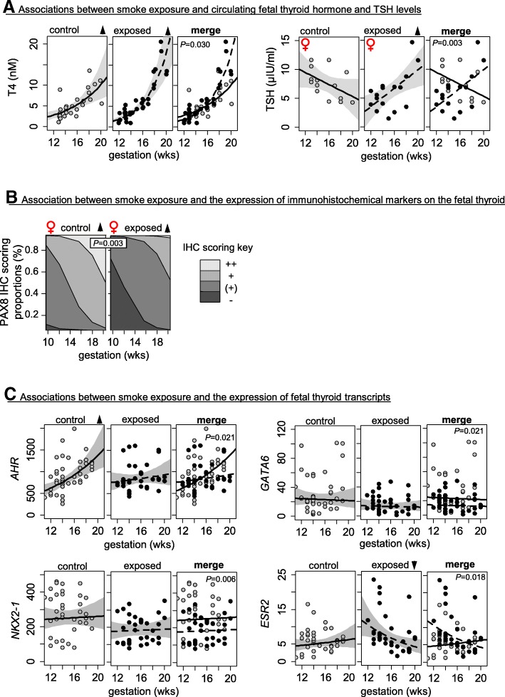
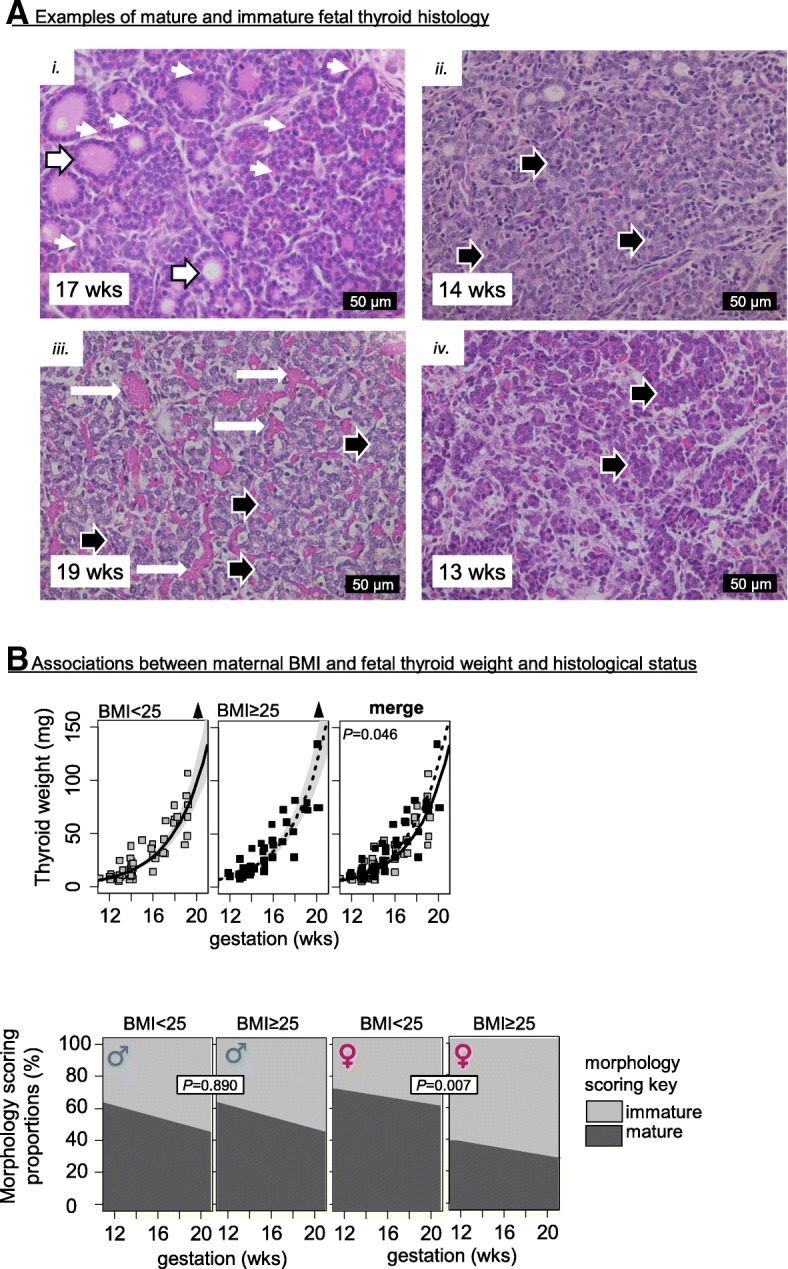
Maternal smoking was associated with significant changes in fetal plasma T4 and TSH levels during the second trimester. Smoke-exposed thyroids had reduced thyroid GATA6 and NKX2-1 transcript levels and altered developmental trajectories for ESR2 and AHR transcript levels. Maternal BMI > 25 was associated with increased fetal thyroid weight, increased plasma TSH levels, and abnormal thyroid histology in female fetuses. Normal developmental changes in AHR and ESR1 transcript expression were also abolished in fetal thyroids from mothers with BMI > 25.
For the first time, we show that maternal smoking and high maternal BMI are associated with disturbed fetal thyroid gland development and endocrine function in a sex-specific manner during the second trimester. These findings suggest that predisposition to post-natal disease is mediated, in part, by altered fetal thyroid gland development.
母体生活方式因素,包括吸烟和体重增加,会增加代谢综合征和不孕等成人疾病的风险。胎儿甲状腺对控制胎儿代谢率、心输出量和大脑发育至关重要。胎儿甲状腺功能改变可能导致成年后疾病发病率增加。在这里,我们研究了母体吸烟和高母体体重对妊娠中期胎儿甲状腺功能的影响。
从妊娠中期(正常进展妊娠)择期终止的胎儿中采集甲状腺和血浆。采用固相萃取-液相色谱-串联质谱法测定血浆总三碘甲状腺原氨酸(T3)和总甲状腺素(T4)。采用人垂体激素的多重分析测定胎儿血浆促甲状腺激素(TSH)水平。在甲状腺切片中检查甲状腺发育标志物的组织学和免疫定位。通过实时 PCR 测定发育、功能、凋亡和解毒标志物的转录水平。使用包含胎儿年龄、性别以及母体吸烟或母体体重指数(BMI)作为协变量的多元线性回归模型进行统计分析。
母体吸烟与妊娠中期胎儿血浆 T4 和 TSH 水平的显著变化有关。暴露于烟雾中的甲状腺组织 GATA6 和 NKX2-1 的转录水平降低,ESR2 和 AHR 的转录水平改变了发育轨迹。母体 BMI>25 与胎儿甲状腺重量增加、血浆 TSH 水平升高以及女性胎儿甲状腺异常组织学有关。母体 BMI>25 的胎儿甲状腺中 AHR 和 ESR1 转录表达的正常发育变化也被消除。
我们首次表明,母体吸烟和高母体 BMI 与妊娠中期胎儿甲状腺发育和内分泌功能的性别特异性紊乱有关。这些发现表明,部分新生儿疾病易感性是由胎儿甲状腺发育改变介导的。