Balconi Michela, Frezza Alessandra, Vanutelli Maria Elide
Department of Psychology, Catholic University of Milan, Milan, Italy.
Research Unit in Affective and Social Neuroscience, Catholic University of Milan, Milan, Italy.
Front Hum Neurosci. 2018 Oct 9;12:395. doi: 10.3389/fnhum.2018.00395. eCollection 2018.
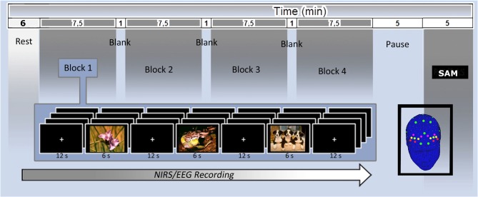
Previous research on Schizophrenia (S) revealed anomalies in brain responsiveness during emotion processing, as shown by neuroimaging and electroencephalography (EEG) measures. Nonetheless preserved capacities to explicitly evaluate the emotional significance of affective stimuli in term of valence have been found. The present study applied functional Near-Infrared Spectroscopy (fNIRS) and EEG to explore the spatial and temporal expressions of emotion processing in the brain before (T0) and after (T2) an emotional Neurofeedback (NF) training of patients, assigned to the control or the experimental group. Explicit measures revealed correct identifications of stimuli emotional valence before (T0) and after (T2) the treatment, while implicit measures (EEG and fNIRS) showed a modulation and increased competencies only after the NF (T2), with more balanced prefrontal activity.
先前对精神分裂症(S)的研究表明,通过神经影像学和脑电图(EEG)测量发现,在情绪处理过程中大脑反应存在异常。尽管如此,研究发现患者在明确评估情感刺激在效价方面的情感意义时仍具备一定能力。本研究应用功能近红外光谱(fNIRS)和脑电图(EEG)来探索,在将患者分为对照组或实验组进行情绪神经反馈(NF)训练之前(T0)和之后(T2),大脑中情绪处理的空间和时间表达。明确测量结果显示,治疗前(T0)和治疗后(T2)对刺激的情感效价都能正确识别,而隐式测量(EEG和fNIRS)仅在神经反馈训练后(T2)显示出调节作用和能力增强,前额叶活动更加平衡。