• 文献检索
  • 文档翻译
  • 深度研究
  • 学术资讯
  • Suppr Zotero 插件Zotero 插件
  • 邀请有礼
  • 套餐&价格
  • 历史记录
应用&插件
Suppr Zotero 插件Zotero 插件浏览器插件Mac 客户端Windows 客户端微信小程序
定价
高级版会员购买积分包购买API积分包
服务
文献检索文档翻译深度研究API 文档MCP 服务
关于我们
关于 Suppr公司介绍联系我们用户协议隐私条款
关注我们

Suppr 超能文献

核心技术专利:CN118964589B侵权必究
粤ICP备2023148730 号-1Suppr @ 2026

文献检索

告别复杂PubMed语法,用中文像聊天一样搜索,搜遍4000万医学文献。AI智能推荐,让科研检索更轻松。

立即免费搜索

文件翻译

保留排版,准确专业,支持PDF/Word/PPT等文件格式,支持 12+语言互译。

免费翻译文档

深度研究

AI帮你快速写综述,25分钟生成高质量综述,智能提取关键信息,辅助科研写作。

立即免费体验

鼠晶状体上皮细胞和纤维中的蛋白质组-转录组分析和蛋白质组重塑。

Proteome-transcriptome analysis and proteome remodeling in mouse lens epithelium and fibers.

机构信息

Departments Ophthalmology and Visual Sciences, Albert Einstein College of Medicine, Bronx, NY, 10461, USA; Genetics, Albert Einstein College of Medicine, Bronx, NY, 10461, USA.

Department of Biochemistry & Molecular Biology, Oregon Health Sciences University, 3181 Southwest Sam Jackson Park Road, Portland, OR, 97239, USA.

出版信息

Exp Eye Res. 2019 Feb;179:32-46. doi: 10.1016/j.exer.2018.10.011. Epub 2018 Oct 22.

DOI:10.1016/j.exer.2018.10.011
PMID:30359574
原文链接:https://pmc.ncbi.nlm.nih.gov/articles/PMC6360118/
Abstract

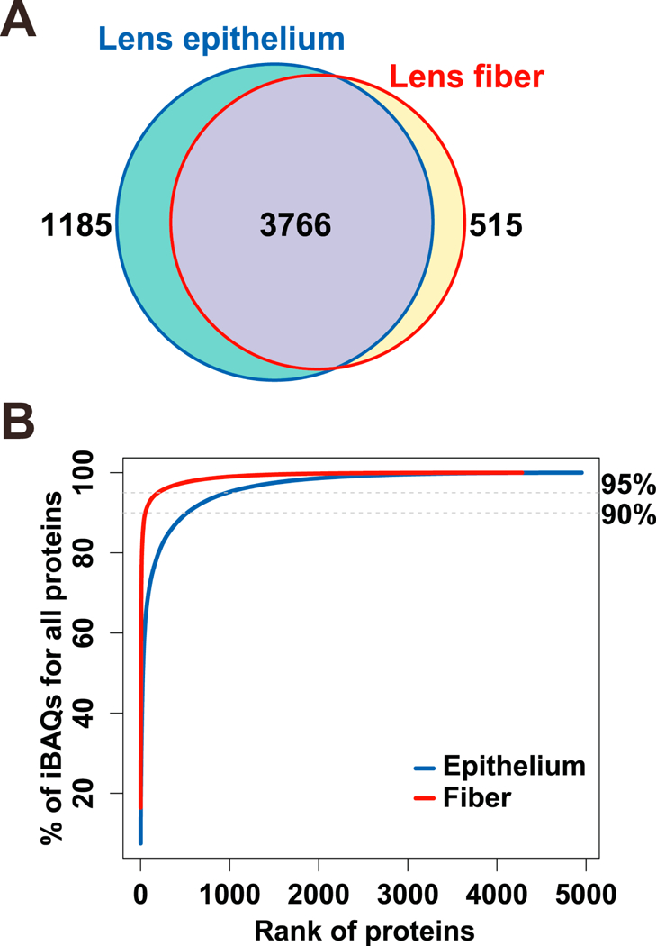
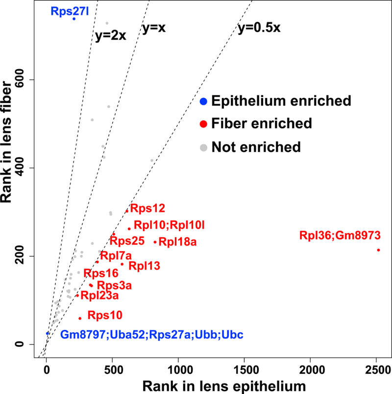
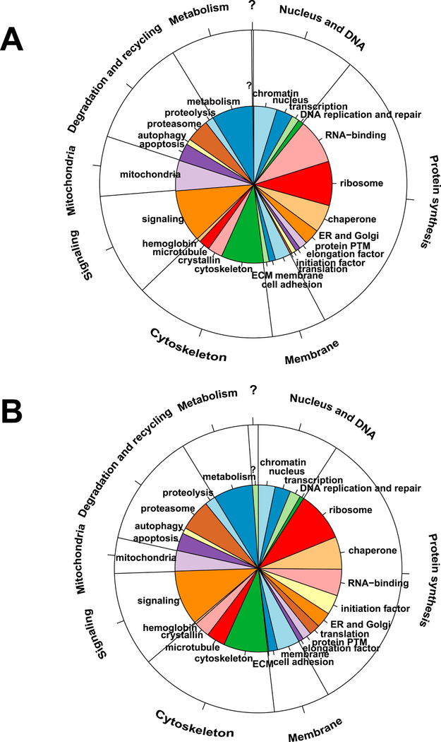
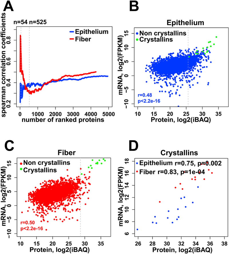
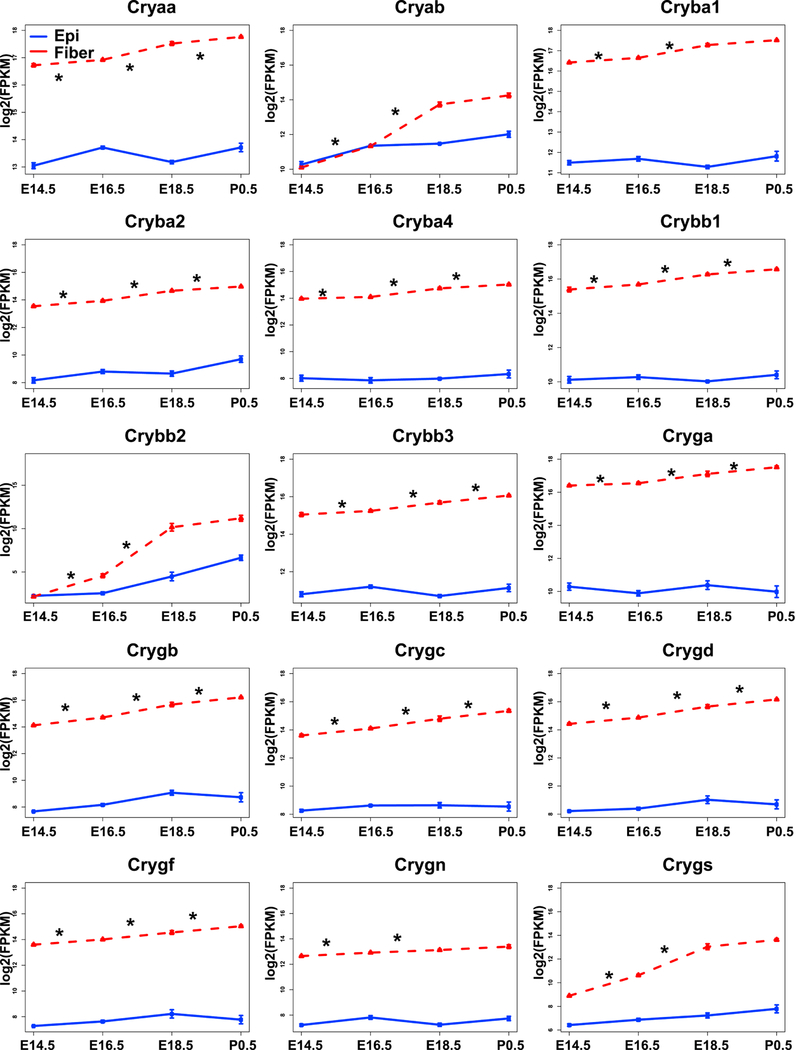
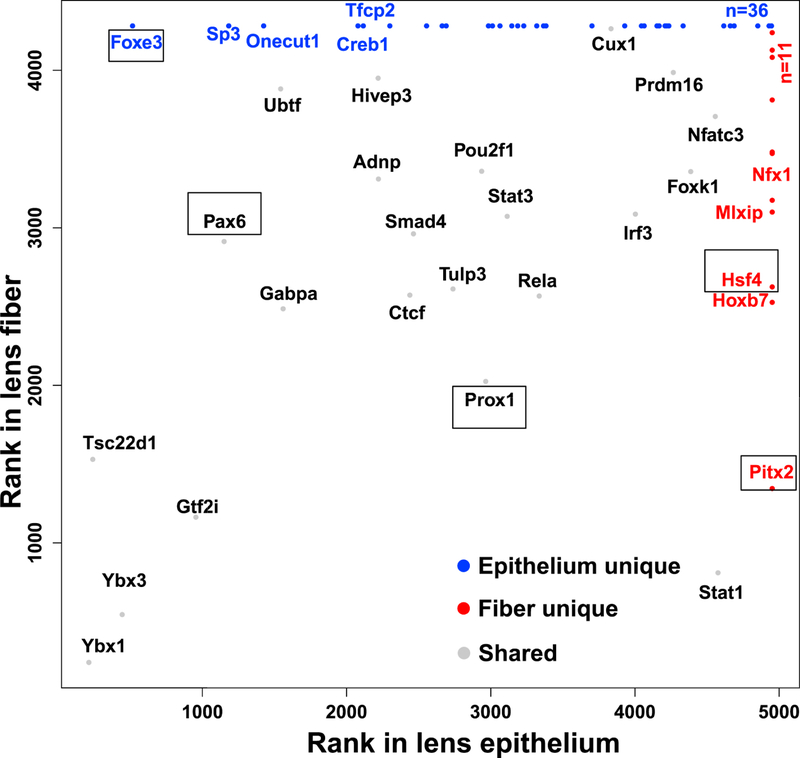
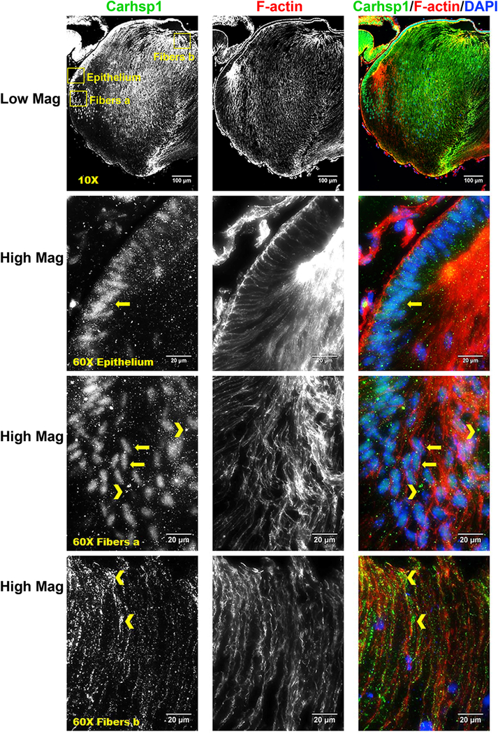
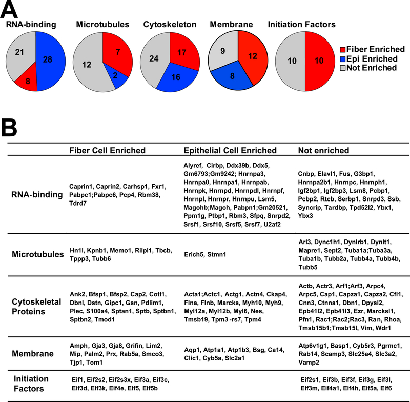
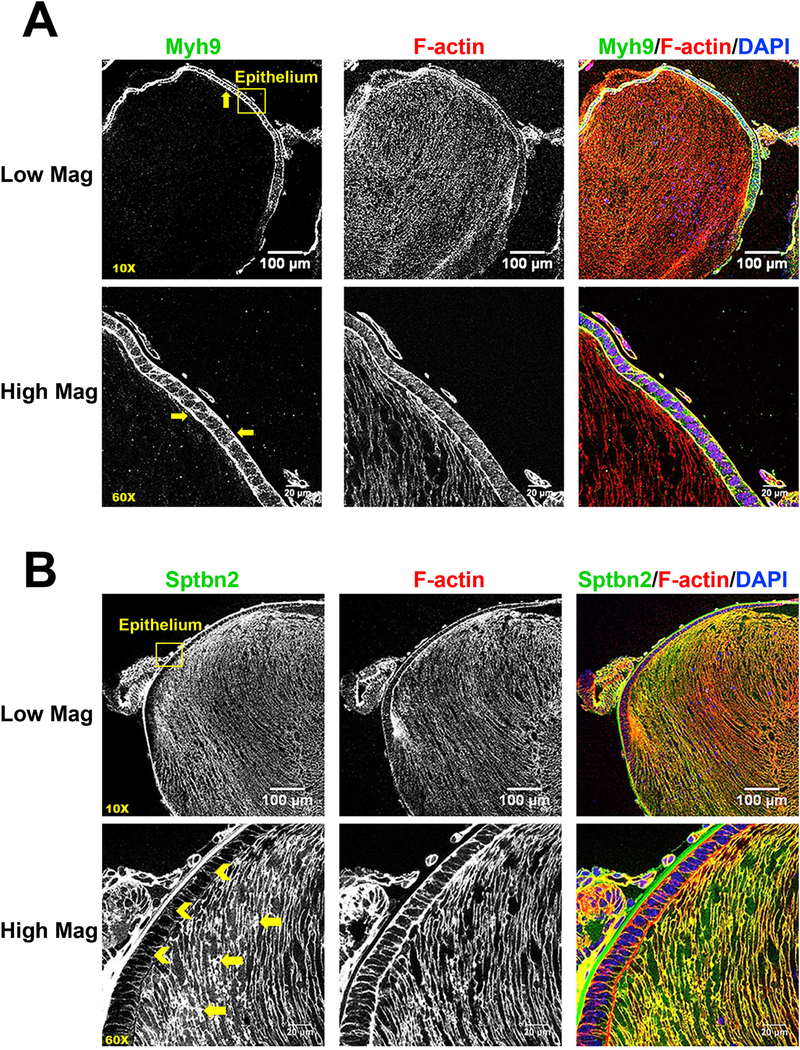
Epithelial cells and differentiated fiber cells represent distinct compartments in the ocular lens. While previous studies have revealed proteins that are preferentially expressed in epithelial vs. fiber cells, a comprehensive proteomics library comparing the molecular compositions of epithelial vs. fiber cells is essential for understanding lens formation, function, disease and regenerative potential, and for efficient differentiation of pluripotent stem cells for modeling of lens development and pathology in vitro. To compare protein compositions between the lens epithelium and fibers, we employed tandem mass spectrometry (2D-LC/MS) analysis of microdissected mouse P0.5 lenses. Functional classifications of the top 525 identified proteins into gene ontology categories by molecular processes and subcellular localizations, were adapted for the lens. Expression levels of both epithelial and fiber proteomes were compared with whole lens proteome and mRNA levels using E14.5, E16.5, E18.5, and P0.5 RNA-Seq data sets. During this developmental time window, multiple complex biosynthetic and catabolic processes generate the molecular and structural foundation for lens transparency. As expected, crystallins showed a high correlation between their mRNA and protein levels. Comprehensive data analysis confirmed and/or predicted roles for transcription factors (TFs), RNA-binding proteins (e.g. Carhsp1), translational apparatus including ribosomal heterogeneity and initiation factors, microtubules, cytoskeletal [e.g. non-muscle myosin IIA heavy chain (Myh9) and βB2-spectrin (Sptbn2)] and membrane proteins in lens formation and maturation. Our data highlighted many proteins with unknown functions in the lens that were preferentially enriched in epithelium or fibers, setting the stage for future studies to further dissect the roles of these proteins in fiber cell differentiation vs. epithelial cell maintenance. In conclusion, the present proteomic datasets represent the first mouse lens epithelium and fiber cell proteomes, establish comparative analyses of protein and RNA-Seq data, and characterize the major proteome remodeling required to form the mature lens fiber cells.

摘要

上皮细胞和分化的纤维细胞代表了眼晶状体中的不同隔室。虽然之前的研究已经揭示了在上皮细胞与纤维细胞中优先表达的蛋白质,但比较上皮细胞与纤维细胞的分子组成的综合蛋白质组学文库对于理解晶状体的形成、功能、疾病和再生潜能以及有效分化多能干细胞用于体外晶状体发育和病理学建模是至关重要的。为了比较晶状体上皮细胞和纤维细胞之间的蛋白质组成,我们采用了串联质谱(2D-LC/MS)分析从小鼠 P0.5 晶状体中微分离的细胞。通过分子过程和亚细胞定位,将前 525 种鉴定出的蛋白质的功能分类归入基因本体论类别,适用于晶状体。使用 E14.5、E16.5、E18.5 和 P0.5 RNA-Seq 数据集比较上皮细胞和纤维细胞蛋白质组的表达水平与整个晶状体蛋白质组和 mRNA 水平。在这个发育时间窗口内,多个复杂的生物合成和分解代谢过程为晶状体的透明度生成了分子和结构基础。正如预期的那样,晶体蛋白的 mRNA 和蛋白质水平之间存在高度相关性。综合数据分析证实和/或预测了转录因子(TFs)、RNA 结合蛋白(例如 Carhsp1)、翻译装置(包括核糖体异质性和起始因子)、微管、细胞骨架(例如非肌肉肌球蛋白 IIA 重链(Myh9)和βB2- spectrin(Sptbn2))和膜蛋白在晶状体形成和成熟中的作用。我们的数据突出了许多在晶状体中具有未知功能的蛋白质,这些蛋白质在上皮细胞或纤维细胞中优先富集,为进一步研究这些蛋白质在纤维细胞分化与上皮细胞维持中的作用奠定了基础。总之,本蛋白质组数据集代表了第一个小鼠晶状体上皮细胞和纤维细胞蛋白质组,建立了蛋白质和 RNA-Seq 数据的比较分析,并描述了形成成熟晶状体纤维细胞所需的主要蛋白质组重排。

https://cdn.ncbi.nlm.nih.gov/pmc/blobs/4c8c/6360118/26b851fbff73/nihms-1511479-f0009.jpg
https://cdn.ncbi.nlm.nih.gov/pmc/blobs/4c8c/6360118/fc137c86639c/nihms-1511479-f0001.jpg
https://cdn.ncbi.nlm.nih.gov/pmc/blobs/4c8c/6360118/5a9a5d5acfd5/nihms-1511479-f0002.jpg
https://cdn.ncbi.nlm.nih.gov/pmc/blobs/4c8c/6360118/6631be60d585/nihms-1511479-f0003.jpg
https://cdn.ncbi.nlm.nih.gov/pmc/blobs/4c8c/6360118/7bc4141f12a1/nihms-1511479-f0004.jpg
https://cdn.ncbi.nlm.nih.gov/pmc/blobs/4c8c/6360118/c7bedc933f6d/nihms-1511479-f0005.jpg
https://cdn.ncbi.nlm.nih.gov/pmc/blobs/4c8c/6360118/245aaa281fd0/nihms-1511479-f0006.jpg
https://cdn.ncbi.nlm.nih.gov/pmc/blobs/4c8c/6360118/8fdcb00e8f89/nihms-1511479-f0007.jpg
https://cdn.ncbi.nlm.nih.gov/pmc/blobs/4c8c/6360118/85838df69b4d/nihms-1511479-f0008.jpg
https://cdn.ncbi.nlm.nih.gov/pmc/blobs/4c8c/6360118/26b851fbff73/nihms-1511479-f0009.jpg

相似文献

1
Proteome-transcriptome analysis and proteome remodeling in mouse lens epithelium and fibers.鼠晶状体上皮细胞和纤维中的蛋白质组-转录组分析和蛋白质组重塑。
Exp Eye Res. 2019 Feb;179:32-46. doi: 10.1016/j.exer.2018.10.011. Epub 2018 Oct 22.
2
A comprehensive spatial-temporal transcriptomic analysis of differentiating nascent mouse lens epithelial and fiber cells.全面的时空转录组分析分化中的新生小鼠晶状体上皮细胞和纤维细胞。
Exp Eye Res. 2018 Oct;175:56-72. doi: 10.1016/j.exer.2018.06.004. Epub 2018 Jun 5.
3
Comparative transcriptome analysis of epithelial and fiber cells in newborn mouse lenses with RNA sequencing.利用RNA测序对新生小鼠晶状体上皮细胞和纤维细胞进行比较转录组分析。
Mol Vis. 2014 Nov 4;20:1491-517. eCollection 2014.
4
Chromatin features, RNA polymerase II and the comparative expression of lens genes encoding crystallins, transcription factors, and autophagy mediators.染色质特征、RNA聚合酶II以及编码晶状体蛋白、转录因子和自噬介质的晶状体基因的比较表达。
Mol Vis. 2015 Aug 28;21:955-73. eCollection 2015.
5
Expression and regulation of alpha-, beta-, and gamma-crystallins in mammalian lens epithelial cells.α-、β-和γ-晶状体蛋白在哺乳动物晶状体上皮细胞中的表达与调控。
Invest Ophthalmol Vis Sci. 2004 Oct;45(10):3608-19. doi: 10.1167/iovs.04-0423.
6
Proteomic analysis of the glutathione-deficient LEGSKO mouse lens reveals activation of EMT signaling, loss of lens specific markers, and changes in stress response proteins.谷胱甘肽缺乏的 LEGSKO 小鼠晶状体的蛋白质组分析揭示了 EMT 信号的激活、晶状体特异性标志物的丢失以及应激反应蛋白的变化。
Free Radic Biol Med. 2017 Dec;113:84-96. doi: 10.1016/j.freeradbiomed.2017.09.019. Epub 2017 Sep 22.
7
Differential protein expression in lens epithelial whole-mounts and lens epithelial cell cultures.晶状体上皮全层及晶状体上皮细胞培养物中的差异蛋白表达
Exp Eye Res. 2003 Jul;77(1):35-49. doi: 10.1016/s0014-4835(03)00090-3.
8
Changes in adhesion complexes define stages in the differentiation of lens fiber cells.黏附复合体的变化定义了晶状体纤维细胞分化的阶段。
Invest Ophthalmol Vis Sci. 2001 Mar;42(3):727-34.
9
Proteome Profiling of Developing Murine Lens Through Mass Spectrometry.通过质谱法对发育中的鼠晶状体进行蛋白质组学分析。
Invest Ophthalmol Vis Sci. 2018 Jan 1;59(1):100-107. doi: 10.1167/iovs.17-21601.
10
Molecular characterization of mouse lens epithelial cell lines and their suitability to study RNA granules and cataract associated genes.小鼠晶状体上皮细胞系的分子特征及其在研究RNA颗粒和白内障相关基因方面的适用性。
Exp Eye Res. 2015 Feb;131:42-55. doi: 10.1016/j.exer.2014.12.011. Epub 2014 Dec 19.

引用本文的文献

1
Artificial Intelligence in Ocular Transcriptomics: Applications of Unsupervised and Supervised Learning.眼科转录组学中的人工智能:无监督学习和监督学习的应用
Cells. 2025 Aug 26;14(17):1315. doi: 10.3390/cells14171315.
2
Human congenital cataract mutation in alters F-actin organization and cell functions.人类先天性白内障突变改变了F-肌动蛋白的组织和细胞功能。
Int J Ophthalmol. 2025 Jun 18;18(6):969-977. doi: 10.18240/ijo.2025.06.01. eCollection 2025.
3
Nucleolar ribosomal RNA synthesis continues in differentiating lens fiber cells until abrupt nuclear degradation required for ocular lens transparency.

本文引用的文献

1
Transcriptional burst fraction and size dynamics during lens fiber cell differentiation and detailed insights into the denucleation process.转录爆发分数和大小动态在晶状体纤维细胞分化过程中,并深入了解去核过程。
J Biol Chem. 2018 Aug 24;293(34):13176-13190. doi: 10.1074/jbc.RA118.001927. Epub 2018 Jun 29.
2
A comprehensive spatial-temporal transcriptomic analysis of differentiating nascent mouse lens epithelial and fiber cells.全面的时空转录组分析分化中的新生小鼠晶状体上皮细胞和纤维细胞。
Exp Eye Res. 2018 Oct;175:56-72. doi: 10.1016/j.exer.2018.06.004. Epub 2018 Jun 5.
3
Deletion of GLUT1 in mouse lens epithelium leads to cataract formation.
核仁核糖体RNA合成在晶状体纤维细胞分化过程中持续进行,直至为实现晶状体透明性所需的细胞核突然降解。
RNA Biol. 2025 Dec;22(1):1-16. doi: 10.1080/15476286.2025.2483118. Epub 2025 Mar 31.
4
Genome-wide DNA methylation and transcriptome sequencing analyses of lens tissue in an age-related mouse cataract model.年龄相关性小鼠白内障模型中晶状体组织的全基因组DNA甲基化和转录组测序分析
PLoS One. 2025 Jan 30;20(1):e0316766. doi: 10.1371/journal.pone.0316766. eCollection 2025.
5
Analysis of mouse lens morphological and proteomic abnormalities following depletion of βB3-crystallin.βB3-晶状体蛋白缺失后小鼠晶状体形态学和蛋白质组学异常分析
bioRxiv. 2024 Dec 30:2024.12.30.630781. doi: 10.1101/2024.12.30.630781.
6
Disease-related non-muscle myosin IIA D1424N rod domain mutation, but not R702C motor domain mutation, disrupts mouse ocular lens fiber cell alignment and hexagonal packing.疾病相关的非肌肉肌球蛋白IIA D1424N杆状结构域突变,而非R702C运动结构域突变,破坏了小鼠眼晶状体纤维细胞的排列和六边形堆积。
Cytoskeleton (Hoboken). 2024 Dec;81(12):789-805. doi: 10.1002/cm.21853. Epub 2024 Mar 22.
7
Integrated single-cell multiomics uncovers foundational regulatory mechanisms of lens development and pathology.单细胞多组学整合揭示了晶状体发育和病变的基础调控机制。
Development. 2024 Jan 1;151(1). doi: 10.1242/dev.202249. Epub 2024 Jan 5.
8
Eye Lens Organoids Made Simple: Characterization of a New Three-Dimensional Organoid Model for Lens Development and Pathology.简易的眼晶状体类器官:用于晶状体发育和病理学的新型三维类器官模型的特征描述。
Cells. 2023 Oct 18;12(20):2478. doi: 10.3390/cells12202478.
9
Spatially Resolved Proteomics Reveals Lens Suture-Related Cell-Cell Junctional Protein Distributions.空间分辨蛋白质组学揭示晶状体缝合线相关细胞-细胞连接蛋白的分布。
Invest Ophthalmol Vis Sci. 2023 Aug 1;64(11):28. doi: 10.1167/iovs.64.11.28.
10
Multiomics Analysis Reveals Novel Genetic Determinants for Lens Differentiation, Structure, and Transparency.多组学分析揭示了晶状体分化、结构和透明度的新遗传决定因素。
Biomolecules. 2023 Apr 19;13(4):693. doi: 10.3390/biom13040693.
GLUT1 在小鼠晶状体上皮细胞中的缺失导致白内障的形成。
Exp Eye Res. 2018 Jul;172:45-53. doi: 10.1016/j.exer.2018.03.021. Epub 2018 Mar 28.
4
The RNA-binding protein Celf1 post-transcriptionally regulates p27Kip1 and Dnase2b to control fiber cell nuclear degradation in lens development.RNA 结合蛋白 Celf1 通过转录后调控 p27Kip1 和 Dnase2b 来控制晶状体发育过程中纤维细胞核的降解。
PLoS Genet. 2018 Mar 22;14(3):e1007278. doi: 10.1371/journal.pgen.1007278. eCollection 2018 Mar.
5
Proteome Profiling of Developing Murine Lens Through Mass Spectrometry.通过质谱法对发育中的鼠晶状体进行蛋白质组学分析。
Invest Ophthalmol Vis Sci. 2018 Jan 1;59(1):100-107. doi: 10.1167/iovs.17-21601.
6
Reconsidering an active role for G-actin in cytoskeletal regulation.重新考虑 G-肌动蛋白在细胞骨架调节中的积极作用。
J Cell Sci. 2018 Jan 10;131(1):jcs203760. doi: 10.1242/jcs.203760.
7
Functional role for stable microtubules in lens fiber cell elongation.稳定微管在晶状体纤维细胞伸长中的功能作用。
Exp Cell Res. 2018 Jan 15;362(2):477-488. doi: 10.1016/j.yexcr.2017.12.012. Epub 2017 Dec 15.
8
The Role of Aquaporins in Ocular Lens Homeostasis.水通道蛋白在眼球晶状体稳态中的作用。
Int J Mol Sci. 2017 Dec 12;18(12):2693. doi: 10.3390/ijms18122693.
9
Light-focusing human micro-lenses generated from pluripotent stem cells model lens development and drug-induced cataract .由多能干细胞生成的光聚焦人类微透镜可模拟晶状体发育及药物诱导性白内障。
Development. 2018 Jan 9;145(1):dev155838. doi: 10.1242/dev.155838.
10
The zebrafish as a model system for analyzing mammalian and native -crystallin promoter function.斑马鱼作为一种用于分析哺乳动物和天然晶状体蛋白启动子功能的模型系统。
PeerJ. 2017 Nov 27;5:e4093. doi: 10.7717/peerj.4093. eCollection 2017.