Department of Histology, Embryology, Genetics and Developmental Biology, Shanghai Jiao Tong University School of Medicine, Shanghai, 200025, China.
Shanghai Key Laboratory for Reproductive Medicine, Shanghai, 200025, China.
Reprod Biol Endocrinol. 2018 Oct 25;16(1):105. doi: 10.1186/s12958-018-0427-x.
Spermatogenesis is a complex process involving the self-renewal and differentiation of spermatogonia into mature spermatids in the seminiferous tubules. During spermatogenesis, germ cells migrate from the basement membrane to cross the blood-testis barrier (BTB) and finally reach the luminal side of the seminiferous epithelium. However, the mechanism for regulating the migration of germ cells remains unclear. In this study, we focused on the expression and function of transcriptional factor EB (TFEB), a master regulator of lysosomal biogenesis, autophagy and endocytosis, in spermatogenesis.
The expression pattern of the TFEB in mouse testes were investigated by Western blotting and immunohistochemistry analyses. Either undifferentiated spermatogonia or differentiating spermatogonia were isolated from testes using magnetic-activated cell sorting based on specific cell surface markers. Differentiation of spermatogonia was induced with 100 nM retinoic acid (RA). shRNA was used to knock down TFEB in cells. TFEB expression was detected by immunofluorescence, qRT-PCR, and Western blotting. Cell migration was determined by both transwell migration assay and wound healing assay applied to a cell line of immortalized spermatogonia, GC-1 cells.
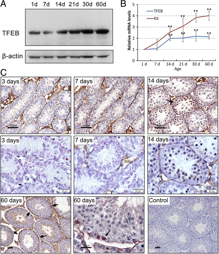
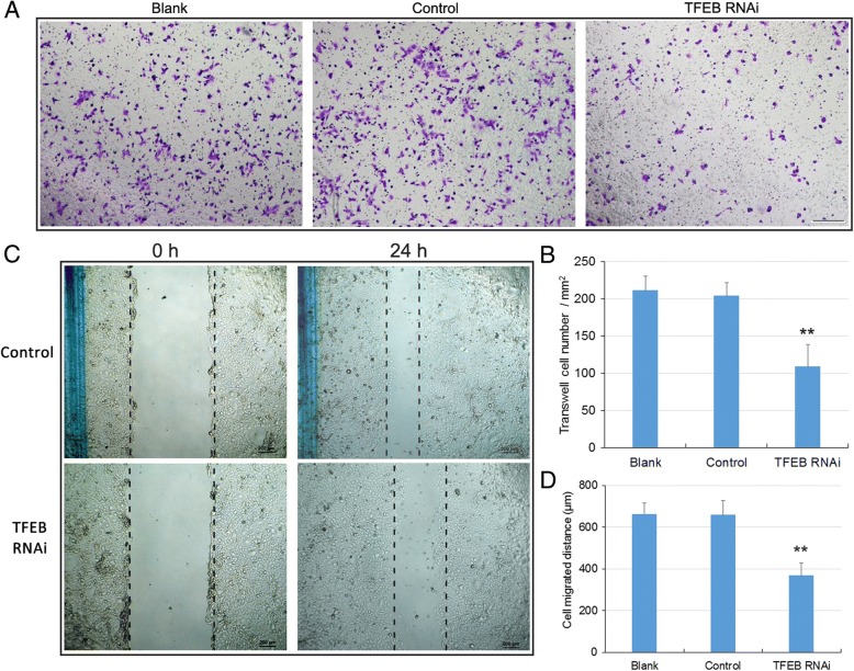
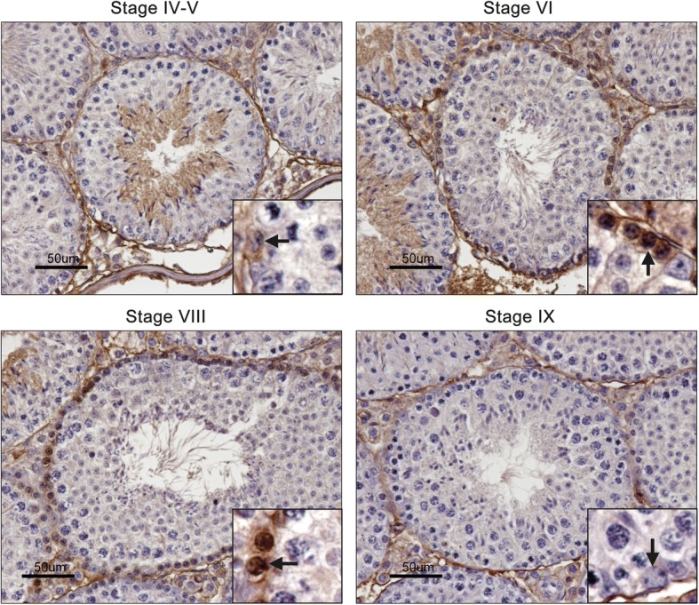
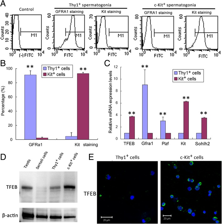
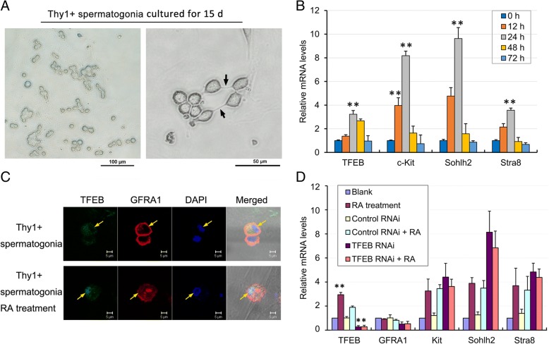
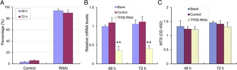
During testicular development, TFEB expression was rapidly increased in the testes at the period of 7 days post-partum (dpp) to 14 dpp, whereas in adult testis, it was predominantly localized in the nucleus of spermatogonia at stages VI to VIII of the seminiferous epithelial cycle. Accordingly, TFEB was observed to be mainly expressed in differentiating spermatogonia and was activated for nuclear translocation by RA treatment. Moreover, knockdown of TFEB expression by RNAi did not affect spermatogonial differentiation, but significantly reduced cell migration in GC-1 cells.
These findings imply that regionally distinct expression and activation of TFEB was strongly associated with RA signaling, and therefore may promote cell migration across the BTB and transport along the seminiferous epithelium.
精子发生是一个复杂的过程,涉及精原细胞在曲细精管内自我更新和分化为成熟精子。在精子发生过程中,生殖细胞从基膜迁移穿过血睾屏障(BTB),最终到达生精上皮的管腔侧。然而,调节生殖细胞迁移的机制尚不清楚。在这项研究中,我们专注于转录因子 EB(TFEB)的表达和功能,TFEB 是溶酶体生物发生、自噬和内吞作用的主要调节剂,在精子发生中。
通过 Western blot 和免疫组织化学分析研究了 TFEB 在小鼠睾丸中的表达模式。使用基于特定细胞表面标志物的磁激活细胞分选技术,从睾丸中分离出未分化的精原细胞或分化的精原细胞。用 100 nM 视黄酸(RA)诱导精原细胞分化。使用 shRNA 敲低细胞中的 TFEB。通过免疫荧光、qRT-PCR 和 Western blot 检测 TFEB 表达。通过 Transwell 迁移实验和划痕愈合实验测定细胞迁移,该实验应用于永生化精原细胞系 GC-1 细胞。
在睾丸发育过程中,TFEB 的表达在产后 7 天至 14 天(dpp)的睾丸中迅速增加,而在成年睾丸中,它主要定位于生精上皮周期 VI 至 VIII 期的精原细胞核内。因此,TFEB 主要在分化的精原细胞中表达,并通过 RA 处理被激活进行核易位。此外,通过 RNAi 敲低 TFEB 表达不会影响精原细胞分化,但会显著减少 GC-1 细胞的迁移。
这些发现表明 TFEB 的区域特异性表达和激活与 RA 信号密切相关,因此可能促进 BTB 穿过和沿生精上皮的运输。