Somsap Kulyada, Chamadol Nittaya, Titapun Attapol, Pairojkul Chawalit, Sangkhamanon Sakkarn
Department of Radiology, Faculty of Medicine, Khon Kaen University, Khon Kaen, Thailand.
Department of Surgery, Faculty of Medicine, Khon Kaen University, Khon Kaen, Thailand.
BJR Case Rep. 2016 Sep 29;3(1):20150242. doi: 10.1259/bjrcr.20150242. eCollection 2017.
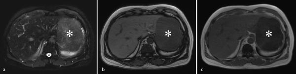
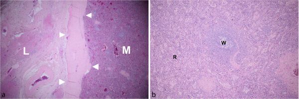
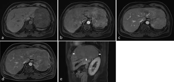
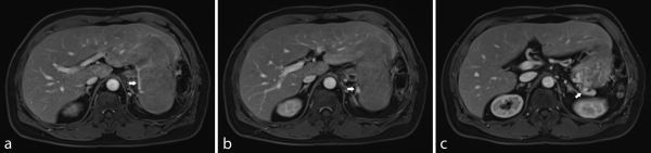
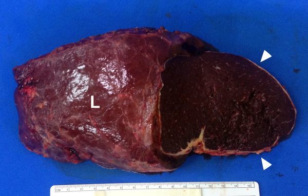
Splenosis refers to the autotransplantation of splenic tissue throughout different anatomic compartments secondary to trauma or splenic surgery. The liver is an uncommon location for splenic implants and imaging findings are often described as non-specific. We report MRI findings of a patient with a large liver mass that was first diagnosed as a malignant tumour but histopathology revealed that it was actually intrahepatic splenosis. The signal characteristics of this mass were low intensity on and high intensity on weighted images; arterial enhancement, which became more homogeneous in the later phases; and a relative hypointensity on the delayed phase images. Because a high level of awareness is necessary for making a correct diagnosis of this condition, one should consider the possibility of intrahepatic splenosis in a patient with a history of abdominal trauma or splenic surgery.
脾组织异位种植是指继发于创伤或脾手术后,脾组织在不同解剖部位的自体移植。肝脏是脾植入物的罕见部位,影像学表现通常被描述为非特异性的。我们报告了一名肝脏巨大肿块患者的MRI表现,该肿块最初被诊断为恶性肿瘤,但组织病理学显示实际上是肝内脾组织异位种植。该肿块的信号特征在T1加权图像上呈低信号,在T2加权图像上呈高信号;动脉期强化,在后期变得更加均匀;延迟期图像上呈相对低信号。由于正确诊断这种情况需要高度的认识,对于有腹部创伤或脾手术史的患者,应考虑肝内脾组织异位种植的可能性。