Molecular Biology, New Mexico State University, Las Cruces, New Mexico.
Chemical & Materials Engineering, New Mexico State University, Las Cruces, New Mexico.
Cytometry A. 2019 Jan;95(1):70-79. doi: 10.1002/cyto.a.23606. Epub 2018 Oct 19.
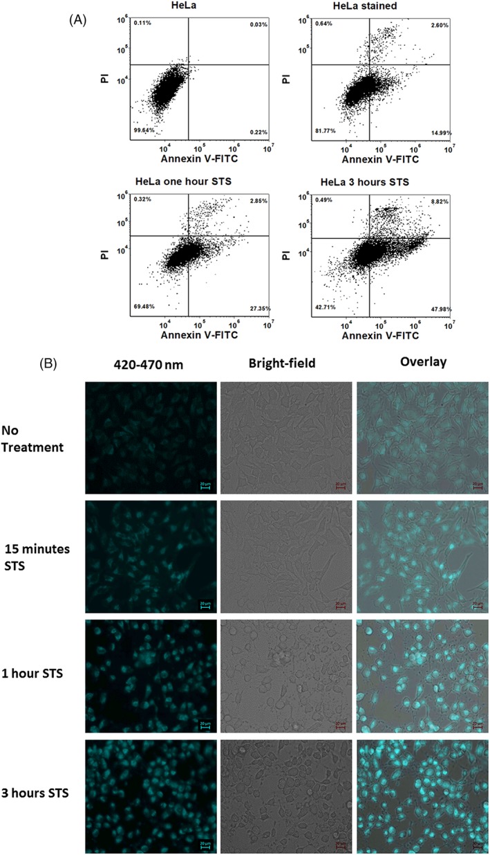
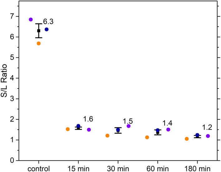
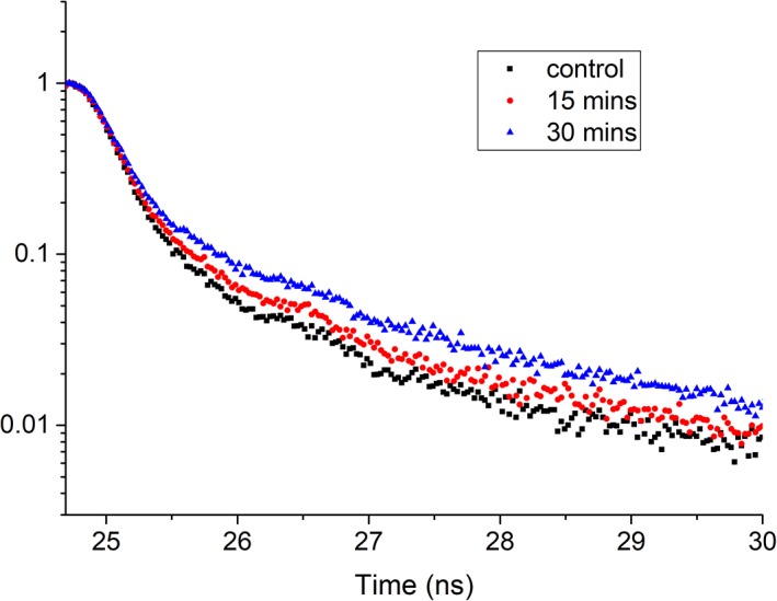
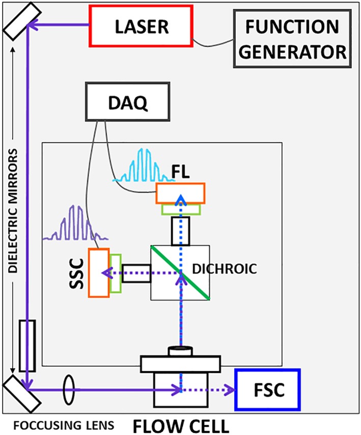
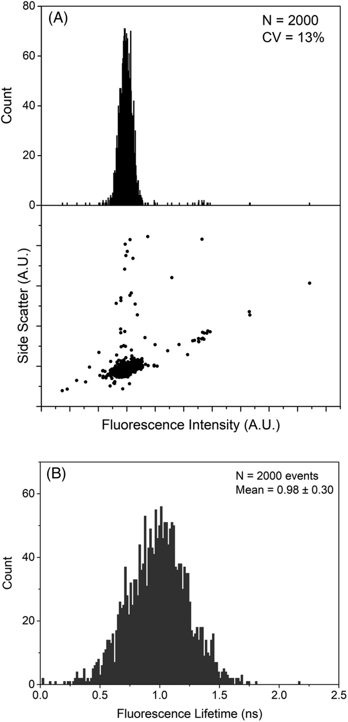
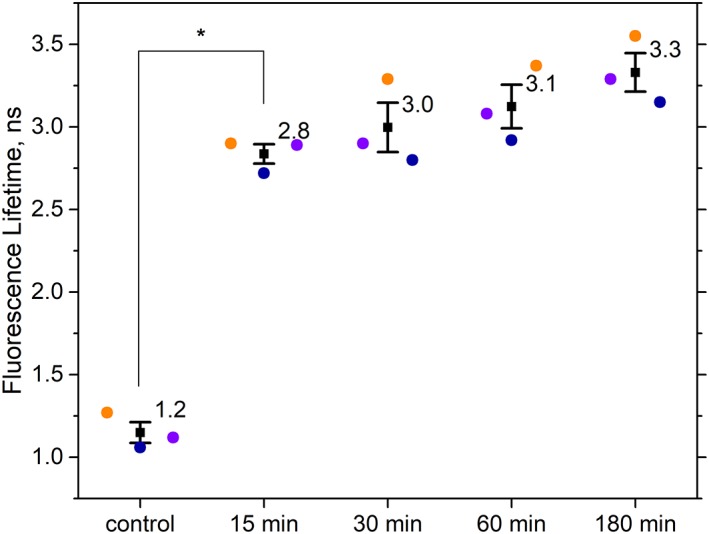
Autofluorescence from the intracellular metabolite, NAD(P)H, is a biomarker that is widely used and known to reliably screen and report metabolic activity as well as metabolic fluctuations within cells. As a ubiquitous endogenous fluorophore, NAD(P)H has a unique rate of fluorescence decay that is altered when bound to coenzymes. In this work we measure the shift in the fluorescence decay, or average fluorescence lifetime (1-3 ns), of NAD(P)H and correlate this shift to changes in metabolism that cells undergo during apoptosis. Our measurements are made with a flow cytometer designed specifically for fluorescence lifetime acquisition within the ultraviolet to violet spectrum. Our methods involved culture, treatment, and preparation of cells for cytometry and microscopy measurements. The evaluation we performed included observations and quantification of the changes in endogenous emission owing to the induction of apoptosis as well as changes in the decay kinetics of the emission measured by flow cytometry. Shifts in NAD(P)H fluorescence lifetime were observed as early as 15 min post-treatment with an apoptosis inducing agent. Results also include a phasor analysis to evaluate free to bound ratios of NAD(P)H at different time points. We defined the free to bound ratios as the ratio of 'short-to-long' (S/L) fluorescence lifetime, where S/L was found to consistently decrease with an increase in apoptosis. With a quantitative framework such as phasor analysis, the short and long lifetime components of NAD(P)H can be used to map the cycling of free and bound NAD(P)H during the early-to-late stages of apoptosis. The combination of lifetime screening and phasor analyses provides the first step in high throughput metabolic profiling of single cells and can be leveraged for screening and sorting for a range of applications in biomedicine. © 2018 The Authors. Cytometry Part A published by Wiley Periodicals, Inc. on behalf of International Society for Advancement of Cytometry.
细胞内代谢物 NAD(P)H 的自发荧光是一种生物标志物,被广泛应用,其能够可靠地筛选和报告细胞代谢活性和代谢波动。作为一种普遍存在的内源性荧光团,NAD(P)H 的荧光衰减率具有独特性,当其与辅酶结合时,荧光衰减率会发生改变。在这项工作中,我们测量了 NAD(P)H 的荧光衰减变化,即平均荧光寿命(1-3ns),并将其与细胞凋亡过程中经历的代谢变化相关联。我们的测量是使用专门设计的荧光寿命分析仪在紫外到紫光光谱范围内进行的。我们的方法包括细胞培养、处理和为细胞计数和显微镜测量做准备。我们进行的评估包括观察和量化由于诱导凋亡而导致的内源性发射的变化,以及通过流式细胞术测量的发射衰减动力学的变化。在用凋亡诱导剂处理 15 分钟后,就观察到 NAD(P)H 荧光寿命的偏移。结果还包括相位分析,以评估不同时间点 NAD(P)H 的自由结合比。我们将自由结合比定义为“短到长”(S/L)荧光寿命的比值,其中 S/L 随着凋亡的增加而持续下降。通过相位分析等定量框架,可以将 NAD(P)H 的短寿命和长寿命分量用于映射凋亡早期到晚期期间自由和结合 NAD(P)H 的循环。寿命筛选和相位分析的结合为高通量单细胞代谢分析提供了第一步,可以用于筛选和分类,在生物医学中有广泛的应用。