Rattanasuwan Kanyawat, Rassameemasmaung Supanee, Kiattavorncharoen Sirichai, Sirikulsathean Anongporn, Thorsuwan Jarinee, Wongsankakorn Wilasinee
Department of Oral Medicine and Periodontology, Faculty of Dentistry, Mahidol University, Bangkok, Thailand.
Department of Oral and Maxillofacial Surgery, Faculty of Dentistry, Mahidol University, Bangkok, Thailand.
Eur J Dent. 2018 Oct-Dec;12(4):469-474. doi: 10.4103/ejd.ejd_255_17.
The aim of this study is to evaluate the effects of platelet-rich plasma (PRP) on the proliferation, migration, and attachment of cultured periodontal ligament (PDL) cells.
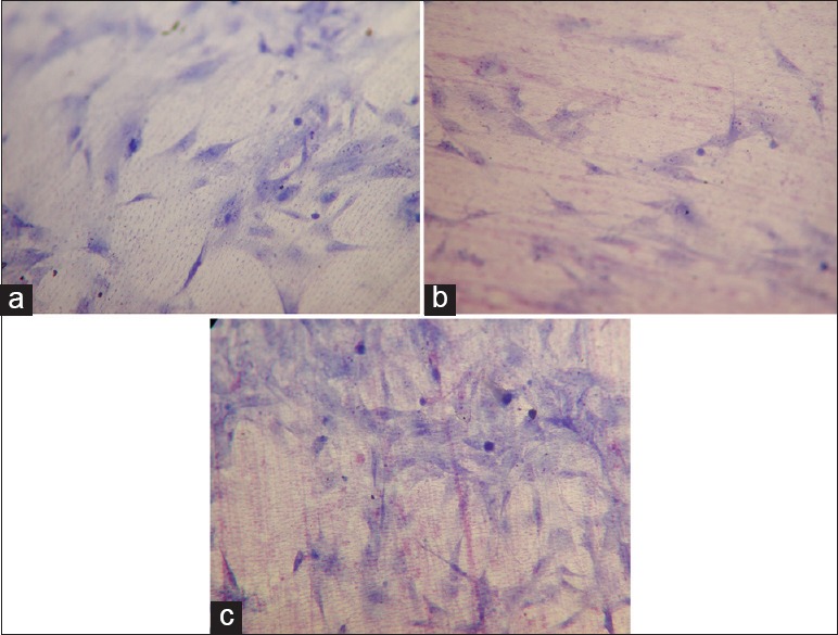
3-(4,5-dimethylthiazole-2-yl)-2,5-diphenyltetrazolium bromide (MTT) assay was used to assess number of PDL cells cultured in medium with or without PRP. Cell migration toward medium with or without PRP was assessed using the Boyden chamber. Cell attachment was assessed by counting cells on PRP or non-PRP coated dentin specimens. Group differences were analyzed using two-way ANOVA at 0.05 significance level.
In the MTT and cell migration assay, the number of cells in 5% and 10% PRP-treated groups were significantly higher than that in the non-PRP-treated group ( < 0.05). In the attachment assay, the number of cells on the dentin specimens in 10% PRP-treated group was significantly higher than that in the non-PRP treated group ( < 0.05).
PRP could stimulate proliferation, migration, and attachment of PDL cells.
本研究旨在评估富血小板血浆(PRP)对培养的牙周膜(PDL)细胞增殖、迁移和黏附的影响。
采用3-(4,5-二甲基噻唑-2)-2,5-二苯基四氮唑溴盐(MTT)法评估在含或不含PRP的培养基中培养的PDL细胞数量。使用博伊登小室评估细胞向含或不含PRP的培养基的迁移情况。通过计数PRP或非PRP包被的牙本质标本上的细胞来评估细胞黏附情况。采用双向方差分析在0.05显著性水平分析组间差异。
在MTT和细胞迁移试验中,5%和10% PRP处理组的细胞数量显著高于非PRP处理组(<0.05)。在黏附试验中,10% PRP处理组牙本质标本上的细胞数量显著高于非PRP处理组(<0.05)。
PRP可刺激PDL细胞的增殖、迁移和黏附。