Pirone Antonella, Alexander Jonathan M, Koenig Jenny B, Cook-Snyder Denise R, Palnati Medha, Wickham Robert J, Eden Lillian, Shrestha Neha, Reijmers Leon, Biederer Thomas, Miczek Klaus A, Dulla Chris G, Jacob Michele H
Department of Neuroscience, Sackler School of Biomedical Sciences, Tufts University School of Medicine, Boston, MA, United States.
Front Synaptic Neurosci. 2018 Oct 12;10:35. doi: 10.3389/fnsyn.2018.00035. eCollection 2018.
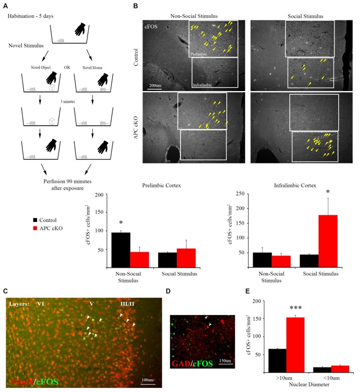
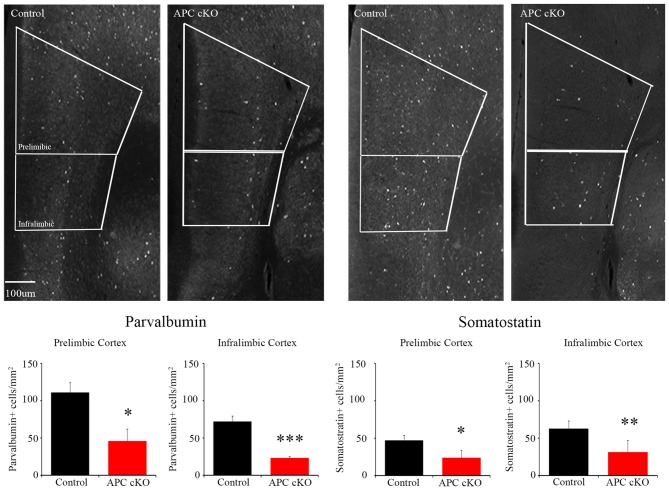
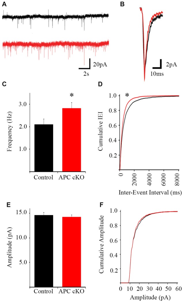
Autism spectrum disorder (ASD) is a highly prevalent and genetically heterogeneous brain disorder. Developing effective therapeutic interventions requires knowledge of the brain regions that malfunction and how they malfunction during ASD-relevant behaviors. Our study provides insights into brain regions activated by a novel social stimulus and how the activation pattern differs between mice that display autism-like disabilities and control littermates. Adenomatous polyposis coli (APC) conditional knockout (cKO) mice display reduced social interest, increased repetitive behaviors and dysfunction of the β-catenin pathway, a convergent target of numerous ASD-linked human genes. Here, we exposed the mice to a novel social vs. non-social stimulus and measured neuronal activation by immunostaining for the protein c-Fos. We analyzed three brain regions known to play a role in social behavior. Compared with control littermates, APC cKOs display excessive activation, as evidenced by an increased number of excitatory pyramidal neurons stained for c-Fos in the medial prefrontal cortex (mPFC), selectively in the infralimbic sub-region. In contrast, two other social brain regions, the medial amygdala and piriform cortex show normal levels of neuron activation. Additionally, APC cKOs exhibit increased frequency of miniature excitatory postsynaptic currents (mEPSCs) in layer 5 pyramidal neurons of the infralimbic sub-region. Further, immunostaining is reduced for the inhibitory interneuron markers parvalbumin (PV) and somatostatin (SST) in the APC cKO mPFC. Our findings suggest aberrant excitatory-inhibitory balance and activation patterns. As β-catenin is a core pathway in ASD, we identify the infralimbic sub-region of the mPFC as a critical brain region for autism-relevant social behavior.
自闭症谱系障碍(ASD)是一种高度流行且具有遗传异质性的脑部疾病。开发有效的治疗干预措施需要了解在与ASD相关行为期间发生功能障碍的脑区以及它们如何发生功能障碍。我们的研究深入探讨了由一种新型社交刺激激活的脑区,以及在表现出自闭症样残疾的小鼠和同窝对照小鼠之间激活模式的差异。腺瘤性息肉病 coli(APC)条件性敲除(cKO)小鼠表现出社交兴趣降低、重复行为增加以及β-连环蛋白通路功能障碍,β-连环蛋白通路是众多与ASD相关的人类基因的共同靶点。在此,我们将小鼠暴露于新型社交与非社交刺激下,并通过对蛋白质c-Fos进行免疫染色来测量神经元激活情况。我们分析了三个已知在社交行为中起作用的脑区。与同窝对照小鼠相比,APC cKO小鼠表现出过度激活,内侧前额叶皮质(mPFC)中,特别是在边缘下亚区域,c-Fos染色的兴奋性锥体神经元数量增加证明了这一点。相比之下,另外两个社交脑区,即内侧杏仁核和梨状皮质,显示出正常水平的神经元激活。此外,APC cKO小鼠在边缘下亚区域第5层锥体神经元中微小兴奋性突触后电流(mEPSCs)的频率增加。此外,APC cKO小鼠mPFC中抑制性中间神经元标志物小白蛋白(PV)和生长抑素(SST)的免疫染色减少。我们的研究结果表明兴奋性 - 抑制性平衡和激活模式异常。由于β-连环蛋白是ASD中的核心通路,我们将mPFC的边缘下亚区域确定为与自闭症相关社交行为的关键脑区。