Division of Brain Sciences, Imperial College London, London, UK.
Department of Neurology, University Heidelberg, Heidelberg, Germany.
J Cachexia Sarcopenia Muscle. 2019 Feb;10(1):54-62. doi: 10.1002/jcsm.12335. Epub 2018 Oct 30.
Stroke can lead to cardiac dysfunction in patients, but the mechanisms underlying the interaction between the injured brain and the heart are poorly understood. The objective of the study is to investigate the effects of experimental murine stroke on cardiac function and molecular signalling in the heart.
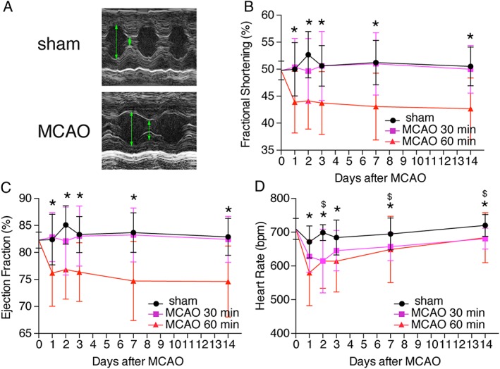
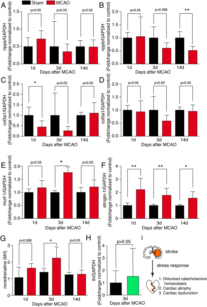
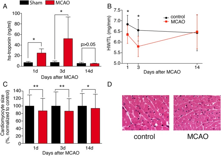
Mice were subjected to filament-induced left middle cerebral artery occlusion for 30 or 60 min or sham surgery and underwent repetitive micro-echocardiography. Left ventricular contractility was reduced early (24-72 h) but not late (2 months) after brain ischaemia. Cardiac dysfunction was accompanied by a release of high-sensitive cardiac troponin (hsTNT (ng/ml): d1: 7.0 ± 1.0 vs. 25.0 ± 3.2*; d3: 7.3 ± 1.1 vs. 52.2 ± 16.7*; d14: 5.7 ± 0.8 vs. 5.2 ± 0.3; sham vs. 60 min. MCAO; mean ± SEM; p < 0.05); reduced heart weight (heart weight/tibia length ratio: d1: 6.9 ± 0.2 vs. 6.4 ± 0.1; d3: 6.7 ± 0.2 vs. 5.8 ± 0.1*; d14: 6.7 ± 0.2 vs. 6.4 ± 03; sham vs. 60 min. MCAO; mean ± SEM; p < 0.05); resulting from cardiomyocyte atrophy (cardiomyocyte size: d1: 12.8% ± 0.002; d3: 13.5% ± 0.002; 14d: 6.3% ± 0.003; 60 min. MCAO vs. sham; mean ± SEM; **p < 0.01; *p < 0.05), accompanied by increased atrogin-1 and the E3 ubiquitin ligase murf-1. Net norepinephrine but not synthesis was increased, suggesting a reduced norepinephrine release or an increase of norepinephrine re-uptake, resulting in a functional denervation. Transcriptome analysis in cardiac tissue identified the transcription factor peroxisome proliferator-activated receptor gamma as a potential mediator of stroke-induced transcriptional dysregulation involved in cardiac atrophy.
Stroke induces a complex molecular response in the heart muscle with immediate but transient cardiac atrophy and dysfunction.
中风可导致患者心脏功能障碍,但受伤大脑与心脏之间相互作用的机制尚不清楚。本研究旨在探讨实验性小鼠中风对心脏功能和心脏内分子信号的影响。
采用线栓法诱导左侧大脑中动脉闭塞 30 或 60 分钟或假手术,重复进行微超声心动图检查。左心室收缩功能在脑缺血后早期(24-72 小时)而非晚期(2 个月)降低。心脏功能障碍伴随着高敏心肌肌钙蛋白(hsTNT(ng/ml):d1:7.0±1.0 与 25.0±3.2*;d3:7.3±1.1 与 52.2±16.7*;d14:5.7±0.8 与 5.2±0.3;假手术与 60 分钟 MCAO 比较;均值±SEM;p<0.05)的释放;心脏重量(心脏重量/胫骨长度比值:d1:6.9±0.2 与 6.4±0.1;d3:6.7±0.2 与 6.4±0.1*;d14:6.7±0.2 与 6.4±0.3;假手术与 60 分钟 MCAO 比较;均值±SEM;p<0.05)减少,这归因于心肌细胞萎缩(心肌细胞大小:d1:12.8%±0.002;d3:13.5%±0.002;d14:6.3%±0.003;60 分钟 MCAO 与假手术比较;均值±SEM;**p<0.01;*p<0.05);伴肌萎缩蛋白-1 和 E3 泛素连接酶 murf-1 的表达增加。去甲肾上腺素的净含量增加,但合成没有增加,这表明去甲肾上腺素释放减少或去甲肾上腺素再摄取增加,导致功能失神经支配。心脏组织的转录组分析鉴定出过氧化物酶体增殖物激活受体γ转录因子作为中风诱导的涉及心肌萎缩的转录失调的潜在介质。
中风会导致心肌发生复杂的分子反应,引起即刻但短暂的心肌萎缩和功能障碍。