Department of Cell and Developmental Biology, University of Michigan Medical School, Ann Arbor, MI.
Department of Biochemistry and Molecular Biology, Bloomberg School of Public Health, Johns Hopkins University, Baltimore, MD.
J Cell Biol. 2018 Dec 3;217(12):4314-4330. doi: 10.1083/jcb.201712130. Epub 2018 Nov 2.
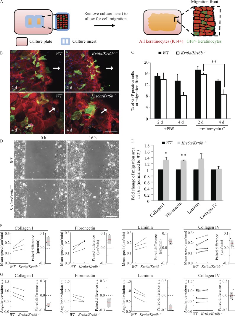
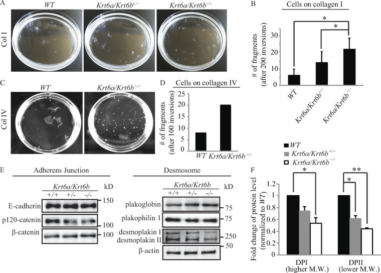
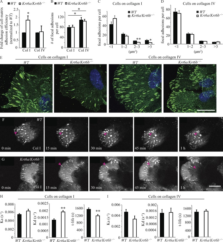
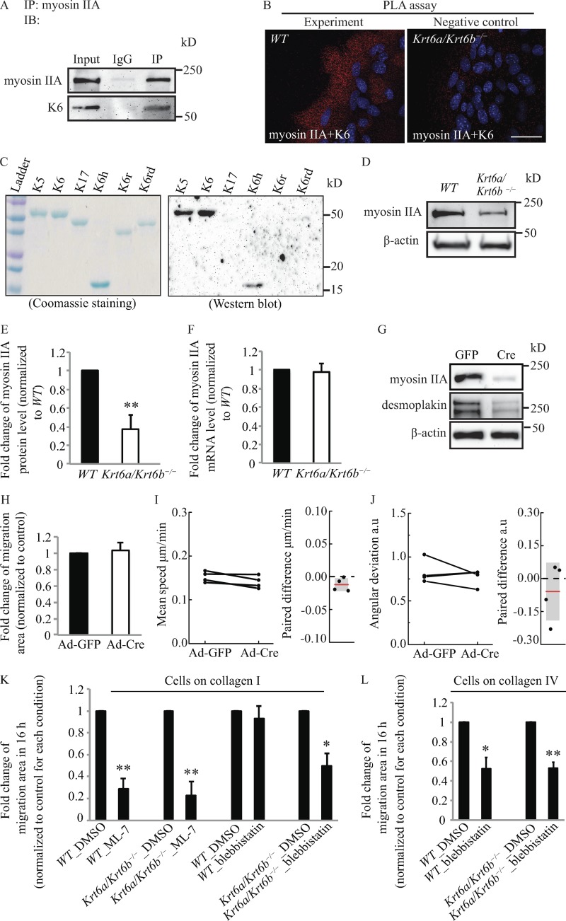
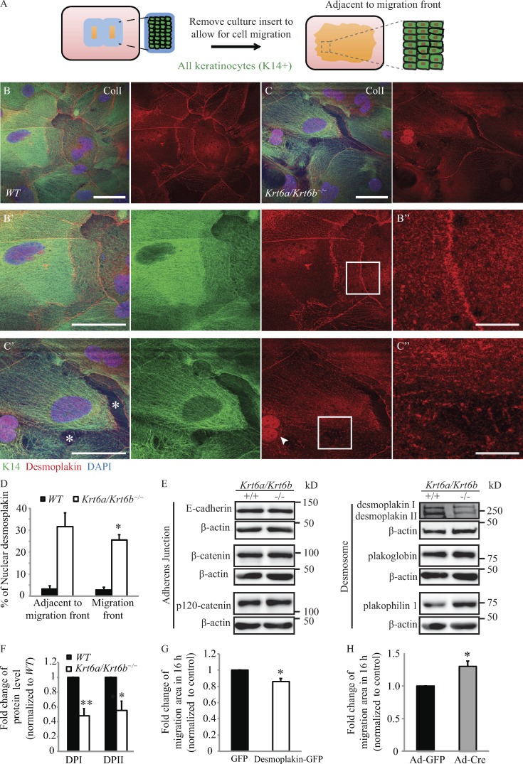
The a and b isoforms of keratin 6 (K6), a type II intermediate filament (IF) protein, are robustly induced upon injury to interfollicular epidermis. We previously showed that complete loss of K6a/K6b stimulates keratinocyte migration, correlating with enhanced Src activity. In this study, we demonstrate that this property is cell autonomous, depends on the ECM, and results from elevated speed, enhanced directionality, and an increased rate of focal adhesion disassembly. We show that myosin IIA interacts with K6a/K6b, that its levels are markedly reduced in -null keratinocytes, and that inhibiting myosin ATPase activity normalizes the enhanced migration potential of -null cells. Desmoplakin, which mediates attachment of IFs to desmosomes, is also expressed at reduced levels and is mislocalized to the nucleus in -null cells, correlating with defects in cell adhesion. These findings reveal that K6a/K6b modulate keratinocyte migration by regulating cell-matrix and cell-cell adhesion and highlight a role for keratins in collective cell migration.
角蛋白 6(K6)的 a 和 b 同工型是一种 II 型中间丝(IF)蛋白,在毛囊间表皮损伤时会强烈诱导。我们之前曾表明,K6a/K6b 的完全缺失会刺激角质形成细胞迁移,这与 Src 活性增强有关。在这项研究中,我们证明了这种特性是细胞自主的,取决于细胞外基质,并且是由于速度加快、方向性增强以及焦点黏附解离速率增加而导致的。我们表明肌球蛋白 IIA 与 K6a/K6b 相互作用,其水平在 -null 角质形成细胞中明显降低,并且抑制肌球蛋白 ATP 酶活性可使 -null 细胞增强的迁移潜能正常化。桥粒中间丝附着到桥粒的连接蛋白桥粒斑蛋白的水平也降低,并且在 -null 细胞中错误定位到核内,这与细胞黏附缺陷相关。这些发现表明 K6a/K6b 通过调节细胞-基质和细胞-细胞黏附来调节角质形成细胞迁移,并强调了角蛋白在细胞集体迁移中的作用。