• 文献检索
  • 文档翻译
  • 深度研究
  • 学术资讯
  • Suppr Zotero 插件Zotero 插件
  • 邀请有礼
  • 套餐&价格
  • 历史记录
应用&插件
Suppr Zotero 插件Zotero 插件浏览器插件Mac 客户端Windows 客户端微信小程序
定价
高级版会员购买积分包购买API积分包
服务
文献检索文档翻译深度研究API 文档MCP 服务
关于我们
关于 Suppr公司介绍联系我们用户协议隐私条款
关注我们

Suppr 超能文献

核心技术专利:CN118964589B侵权必究
粤ICP备2023148730 号-1Suppr @ 2026

文献检索

告别复杂PubMed语法,用中文像聊天一样搜索,搜遍4000万医学文献。AI智能推荐,让科研检索更轻松。

立即免费搜索

文件翻译

保留排版,准确专业,支持PDF/Word/PPT等文件格式,支持 12+语言互译。

免费翻译文档

深度研究

AI帮你快速写综述,25分钟生成高质量综述,智能提取关键信息,辅助科研写作。

立即免费体验

无损、多重三维映射核心针活检中的免疫浸润。

Nondestructive, multiplex three-dimensional mapping of immune infiltrates in core needle biopsy.

机构信息

Department of Molecular Genetics and Cell Biology, The University of Chicago, Chicago, IL, USA.

Department of Biopharmaceutical Sciences, The University of Illinois at Chicago, Chicago, IL, USA.

出版信息

Lab Invest. 2019 Sep;99(9):1400-1413. doi: 10.1038/s41374-018-0156-y. Epub 2018 Nov 6.

DOI:10.1038/s41374-018-0156-y
PMID:30401959
原文链接:https://pmc.ncbi.nlm.nih.gov/articles/PMC6502706/
Abstract

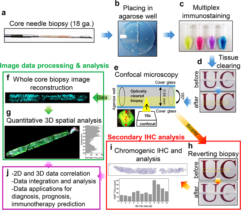
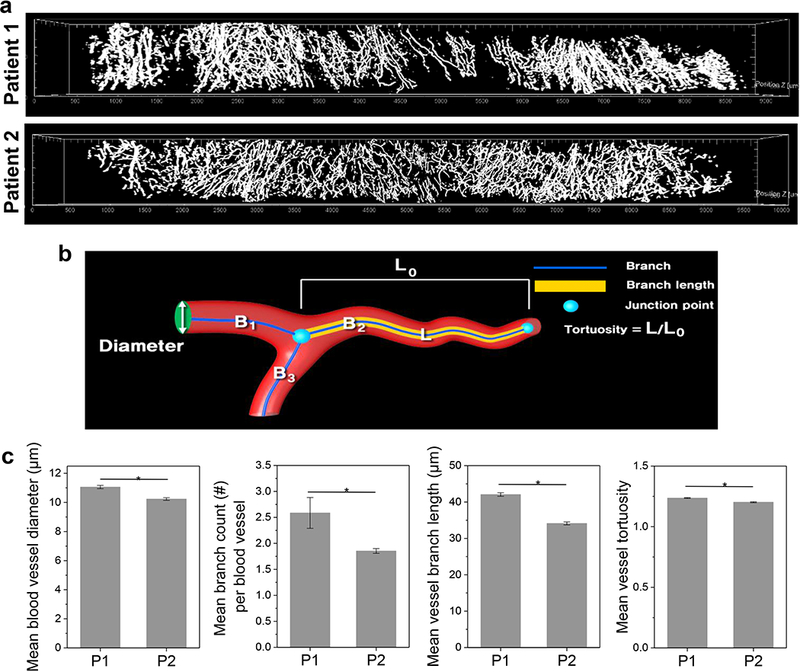
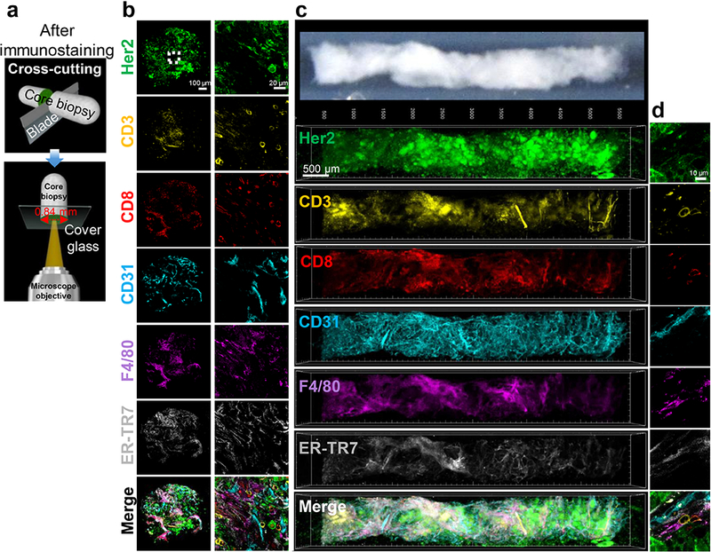
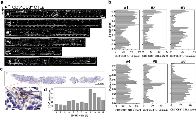
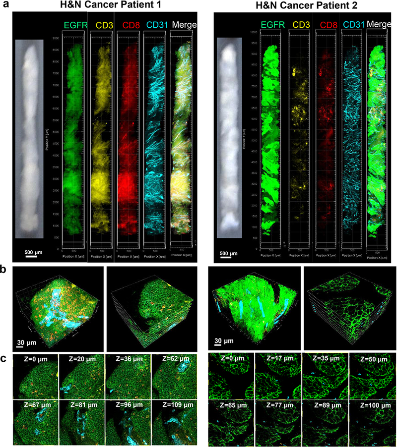
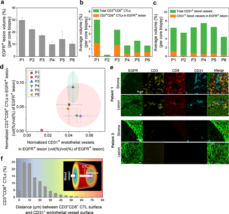
Enumeration of tumor-infiltrating lymphocytes (TILs) in H&E stained tissue sections has demonstrated limited value in predicting immune responses to cancer immunotherapy, likely reflecting the diversity of cell types and immune activation states among tumor infiltrates. Multiparametric flow cytometry enables robust phenotypic and functional analysis to distinguish suppression from activation, but tissue dissociation eliminates spatial context. Multiplex methods for immunohistochemistry (IHC) are emerging, but these interrogate only a single tissue section at a time. Here, we report transparent tissue tomography (T3) as a tool for three-dimensional (3D) imaging cytometry in the complex architecture of the tumor microenvironment, demonstrating multiplexed immunofluorescent analysis in core needle biopsies. Using T3 imaging, image processing and machine learning to map CD3CD8 cytotoxic T cells (CTLs) in whole core needle biopsies from Her2 murine mammary tumors and human head and neck surgical specimens revealed marked inhomogeneity within single needle cores, confirmed by serial section IHC. Applying T3 imaging cytometry, we discovered a strong spatial correlation between CD3CD8 CTLs and microvasculature in the EGFR parenchyma, revealing significant differences among head and neck cancer patients. These results show that T3 offers simple and rapid access to three-dimensional and quantitative maps of the tumor microenvironment and immune infiltrate, offering a new diagnostic tool for personalized cancer immunotherapy.

摘要

在 H&E 染色组织切片中对肿瘤浸润淋巴细胞(TILs)进行计数,其预测癌症免疫治疗反应的价值有限,这可能反映了肿瘤浸润物中细胞类型和免疫激活状态的多样性。多参数流式细胞术能够进行强大的表型和功能分析,以区分抑制和激活,但组织解离消除了空间背景。免疫组化(IHC)的多重方法正在出现,但这些方法一次只能检测单个组织切片。在这里,我们报告透明组织断层成像术(T3)作为肿瘤微环境复杂结构中三维(3D)成像细胞术的工具,在核心针活检中证明了多重免疫荧光分析。使用 T3 成像、图像处理和机器学习来绘制 Her2 鼠乳腺肿瘤和人类头颈部手术标本的整个核心针活检中的 CD3CD8 细胞毒性 T 细胞(CTLs),发现单个针芯内存在明显的不均匀性,通过连续切片 IHC 得到证实。应用 T3 成像细胞术,我们发现 CD3CD8 CTLs 与 EGFR 实质中的微血管之间存在很强的空间相关性,揭示了头颈部癌症患者之间的显著差异。这些结果表明,T3 为肿瘤微环境和免疫浸润的三维和定量图谱提供了简单快速的获取途径,为个性化癌症免疫治疗提供了一种新的诊断工具。

https://cdn.ncbi.nlm.nih.gov/pmc/blobs/23b2/6502706/83a93305ea9d/nihms-1509068-f0006.jpg
https://cdn.ncbi.nlm.nih.gov/pmc/blobs/23b2/6502706/e3502b152071/nihms-1509068-f0001.jpg
https://cdn.ncbi.nlm.nih.gov/pmc/blobs/23b2/6502706/11528fb208ab/nihms-1509068-f0002.jpg
https://cdn.ncbi.nlm.nih.gov/pmc/blobs/23b2/6502706/38fd69d93572/nihms-1509068-f0003.jpg
https://cdn.ncbi.nlm.nih.gov/pmc/blobs/23b2/6502706/37813fbc2441/nihms-1509068-f0004.jpg
https://cdn.ncbi.nlm.nih.gov/pmc/blobs/23b2/6502706/447d5dd64c1a/nihms-1509068-f0005.jpg
https://cdn.ncbi.nlm.nih.gov/pmc/blobs/23b2/6502706/83a93305ea9d/nihms-1509068-f0006.jpg

相似文献

1
Nondestructive, multiplex three-dimensional mapping of immune infiltrates in core needle biopsy.无损、多重三维映射核心针活检中的免疫浸润。
Lab Invest. 2019 Sep;99(9):1400-1413. doi: 10.1038/s41374-018-0156-y. Epub 2018 Nov 6.
2
Multiplex three-dimensional optical mapping of tumor immune microenvironment.肿瘤免疫微环境的多重三维光学图谱
Sci Rep. 2017 Dec 5;7(1):17031. doi: 10.1038/s41598-017-16987-x.
3
Kv1.3 Channels Mark Functionally Competent CD8+ Tumor-Infiltrating Lymphocytes in Head and Neck Cancer.Kv1.3通道标记头颈部癌中功能成熟的CD8 +肿瘤浸润淋巴细胞。
Cancer Res. 2017 Jan 1;77(1):53-61. doi: 10.1158/0008-5472.CAN-16-2372. Epub 2016 Nov 4.
4
Expression of immune response biomarkers (PD‑L1, p16, CD3+ and CD8+ TILs) in recurrent head and neck squamous cell carcinoma within previously irradiated areas.免疫反应生物标志物(PD-L1、p16、CD3+和 CD8+TILs)在既往照射区域内复发性头颈部鳞状细胞癌中的表达。
Oncol Rep. 2021 Mar;45(3):1273-1283. doi: 10.3892/or.2021.7928. Epub 2021 Jan 7.
5
Differences in TCR repertoire and T cell activation underlie the divergent outcomes of antitumor immune responses in tumor-eradicating versus tumor-progressing hosts.TCR repertoire 和 T 细胞激活的差异是抗肿瘤免疫反应在清除肿瘤宿主和进展肿瘤宿主中产生不同结果的基础。
J Immunother Cancer. 2021 Jan;9(1). doi: 10.1136/jitc-2020-001615.
6
IFNγ PET Imaging as a Predictive Tool for Monitoring Response to Tumor Immunotherapy.IFNγ PET 成像作为监测肿瘤免疫治疗反应的预测工具。
Cancer Res. 2018 Oct 1;78(19):5706-5717. doi: 10.1158/0008-5472.CAN-18-0253. Epub 2018 Aug 16.
7
Quantitative image analysis for CD8 score in lung small biopsies and cytology cell-blocks.肺小活检组织和细胞学细胞块中CD8评分的定量图像分析。
Cytopathology. 2020 Sep;31(5):393-401. doi: 10.1111/cyt.12812. Epub 2020 Apr 6.
8
Robust Cell Detection and Segmentation for Image Cytometry Reveal Th17 Cell Heterogeneity.稳健的细胞检测和分割技术在图像细胞计量术中的应用揭示了 Th17 细胞的异质性。
Cytometry A. 2019 Apr;95(4):389-398. doi: 10.1002/cyto.a.23726. Epub 2019 Feb 4.
9
Multispectral quantitative immunohistochemical analysis of tumor-infiltrating lymphocytes in relation to programmed death-ligand 1 expression in triple-negative breast cancer.三阴性乳腺癌中与程序性死亡配体1表达相关的肿瘤浸润淋巴细胞的多光谱定量免疫组织化学分析
Breast Cancer. 2020 Jul;27(4):519-526. doi: 10.1007/s12282-020-01110-2. Epub 2020 May 23.
10
Improving function of cytotoxic T-lymphocytes by transforming growth factor-β inhibitor in oral squamous cell carcinoma.转化生长因子-β抑制剂改善口腔鳞状细胞癌细胞毒性 T 淋巴细胞的功能。
Cancer Sci. 2021 Oct;112(10):4037-4049. doi: 10.1111/cas.15081. Epub 2021 Aug 2.

引用本文的文献

1
Correlative multiscale 3D imaging of mouse primary and metastatic tumors by sequential light sheet and confocal fluorescence microscopy.通过顺序光片和共聚焦荧光显微镜对小鼠原发性和转移性肿瘤进行相关多尺度3D成像。
iScience. 2025 Feb 3;28(3):111934. doi: 10.1016/j.isci.2025.111934. eCollection 2025 Mar 21.
2
Increased Multiplexity in Optical Tissue Clearing-Based Three-Dimensional Immunofluorescence Microscopy of the Tumor Microenvironment by Light-Emitting Diode Photobleaching.通过发光二极管光漂白提高基于光学组织通透的三维免疫荧光显微镜检测肿瘤微环境的多重性。
Lab Invest. 2024 Jun;104(6):102072. doi: 10.1016/j.labinv.2024.102072. Epub 2024 Apr 26.
3

本文引用的文献

1
Whole-tissue biopsy phenotyping of three-dimensional tumours reveals patterns of cancer heterogeneity.三维肿瘤的全组织活检表型分析揭示了癌症异质性模式。
Nat Biomed Eng. 2017 Oct;1(10):796-806. doi: 10.1038/s41551-017-0139-0. Epub 2017 Oct 2.
2
Light-sheet microscopy for slide-free non-destructive pathology of large clinical specimens.用于大型临床标本的无玻片非破坏性病理学的光片显微镜技术。
Nat Biomed Eng. 2017 Jul;1(7). doi: 10.1038/s41551-017-0084. Epub 2017 Jun 26.
3
Enhancing cancer immunotherapy using antiangiogenics: opportunities and challenges.
Direct three-dimensional segmentation of prostate glands with nnU-Net.
使用nnU-Net对前列腺进行直接三维分割。
J Biomed Opt. 2024 Mar;29(3):036001. doi: 10.1117/1.JBO.29.3.036001. Epub 2024 Mar 1.
4
Multiresolution 3D Optical Mapping of Immune Cell Infiltrates in Mouse Asthmatic Lung.多分辨率 3D 光学成像技术在小鼠哮喘肺部中免疫细胞浸润的研究。
Am J Respir Cell Mol Biol. 2023 Jul;69(1):13-21. doi: 10.1165/rcmb.2022-0353MA.
5
Deep-learning based breast cancer detection for cross-staining histopathology images.基于深度学习的跨染色组织病理学图像乳腺癌检测
Heliyon. 2023 Jan 21;9(2):e13171. doi: 10.1016/j.heliyon.2023.e13171. eCollection 2023 Feb.
6
Multidimensional Imaging of Breast Cancer.乳腺癌的多维成像。
Cold Spring Harb Perspect Med. 2023 May 2;13(5):a041330. doi: 10.1101/cshperspect.a041330.
7
3D imaging for driving cancer discovery.三维成像助力癌症研究。
EMBO J. 2022 May 16;41(10):e109675. doi: 10.15252/embj.2021109675. Epub 2022 Apr 11.
8
Prostate Cancer Risk Stratification via Nondestructive 3D Pathology with Deep Learning-Assisted Gland Analysis.基于深度学习辅助腺体分析的非破坏性 3D 病理学前列腺癌风险分层。
Cancer Res. 2022 Jan 15;82(2):334-345. doi: 10.1158/0008-5472.CAN-21-2843. Epub 2021 Dec 1.
9
Multiplexed Tissue Tomography.多重组织断层成像技术。
Methods Mol Biol. 2021;2350:77-93. doi: 10.1007/978-1-0716-1593-5_6.
10
Harnessing non-destructive 3D pathology.利用非破坏性三维病理学
Nat Biomed Eng. 2021 Mar;5(3):203-218. doi: 10.1038/s41551-020-00681-x. Epub 2021 Feb 15.
利用抗血管生成药物增强癌症免疫治疗:机遇与挑战。
Nat Rev Clin Oncol. 2018 May;15(5):325-340. doi: 10.1038/nrclinonc.2018.29. Epub 2018 Mar 6.
4
Immunotherapy for Head and Neck Squamous Cell Carcinoma.头颈部鳞状细胞癌的免疫治疗。
Curr Oncol Rep. 2018 Mar 3;20(2):22. doi: 10.1007/s11912-018-0654-5.
5
Seeing is believing: anti-PD-1/PD-L1 monoclonal antibodies in action for checkpoint blockade tumor immunotherapy.眼见为实:抗 PD-1/PD-L1 单克隆抗体在检查点阻断肿瘤免疫治疗中的作用。
Signal Transduct Target Ther. 2016 Nov 25;1:16029. doi: 10.1038/sigtrans.2016.29. eCollection 2016.
6
Multiplex three-dimensional optical mapping of tumor immune microenvironment.肿瘤免疫微环境的多重三维光学图谱
Sci Rep. 2017 Dec 5;7(1):17031. doi: 10.1038/s41598-017-16987-x.
7
Migrating into the Tumor: a Roadmap for T Cells.迁移至肿瘤:T细胞的路线图
Trends Cancer. 2017 Nov;3(11):797-808. doi: 10.1016/j.trecan.2017.09.006. Epub 2017 Nov 6.
8
Validation of multiplex immunofluorescence panels using multispectral microscopy for immune-profiling of formalin-fixed and paraffin-embedded human tumor tissues.利用多光谱显微镜对福尔马林固定石蜡包埋的人类肿瘤组织进行免疫分析的多重免疫荧光试剂盒验证。
Sci Rep. 2017 Oct 17;7(1):13380. doi: 10.1038/s41598-017-13942-8.
9
Three-dimensional Imaging and Scanning: Current and Future Applications for Pathology.三维成像与扫描:病理学的当前及未来应用
J Pathol Inform. 2017 Sep 7;8:36. doi: 10.4103/jpi.jpi_32_17. eCollection 2017.
10
CUBIC pathology: three-dimensional imaging for pathological diagnosis.立方病理:用于病理诊断的三维成像。
Sci Rep. 2017 Aug 24;7(1):9269. doi: 10.1038/s41598-017-09117-0.