• 文献检索
  • 文档翻译
  • 深度研究
  • 学术资讯
  • Suppr Zotero 插件Zotero 插件
  • 邀请有礼
  • 套餐&价格
  • 历史记录
应用&插件
Suppr Zotero 插件Zotero 插件浏览器插件Mac 客户端Windows 客户端微信小程序
定价
高级版会员购买积分包购买API积分包
服务
文献检索文档翻译深度研究API 文档MCP 服务
关于我们
关于 Suppr公司介绍联系我们用户协议隐私条款
关注我们

Suppr 超能文献

核心技术专利:CN118964589B侵权必究
粤ICP备2023148730 号-1Suppr @ 2026

文献检索

告别复杂PubMed语法,用中文像聊天一样搜索,搜遍4000万医学文献。AI智能推荐,让科研检索更轻松。

立即免费搜索

文件翻译

保留排版,准确专业,支持PDF/Word/PPT等文件格式,支持 12+语言互译。

免费翻译文档

深度研究

AI帮你快速写综述,25分钟生成高质量综述,智能提取关键信息,辅助科研写作。

立即免费体验

急性 ST 段抬高型心肌梗死患者行直接经皮冠状动脉介入治疗后心血管磁共振检测左心室血栓的发生率及预测因素:荟萃分析。

Incidence and predictors of left ventricular thrombus by cardiovascular magnetic resonance in acute ST-segment elevation myocardial infarction treated by primary percutaneous coronary intervention: a meta-analysis.

机构信息

Norfolk and Norwich University Hospital, Colney Lane, Norwich, NR4 7UY, UK.

The Hatter Cardiovascular Institute, Institute of Cardiovascular Science, University College London, London, UK.

出版信息

J Cardiovasc Magn Reson. 2018 Nov 8;20(1):72. doi: 10.1186/s12968-018-0494-3.

DOI:10.1186/s12968-018-0494-3
PMID:30404623
原文链接:https://pmc.ncbi.nlm.nih.gov/articles/PMC6222991/
Abstract

INTRODUCTION

The incidence of left ventricular (LV) thrombus formation in ST-segment elevation myocardial infarction (STEMI) patients in the current era of primary percutaneous coronary intervention (PCI) is not well established. We performed a meta-analysis to assess the actual incidence and predictors of LV thrombus by cardiovascular magnetic resonance (CMR) in STEMI treated by primary PCI.

METHODS

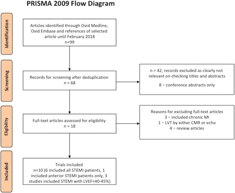
We searched MEDLINE and EMBASE databases up to February 2018. We included all studies published as a full-text article, reporting the incidence of LV thrombus by CMR within 1 month following acute STEMI in patients treated by primary PCI. A binary random-effects model was used to estimate the pooled incidence of LV thrombus. The diagnostic performance of transthoracic echocardiography (TTE) as compared with CMR was pooled to obtain the sensitivity and specificity of TTE with CMR as the gold standard. Embolic and bleeding complications of LV thrombus were also evaluated.

RESULTS

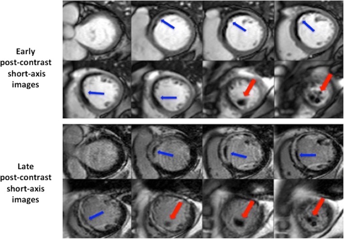
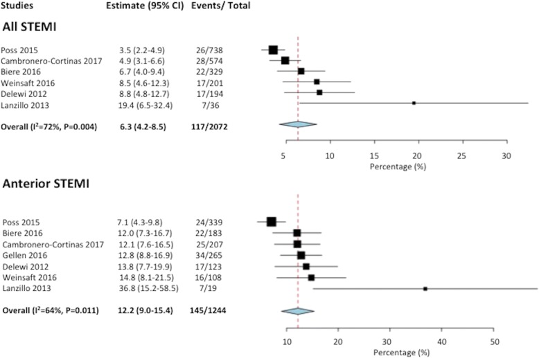
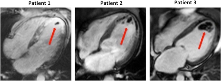
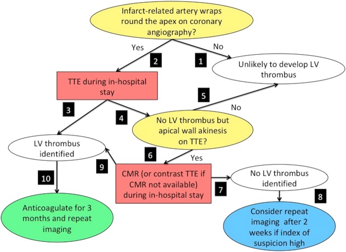
Ten studies were included in the meta-analysis. The incidence of LV thrombus by CMR in all-comer STEMI patients (n = 2072) was 6.3% with 96% of LV thrombus occurring in those with anterior STEMI (12.2% incidence). When only anterior STEMI with LVEF< 50% were considered (n = 447), the incidence of LV thrombus was 19.2%. Compared with CMR, the sensitivity of TTE to detect LV thrombus was 29% with a specificity of 98%. The sensitivity of TTE increased to 70% in those with anterior STEMI and reduced LVEF. LV thrombus resolved in 88% of cases by 3 to 6 months. After 1-2 years follow-up, the embolic complication rate was similar at 1.5% (P = 0.25) but the bleeding complication rate was significantly higher (8.8% versus 0.5%, P < 0.001) in the LV thrombus group on triple therapy when compared to the no LV thrombus group on dual antiplatelet therapy.

CONCLUSION

In the primary PCI era, CMR detection of an LV thrombus post-STEMI remains high with incidence of nearly 20% in anterior STEMI with depressed LVEF. Patients with LV thrombus treated by triple therapy had similar embolic complications but higher bleeding complications than those with no LV thrombus treated with dual antiplatelet therapy. A 3 month follow-up CMR scan to guide anticoagulation duration might help mitigate bleeding risk.

摘要

简介

在直接经皮冠状动脉介入治疗(PCI)的时代,ST 段抬高型心肌梗死(STEMI)患者左心室(LV)血栓形成的发生率尚不清楚。我们进行了一项荟萃分析,以评估通过心血管磁共振(CMR)在直接 PCI 治疗后的 STEMI 患者中 LV 血栓的实际发生率和预测因素。

方法

我们检索了 MEDLINE 和 EMBASE 数据库,截至 2018 年 2 月。我们纳入了所有以全文形式发表的研究,报告了在直接 PCI 治疗后的急性 STEMI 后 1 个月内通过 CMR 检测到的 LV 血栓的发生率。采用二项随机效应模型估计 LV 血栓的合并发生率。以 CMR 为金标准,汇总经胸超声心动图(TTE)与 CMR 相比的诊断性能,以获得 TTE 的敏感性和特异性。还评估了 LV 血栓的栓塞和出血并发症。

结果

共有 10 项研究纳入荟萃分析。所有 STEMI 患者(n=2072)中,CMR 检测到的 LV 血栓发生率为 6.3%,前壁 STEMI 患者中有 96%发生 LV 血栓(发生率为 12.2%)。当仅考虑前壁 STEMI 且 LVEF<50%时(n=447),LV 血栓的发生率为 19.2%。与 CMR 相比,TTE 检测 LV 血栓的敏感性为 29%,特异性为 98%。在前壁 STEMI 且 LVEF 降低的患者中,TTE 的敏感性增加至 70%。3 至 6 个月时,88%的 LV 血栓得到溶解。在 1-2 年的随访中,LV 血栓组的栓塞并发症发生率相似,为 1.5%(P=0.25),但在 LV 血栓组接受三联治疗时,出血并发症发生率明显更高(8.8%比 0.5%,P<0.001),而在无 LV 血栓组接受双联抗血小板治疗时。

结论

在直接 PCI 时代,CMR 检测到 STEMI 后 LV 血栓的发生率仍然很高,在前壁 STEMI 且 LVEF 降低的患者中,LV 血栓的发生率接近 20%。接受三联治疗的 LV 血栓患者的栓塞并发症发生率相似,但出血并发症发生率高于接受双联抗血小板治疗的无 LV 血栓患者。进行 3 个月的 CMR 随访以指导抗凝持续时间可能有助于降低出血风险。

https://cdn.ncbi.nlm.nih.gov/pmc/blobs/2a78/6222991/5b6e0dc591a7/12968_2018_494_Fig5_HTML.jpg
https://cdn.ncbi.nlm.nih.gov/pmc/blobs/2a78/6222991/f0a42af3a8f3/12968_2018_494_Fig1_HTML.jpg
https://cdn.ncbi.nlm.nih.gov/pmc/blobs/2a78/6222991/efb0f4da93b7/12968_2018_494_Fig2_HTML.jpg
https://cdn.ncbi.nlm.nih.gov/pmc/blobs/2a78/6222991/451e5dfea915/12968_2018_494_Fig3_HTML.jpg
https://cdn.ncbi.nlm.nih.gov/pmc/blobs/2a78/6222991/4dec4aedd583/12968_2018_494_Fig4_HTML.jpg
https://cdn.ncbi.nlm.nih.gov/pmc/blobs/2a78/6222991/5b6e0dc591a7/12968_2018_494_Fig5_HTML.jpg

相似文献

1
Incidence and predictors of left ventricular thrombus by cardiovascular magnetic resonance in acute ST-segment elevation myocardial infarction treated by primary percutaneous coronary intervention: a meta-analysis.急性 ST 段抬高型心肌梗死患者行直接经皮冠状动脉介入治疗后心血管磁共振检测左心室血栓的发生率及预测因素:荟萃分析。
J Cardiovasc Magn Reson. 2018 Nov 8;20(1):72. doi: 10.1186/s12968-018-0494-3.
2
Incidence and Predictors of Left Ventricular Thrombus After Primary Percutaneous Coronary Intervention for Anterior ST-Segment Elevation Myocardial Infarction.前壁ST段抬高型心肌梗死直接经皮冠状动脉介入治疗后左心室血栓的发生率及预测因素
Clin Cardiol. 2015 Oct;38(10):590-7. doi: 10.1002/clc.22450. Epub 2015 Sep 10.
3
Incidence and Risk Factors of Left Ventricular Thrombus in Acute ST-Segment Elevation Myocardial Infarction Treated by Primary Percutaneous Coronary Intervention: A Meta-Analysis.急性 ST 段抬高型心肌梗死患者行直接经皮冠状动脉介入治疗后左心室血栓形成的发生率及相关危险因素的荟萃分析。
Med Princ Pract. 2022;31(5):415-423. doi: 10.1159/000525943. Epub 2022 Jul 7.
4
Association between cardiac magnetic resonance ventricular strain and left ventricular thrombus in patients with ST-segment elevation myocardial infarction.ST 段抬高型心肌梗死患者心脏磁共振心室应变与左心室血栓的相关性。
Int J Cardiovasc Imaging. 2024 Aug;40(8):1735-1744. doi: 10.1007/s10554-024-03163-2. Epub 2024 Jun 17.
5
Left ventricular thrombus after acute ST-segment elevation myocardial infarction: multi-parametric cardiac magnetic resonance imaging with long-term outcomes.急性 ST 段抬高型心肌梗死患者左心室血栓:多参数心脏磁共振成像与长期预后。
Int J Cardiovasc Imaging. 2022 Nov;38(11):2373-2384. doi: 10.1007/s10554-022-02598-9. Epub 2022 Mar 27.
6
Association between inflammation and left ventricular thrombus formation following ST-elevation myocardial infarction.炎症与 ST 段抬高型心肌梗死后左心室血栓形成的关系。
Int J Cardiol. 2022 Aug 15;361:1-6. doi: 10.1016/j.ijcard.2022.05.009. Epub 2022 May 6.
7
Prophylactic warfarin post anterior ST-elevation myocardial infarction: A systematic review and meta-analysis.前壁ST段抬高型心肌梗死后预防性使用华法林:一项系统评价和荟萃分析。
Cardiovasc Revasc Med. 2017 Dec;18(8):559-564. doi: 10.1016/j.carrev.2017.05.002. Epub 2017 May 9.
8
Left Ventricular Hypertrophy Is Associated With Increased Infarct Size and Decreased Myocardial Salvage in Patients With ST-Segment Elevation Myocardial Infarction Undergoing Primary Percutaneous Coronary Intervention.在接受直接经皮冠状动脉介入治疗的ST段抬高型心肌梗死患者中,左心室肥厚与梗死面积增加及心肌挽救率降低相关。
J Am Heart Assoc. 2017 Jan 9;6(1):e004823. doi: 10.1161/JAHA.116.004823.
9
Defining left ventricular remodeling following acute ST-segment elevation myocardial infarction using cardiovascular magnetic resonance.使用心血管磁共振成像定义急性ST段抬高型心肌梗死后的左心室重构。
J Cardiovasc Magn Reson. 2017 Mar 13;19(1):26. doi: 10.1186/s12968-017-0343-9.
10
Effect of Early Metoprolol During ST-Segment Elevation Myocardial Infarction on Left Ventricular Strain: Feature-Tracking Cardiovascular Magnetic Resonance Substudy From the METOCARD-CNIC Trial.ST 段抬高型心肌梗死早期美托洛尔对左心室应变的影响:来自 METOCARD-CNIC 试验的特征追踪心血管磁共振子研究。
JACC Cardiovasc Imaging. 2019 Jul;12(7 Pt 1):1188-1198. doi: 10.1016/j.jcmg.2018.07.019. Epub 2018 Sep 12.

引用本文的文献

1
Beyond cardiac embolism and cryptogenic stroke: unveiling the mechanisms of cerebrovascular events in Chagas disease.超越心脏栓塞和隐源性卒中:揭示恰加斯病脑血管事件的机制
Lancet Reg Health Am. 2025 Aug 8;50:101203. doi: 10.1016/j.lana.2025.101203. eCollection 2025 Oct.
2
Rivaroxaban vs Warfarin in Acute Left Ventricular Thrombus Following Myocardial Infarction: RIVAWAR, An Open-Label RCT.利伐沙班与华法林治疗心肌梗死后急性左心室血栓的对比:RIVAWAR,一项开放标签随机对照试验
JACC Adv. 2025 Jul 23;4(8):101978. doi: 10.1016/j.jacadv.2025.101978.
3
Management of Biventricular and Left Atrium Thrombi in a Patient With Acute Myocardial Infarction.

本文引用的文献

1
2017 ESC Guidelines for the management of acute myocardial infarction in patients presenting with ST-segment elevation: The Task Force for the management of acute myocardial infarction in patients presenting with ST-segment elevation of the European Society of Cardiology (ESC).2017年欧洲心脏病学会(ESC)ST段抬高型急性心肌梗死患者管理指南:欧洲心脏病学会(ESC)ST段抬高型急性心肌梗死患者管理工作组
Eur Heart J. 2018 Jan 7;39(2):119-177. doi: 10.1093/eurheartj/ehx393.
2
Incidence, Outcomes, and Predictors of Ventricular Thrombus after Reperfused ST-Segment-Elevation Myocardial Infarction by Using Sequential Cardiac MR Imaging.应用序贯心脏磁共振成像评估再灌注治疗的 ST 段抬高型心肌梗死患者并发心室血栓的发生率、转归和预测因素。
Radiology. 2017 Aug;284(2):372-380. doi: 10.1148/radiol.2017161898. Epub 2017 Mar 2.
3
急性心肌梗死患者双心室及左心房血栓的管理
JACC Case Rep. 2025 Jun 18;30(15):103770. doi: 10.1016/j.jaccas.2025.103770.
4
Man With Chest Pain and Unilateral Weakness.胸痛伴单侧肌无力的男性。
J Am Coll Emerg Physicians Open. 2025 May 25;6(4):100186. doi: 10.1016/j.acepjo.2025.100186. eCollection 2025 Aug.
5
Predictive value of lymphocyte-to-C-reactive protein ratio for left ventricular thrombus in patients with ST-segment elevation myocardial infarction.淋巴细胞与C反应蛋白比值对ST段抬高型心肌梗死患者左心室血栓的预测价值
Front Cardiovasc Med. 2025 Apr 24;12:1465350. doi: 10.3389/fcvm.2025.1465350. eCollection 2025.
6
Intraventricular Thrombosis After Myocardial Infarction: Prognostic Evaluation in Relation to Microvascular Obstruction Extent by CMR.心肌梗死后脑室内血栓形成:基于心脏磁共振成像对微血管阻塞程度的预后评估
J Clin Med. 2025 Apr 13;14(8):2658. doi: 10.3390/jcm14082658.
7
Myocardial strain characteristics in ST-segment elevation myocardial infarction patients with left ventricular thrombus: a cardiac magnetic resonance imaging study.ST段抬高型心肌梗死合并左心室血栓患者的心肌应变特征:一项心脏磁共振成像研究
Quant Imaging Med Surg. 2025 Apr 1;15(4):3237-3247. doi: 10.21037/qims-24-1951. Epub 2025 Mar 28.
8
Soluble suppression of tumorigenicity 2 associated with left ventricular thrombosis in patients with ST-segment elevation myocardial infarction.可溶性致瘤性抑制因子2与ST段抬高型心肌梗死患者左心室血栓形成有关。
BMC Cardiovasc Disord. 2025 Mar 20;25(1):204. doi: 10.1186/s12872-025-04667-y.
9
Left Ventricular Thrombosis in Ischemic and Non-Ischemic Cardiomyopathies: Focus on Evidence-Based Treatment.缺血性和非缺血性心肌病中的左心室血栓形成:聚焦循证治疗
J Clin Med. 2025 Feb 27;14(5):1615. doi: 10.3390/jcm14051615.
10
Feasibility and Role of Cardiac Magnetic Resonance in Intensive and Acute Cardiovascular Care.心脏磁共振成像在重症及急性心血管护理中的可行性及作用
J Clin Med. 2025 Feb 9;14(4):1112. doi: 10.3390/jcm14041112.
Timing of Cardiac Magnetic Resonance Imaging Impacts on the Detection Rate of Left Ventricular Thrombus After Myocardial Infarction.心肌梗死后心脏磁共振成像的时机对左心室血栓检出率的影响
JACC Cardiovasc Imaging. 2017 Nov;10(11):1404-1405. doi: 10.1016/j.jcmg.2016.12.006. Epub 2017 Feb 15.
4
First pass perfusion imaging to improve the assessment of left ventricular thrombus following a myocardial infarction.首次通过灌注成像改善心肌梗死后左心室血栓的评估。
Eur J Radiol. 2016 Sep;85(9):1532-7. doi: 10.1016/j.ejrad.2016.05.017. Epub 2016 Jun 4.
5
Left ventricular thrombi after STEMI in the primary PCI era: A systematic review and meta-analysis.直接经皮冠状动脉介入治疗时代ST段抬高型心肌梗死后左心室血栓形成:一项系统评价与荟萃分析
Int J Cardiol. 2016 Oct 15;221:554-9. doi: 10.1016/j.ijcard.2016.07.069. Epub 2016 Jul 6.
6
Left Ventricular Thrombus Formation After ST-Segment-Elevation Myocardial Infarction: Insights From a Cardiac Magnetic Resonance Multicenter Study.左心室血栓形成后的 ST 段抬高型心肌梗死:来自心脏磁共振多中心研究的见解。
Circ Cardiovasc Imaging. 2015 Oct;8(10):e003417. doi: 10.1161/CIRCIMAGING.115.003417.
7
Echocardiographic Algorithm for Post-Myocardial Infarction LV Thrombus: A Gatekeeper for Thrombus Evaluation by Delayed Enhancement CMR.心肌梗死后左心室血栓的超声心动图算法:延迟强化心脏磁共振成像评估血栓的守门人
JACC Cardiovasc Imaging. 2016 May;9(5):505-15. doi: 10.1016/j.jcmg.2015.06.017. Epub 2015 Oct 14.
8
Incidence, diagnostic methods, and evolution of left ventricular thrombus in patients with anterior myocardial infarction and low left ventricular ejection fraction: a prospective multicenter study.前壁心肌梗死合并左心室射血分数降低患者左心室血栓的发生率、诊断方法及演变:一项前瞻性多中心研究
Am Heart J. 2015 Aug;170(2):256-62. doi: 10.1016/j.ahj.2015.04.029. Epub 2015 May 2.
9
Thrombus formation in the left ventricle after large myocardial infarction – assessment with cardiac magnetic resonance imaging.大面积心肌梗死后左心室血栓形成——心脏磁共振成像评估
Swiss Med Wkly. 2015 Jun 22;145:w14122. doi: 10.4414/smw.2015.14122. eCollection 2015.
10
Usefulness of the Left Anterior Descending Artery Wrapping Around the Left Ventricular Apex to Predict Adverse Clinical Outcomes in Patients With Anterior Wall ST-Segment Elevation Myocardial Infarction (an INFUSE-AMI Substudy).前降支动脉环绕左心室心尖对预测前壁ST段抬高型心肌梗死患者不良临床结局的价值(一项INFUSE-AMI亚研究)
Am J Cardiol. 2015 May 15;115(10):1389-95. doi: 10.1016/j.amjcard.2015.02.034. Epub 2015 Feb 19.