Tominaga Yoko, Taketoshi Makiko, Tominaga Takashi
Laboratory for Neural Circuit Systems, Institute of Neuroscience, Tokushima Bunri University, Sanuki, Japan.
Front Cell Neurosci. 2018 Oct 24;12:389. doi: 10.3389/fncel.2018.00389. eCollection 2018.
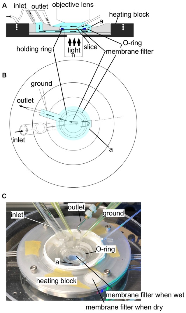
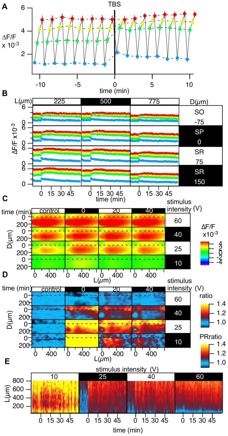
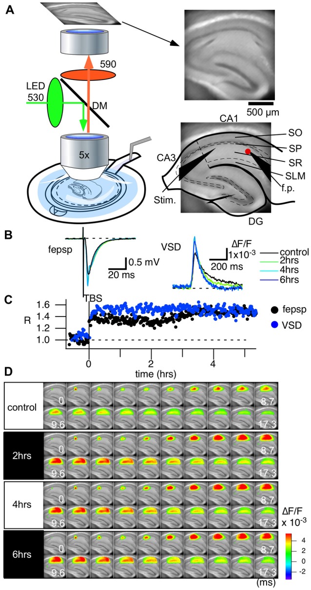
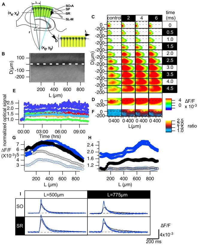
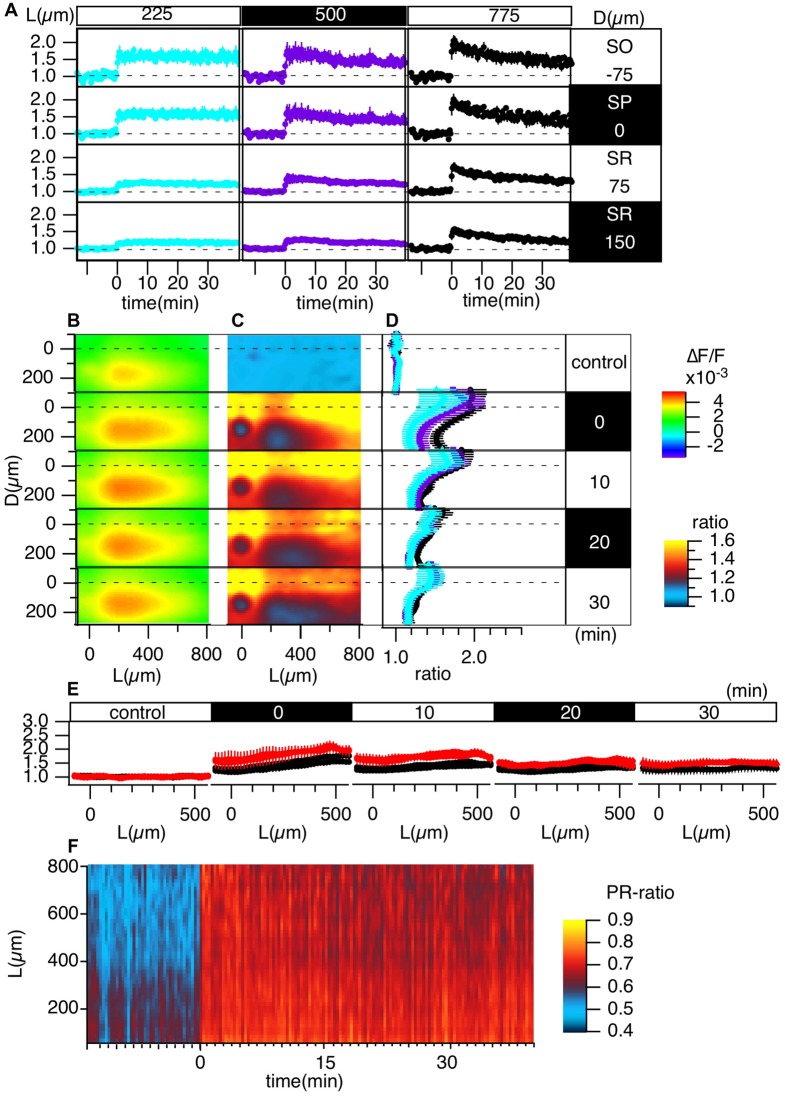
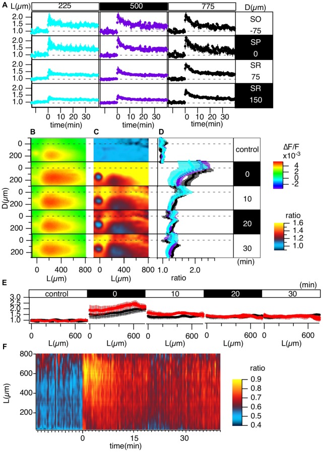
Activity-dependent changes in the input-output (I-O) relationship of a neural circuit are central in the learning and memory function of the brain. To understand circuit-wide adjustments, optical imaging techniques to probe the membrane potential at every component of neurons, such as dendrites, axons and somas, in the circuit are essential. We have been developing fast voltage-sensitive dye (VSD) imaging methods for quantitative measurements, especially for single-photon wide-field optical imaging. The long-term continuous measurements needed to evaluate circuit-wide modifications require stable and quantitative long-term recordings. Here, we show that VSD imaging (VSDI) can be used to record changes in circuit activity in association with theta-burst stimulation (TBS)-induced long-term potentiation (LTP) of synaptic strength in the CA1 area. Our optics, together with the fast imaging system, enabled us to measure neuronal signals from the entire CA1 area at a maximum frame speed of 0.1 ms/frame every 60 s for over 12 h. We also introduced a method to evaluate circuit activity changes by mapping the variation in recordings from the CA1 area to coordinates defined by the morphology of CA1 pyramidal cells. The results clearly showed two types of spatial heterogeneity in LTP induction. The first heterogeneity is that LTP increased with distance from the stimulation site. The second heterogeneity is that LTP is higher in the stratum pyramidale (SP)-oriens region than in the stratum radiatum (SR). We also showed that the pattern of the heterogeneity changed according to the induction protocol, such as induction by TBS or high-frequency stimulation (HFS). We further demonstrated that part of the heterogeneity depends on the I-O response of the circuit elements. The results show the usefulness of VSDI in probing the function of hippocampal circuits.
神经回路输入-输出(I-O)关系中依赖活动的变化是大脑学习和记忆功能的核心。为了理解全回路的调整,用于探测回路中神经元每个组成部分(如树突、轴突和胞体)膜电位的光学成像技术至关重要。我们一直在开发用于定量测量的快速电压敏感染料(VSD)成像方法,特别是用于单光子宽场光学成像。评估全回路修饰所需的长期连续测量需要稳定且定量的长期记录。在这里,我们表明VSD成像(VSDI)可用于记录与CA1区突触强度的θ波爆发刺激(TBS)诱导的长期增强(LTP)相关的回路活动变化。我们的光学系统与快速成像系统一起,使我们能够以每60秒0.1毫秒/帧的最大帧速,对整个CA1区的神经元信号进行超过12小时的测量。我们还引入了一种方法,通过将CA1区记录的变化映射到由CA1锥体细胞形态定义的坐标来评估回路活动变化。结果清楚地显示了LTP诱导中的两种空间异质性。第一种异质性是LTP随着与刺激位点距离的增加而增加。第二种异质性是LTP在锥体层(SP)-始层区域比在辐射层(SR)更高。我们还表明,异质性模式根据诱导方案而变化,例如TBS或高频刺激(HFS)诱导。我们进一步证明,部分异质性取决于回路元件的I-O反应。结果表明VSDI在探测海马回路功能方面的有用性。