Kajiwara Riichi, Tominaga Yoko, Tominaga Takashi
Department of Electronics and Bioinformatics, School of Science and Technology, Meiji University, Kawasaki, Japan.
Laboratory for Neural Circuit Systems, Institute of Neuroscience, Tokushima Bunri University, Sanuki, Japan.
Front Cell Neurosci. 2019 Feb 6;13:20. doi: 10.3389/fncel.2019.00020. eCollection 2019.
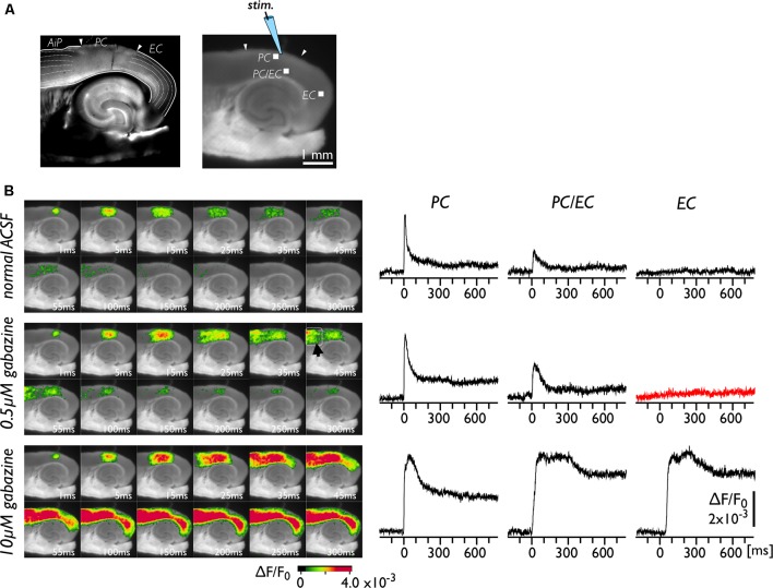
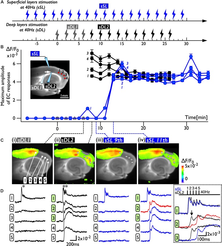
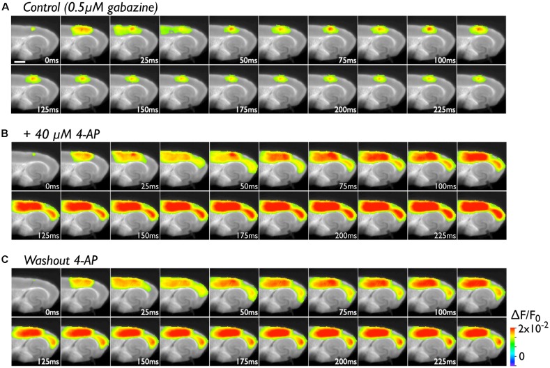
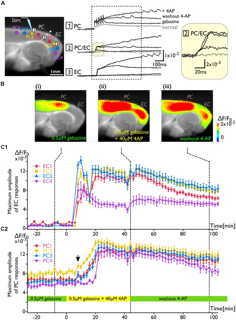
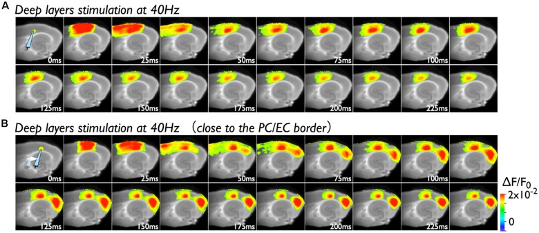
The rhinal cortices, such as the perirhinal cortex (PC) and the entorhinal cortex (EC), are located within the bidirectional pathway between the neocortex and the hippocampus. Physiological studies indicate that the perirhinal transmission of neocortical inputs to the EC occurs at an extremely low probability, though many anatomical studies indicated strong connections exist in the pathway. Our previous study in rat brain slices indicated that an increase in excitability in deep layers of the PC/EC border initiated the neural activity transfer from the PC to the EC. In the present study, we hypothesized that such changes in network dynamics are not incidental observations but rather due to the plastic features of the perirhinal network, which links with the EC. To confirm this idea, we analyzed the network properties of neural transmission throughout the rhinal cortices and the plastic behavior of the network by performing a single-photon wide-field optical recording technique with a voltage-sensitive dye (VSD) in mouse brain slices of the PC, the EC, and the hippocampus. The low concentration of 4-aminopyridine (4-AP; 40 μM) enhanced neural activity in the PC, which eventually propagated to the EC the deep layers of the PC/EC border. Interestingly, washout of 4-AP was unable to reverse entorhinal activation to the previous state. This change in the network property persisted for more than 1 h. This observation was not limited to the application of 4-AP. Burst stimulation to neurons in the perirhinal deep layers also induced the same change of network property. These results indicate the long-lasting modification of physiological connection between the PC and the EC, suggesting the existence of plasticity in the perirhinal-entorhinal network.
嗅周皮质,如嗅周皮层(PC)和内嗅皮层(EC),位于新皮层和海马体之间的双向通路内。生理学研究表明,新皮层输入到EC的嗅周传递发生的概率极低,尽管许多解剖学研究表明该通路中存在强连接。我们之前对大鼠脑片的研究表明,PC/EC边界深层兴奋性的增加启动了神经活动从PC向EC的传递。在本研究中,我们假设这种网络动力学的变化并非偶然观察结果,而是由于与EC相连的嗅周网络的可塑性特征所致。为了证实这一观点,我们通过在PC、EC和海马体的小鼠脑片中使用电压敏感染料(VSD)进行单光子宽场光学记录技术,分析了整个嗅周皮质的神经传递网络特性以及网络的可塑性行为。低浓度的4-氨基吡啶(4-AP;40μM)增强了PC中的神经活动,最终传播到PC/EC边界的深层EC。有趣的是,洗脱4-AP无法将内嗅激活恢复到先前状态。这种网络特性的变化持续了超过1小时。这一观察结果并不局限于4-AP的应用。对嗅周深层神经元的爆发性刺激也诱导了相同的网络特性变化。这些结果表明PC和EC之间生理连接的长期改变,提示嗅周-内嗅网络中存在可塑性。