Department of Radiology, University of Washington, Seattle, USA.
Applied Physics Laboratory, University of Washington, Seattle, USA.
Cancer Imaging. 2018 Nov 8;18(1):41. doi: 10.1186/s40644-018-0172-6.
Pancreatic ductal adenocarcinoma (PDA) is a fatal disease with very poor prognosis. Development of sensitive and noninvasive methods to monitor tumor progression in PDA is a critical and unmet need. Magnetic resonance imaging (MRI) can noninvasively provide information regarding underlying pathophysiological processes such as necrosis, inflammatory changes and fibrotic tissue deposition.
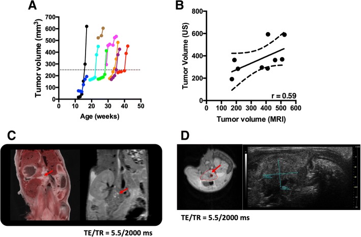
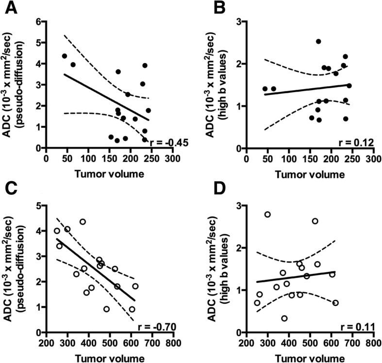
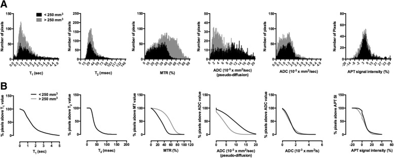
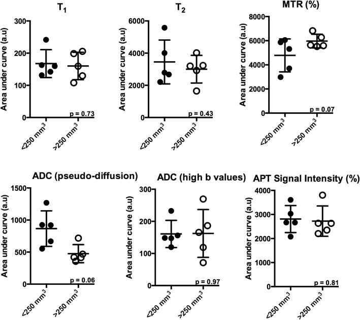
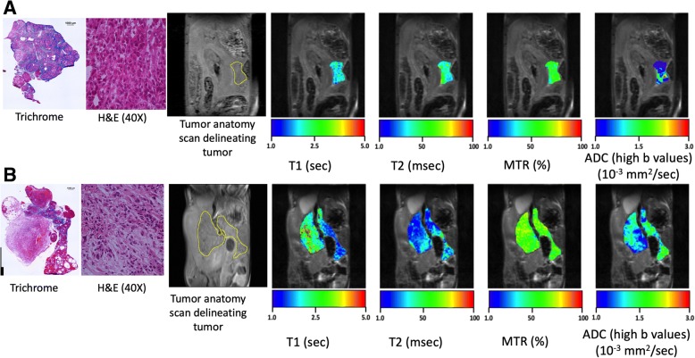
A genetically engineered KPC mouse model that recapitulates human PDA was used to characterize disease progression. MR measures of T and T relaxation times, magnetization transfer ratio (MTR), diffusion and chemical exchange saturation transfer were compared in two separate phases i.e. slow and rapid growth phase of tumor. Fibrotic tissue accumulation was assessed histologically using Masson's trichrome staining. Pearson correlation coefficient (r) was computed to assess the relationship between the fibrotic tissue accumulation and different MR parameters.
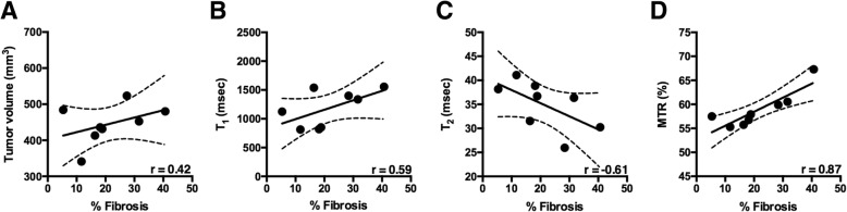
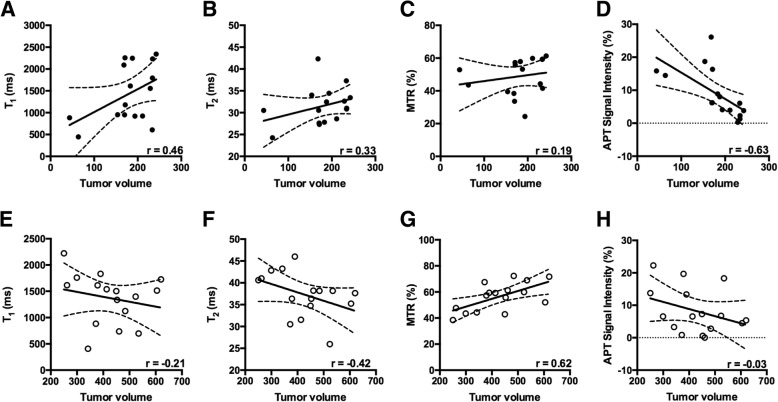
There was a negative correlation between amide proton transfer signal intensity and tumor volume (r = - 0.63, p = 0.003) in the slow growth phase of the tumor development. In the terminal stage of rapid growth phase of the tumor development MTR was strongly correlated with tumor volume (r = 0.62, p = 0.008). Finally, MTR was significantly correlated with % fibrosis (r = 0.87; p < 0.01), followed by moderate correlation between tumor volume (r = 0.42); T (r = - 0.61), T (r = - 0.61) and accumulation of fibrotic tissue.
Here we demonstrated, using multi-parametric MRI (mp-MRI), that MRI parameters changed with tumor progression in a mouse model of PDA. Use of mp-MRI may have the potential to monitor the dynamic changes of tumor microenvironment with increase in tumor size in the transgenic KPC mouse model of pancreatic tumor.
胰腺导管腺癌(PDA)是一种预后极差的致命疾病。开发敏感且非侵入性的方法来监测 PDA 中的肿瘤进展是一项至关重要且尚未满足的需求。磁共振成像(MRI)可以无创地提供有关潜在病理生理过程的信息,例如坏死、炎症变化和纤维组织沉积。
使用基因工程 KPC 小鼠模型来模拟人类 PDA,以对疾病进展进行特征描述。在肿瘤生长的缓慢和快速两个阶段,比较了 T 和 T 弛豫时间、磁化转移率(MTR)、扩散和化学交换饱和转移的 MR 测量值。使用 Masson 三色染色法对纤维组织的积累进行组织学评估。计算 Pearson 相关系数(r)来评估纤维组织积累与不同 MR 参数之间的关系。
在肿瘤生长的缓慢阶段,酰胺质子转移信号强度与肿瘤体积之间存在负相关(r=-0.63,p=0.003)。在肿瘤生长的快速阶段末期,MTR 与肿瘤体积呈强相关性(r=0.62,p=0.008)。最后,MTR 与纤维化百分比显著相关(r=0.87;p<0.01),其次是肿瘤体积(r=0.42)、T1(r=-0.61)和 T2(r=-0.61)与纤维组织积累之间存在中度相关性。
本研究通过多参数 MRI(mp-MRI)显示,在 PDA 的小鼠模型中,MRI 参数随肿瘤进展而变化。在胰腺肿瘤的转基因 KPC 小鼠模型中,mp-MRI 的使用可能具有监测肿瘤大小增加时肿瘤微环境动态变化的潜力。