Hematology and Transfusion Medicine Center-University of Campinas/Hemocentro-UNICAMP, Instituto Nacional de Ciencia e Tecnologia do Sangue, Rua Carlos Chagas, 480, CEP, Campinas, SP, 13083-878, Brazil.
Clin Epigenetics. 2018 Nov 8;10(1):139. doi: 10.1186/s13148-018-0563-3.
In the present study, we investigated the molecular mechanisms underlying the pro-apoptotic effects of quercetin (Qu) by evaluating the effect of Qu treatment on DNA methylation and posttranslational histone modifications of genes related to the apoptosis pathway. This study was performed in vivo in two human xenograft acute myeloid leukemia (AML) models and in vitro using HL60 and U937 cell lines.
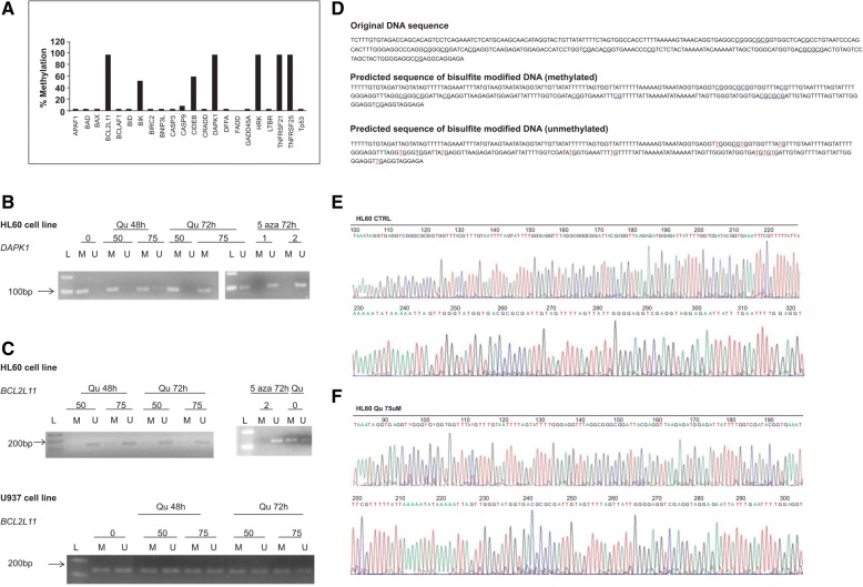
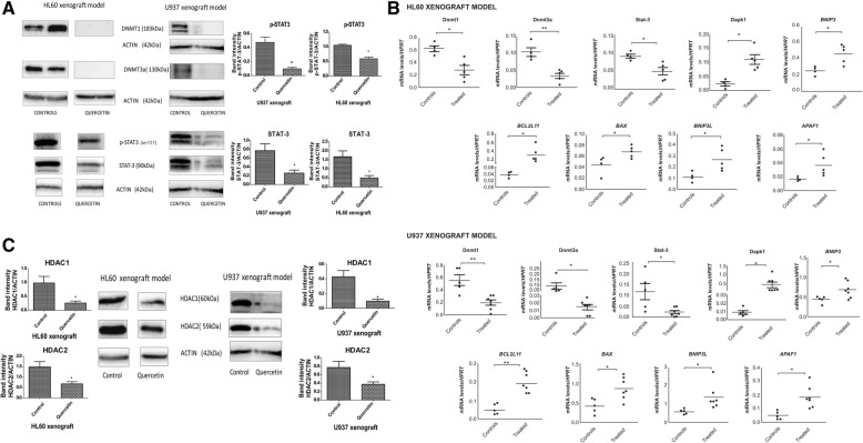
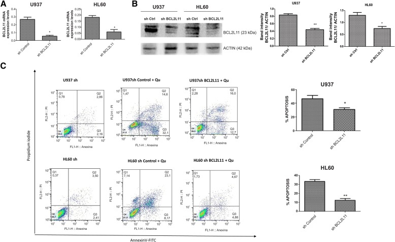
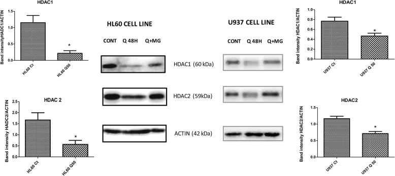
Qu treatment almost eliminates DNMT1 and DNMT3a expression, and this regulation was in part STAT-3 dependent. The treatment also downregulated class I HDACs. Furthermore, treatment of the cell lines with the proteasome inhibitor, MG132, together with Qu prevented degradation of class I HDACs compared to cells treated with Qu alone, indicating increased proteasome degradation of class I HDACS by Qu. Qu induced demethylation of the pro-apoptotic BCL2L11, DAPK1 genes, in a dose- and time-dependent manner. Moreover, Qu (50 μmol/L) treatment of cell lines for 48 h caused accumulation of acetylated histone 3 and histone 4, resulting in three- to ten fold increases in the promoter region of DAPK1, BCL2L11, BAX, APAF1, BNIP3, and BNIP3L. In addition, Qu treatment significantly increased the mRNA levels of all these genes, when compared to cells treated with vehicle only (control cells) (*p < 0.05).
In summary, our results showed that enhanced apoptosis, induced by Qu, might be caused in part by its DNA demethylating activity, by HDAC inhibition, and by the enrichment of H3ac and H4ac in the promoter regions of genes involved in the apoptosis pathway, leading to their transcription activation.
在本研究中,我们通过评估 Qu 处理对与凋亡途径相关的基因的 DNA 甲基化和翻译后组蛋白修饰的影响,研究了槲皮素(Qu)促凋亡作用的分子机制。这项研究在两个人类异种移植物急性髓细胞白血病(AML)模型中进行了体内研究,并在 HL60 和 U937 细胞系中进行了体外研究。
Qu 处理几乎消除了 DNMT1 和 DNMT3a 的表达,这种调节部分依赖于 STAT-3。该处理还下调了 I 类组蛋白去乙酰化酶。此外,与单独用 Qu 处理的细胞相比,用蛋白酶体抑制剂 MG132 与 Qu 一起处理细胞系可防止 I 类组蛋白去乙酰化酶降解,表明 Qu 增加了 I 类组蛋白去乙酰化酶的蛋白酶体降解。Qu 以剂量和时间依赖的方式诱导促凋亡 BCL2L11、DAPK1 基因去甲基化。此外,Qu(50μmol/L)处理细胞系 48 小时导致乙酰化组蛋白 3 和组蛋白 4 积累,导致 DAPK1、BCL2L11、BAX、APAF1、BNIP3 和 BNIP3L 的启动子区域增加 3 到 10 倍。此外,与仅用载体(对照细胞)处理的细胞相比,Qu 处理显著增加了这些基因的 mRNA 水平(*p<0.05)。
总之,我们的结果表明,Qu 诱导的凋亡增强可能部分是由其 DNA 去甲基化活性、HDAC 抑制以及参与凋亡途径的基因启动子区域 H3ac 和 H4ac 的富集引起的,从而导致它们的转录激活。