Department of Biomedical and Pharmaceutical Sciences, Center for Targeted Drug Delivery, Chapman University, Irvine, CA 92618-1908, USA; Department of Bioengineering, University of Illinois, 851 South Morgan Street, Chicago, IL 60607-7052, USA.
Istituto di Struttura della Materia, Consiglio Nazionale delle Ricerche (ISM-CNR), Via del Fosso del Cavaliere, 100-00133 Rome, Italy.
Mater Sci Eng C Mater Biol Appl. 2019 Jan 1;94:798-810. doi: 10.1016/j.msec.2018.10.028. Epub 2018 Oct 5.
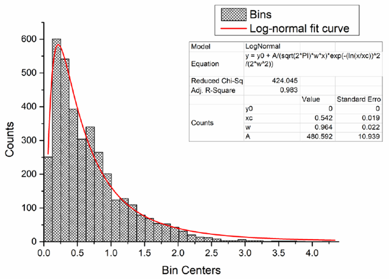
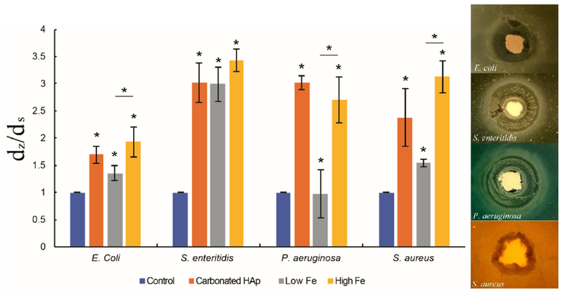
Self-hardening calcium phosphate cements present ideal bone tissue substitutes from the standpoints of bioactivity and biocompatibility, yet they suffer from (a) weak mechanical properties, (b) negligible osteoinduction without the use of exogenous growth factors, and (c) a lack of intrinsic antibacterial activity. Here we attempt to improve on these deficiencies by studying the properties of self-setting Fe-doped bone-integrative cements containing two different concentrations of the dopant: 0.49 and 1.09 wt% Fe. The hardening process, which involved the transformation of Fe-doped β-tricalcium phosphate (Fe-TCP) to nanocrystalline brushite, was investigated in situ by continuously monitoring the cements using the Energy Dispersive X-Ray Diffraction technique. The setting time was 20 min and the hardening time 2 h, but it took 50 h for the cement to completely stabilize compositionally and mechanically. Still, compared to other similar systems, the phase transformation during hardening was relatively fast and it also followed a relatively simple reaction path, virtually free of complex intermediates and noisy background. Mössbauer spectrometry demonstrated that Fe atoms in Fe-TCP were located in two non-equivalent crystallographic sites and distributed over positions with a strong crystal distortion. The pronounced presence of ultrafine crystals in the final, brushite phase contributed to the reduction of the porosity and thereby to the enhancement of the mechanical properties. The compressive strength of the hardened TCP cements increased by more than twofold when Fe was added as a dopant, i.e., from 11.5 ± 0.5 to 24.5 ± 2.0 MPa. The amount of iron released from the cements in physiological media steadied after 10 days and was by an order of magnitude lower than the clinical threshold that triggers the toxic response. The cements exhibited osteoinductive activity, as observed from the elevated levels of expression of genes encoding for osteocalcin and Runx2 in both undifferentiated and differentiated MC3T3-E1 cells challenged with the cements. The osteoinductive effect was inversely proportional to the content of Fe ions in the cements, indicating that an excessive amount of iron can have a detrimental effect on the induction of bone growth by osteoblasts in contact with the cement. In contrast, the antibacterial activity of the cement in the agar assay increased against all four bacterial species analysed (E. coli, S. enteritidis, P. aeruginosa, S. aureus) in direct proportion with the concentration of Fe ions in it, indicating their key effect on the promotion of the antibacterial effect in this material. This effect was less pronounced in broth assays. Experiments involving co-incubation of cements with cells in an alternate magnetic radiofrequency field for 30 min demonstrated a good potential for the use of these magnetic cements in hyperthermia cancer therapies. Specifically, the population of human glioblastoma cells decreased six-fold at the 24 h time point following the end of the magnetic field treatment, while the population of the bone cancer cells dropped approximately twofold. The analysis of the MC3T3-E1 cell/cement interaction reiterated the effects of iron in the cement on the bone growth marker expression by showing signs of adverse effects on the cell morphology and proliferation only for the cement containing the higher concentration of Fe ions (1.09 wt%). Biological testing concluded that the effects of iron are beneficial from the perspective of a magnetic hyperthermia therapy and antibacterial prophylaxis, but its concentration in the material must be carefully optimized to avoid the adverse effects induced above a certain level of iron concentrations.
自硬磷酸钙骨水泥在生物活性和生物相容性方面具有理想的骨组织替代物特性,但它们存在以下问题:(a) 机械性能较弱;(b) 在不使用外源性生长因子的情况下,成骨诱导作用可忽略不计;(c) 缺乏内在的抗菌活性。在这里,我们试图通过研究含有两种不同浓度掺杂剂的自凝固铁掺杂骨整合水泥的特性来改善这些缺陷:0.49 和 1.09 wt% Fe。通过连续使用能量色散 X 射线衍射技术监测水泥,原位研究了包含掺杂β-磷酸三钙(Fe-TCP)的水泥的凝固过程,转变为纳米晶水合羟磷灰石。凝固时间为 20 分钟,硬化时间为 2 小时,但水泥需要 50 小时才能完全稳定的化学成分和机械性能。尽管如此,与其他类似体系相比,硬化过程中的相变相对较快,而且遵循相对简单的反应途径,几乎没有复杂的中间体和嘈杂的背景。穆斯堡尔光谱表明,Fe-TCP 中的 Fe 原子位于两个非等效的晶格格位上,并分布在具有强烈晶体变形的位置上。在最终的水合羟磷灰石相中存在大量的超细晶体,有助于降低孔隙率,从而提高机械性能。当 Fe 作为掺杂剂添加时,TCP 水泥的抗压强度增加了两倍以上,即从 11.5 ± 0.5 增加到 24.5 ± 2.0 MPa。在生理介质中从水泥中释放的铁量在 10 天后稳定下来,释放量比触发毒性反应的临床阈值低一个数量级。水泥表现出成骨诱导活性,这从未分化和分化的 MC3T3-E1 细胞用水泥刺激后编码骨钙素和 Runx2 的基因表达水平升高可以看出。成骨诱导作用与水泥中 Fe 离子的含量成反比,表明与与水泥接触的成骨细胞的骨生长诱导相比,过量的铁可能会产生不利影响。相比之下,水泥在琼脂试验中的抗菌活性与水泥中 Fe 离子的浓度成正比,这表明它们对促进该材料中抗菌作用的关键作用。在肉汤试验中,这种效果不那么明显。涉及在交变磁射频场中交替孵育水泥和细胞 30 分钟的实验表明,这些磁性水泥在高热癌症治疗中具有良好的应用潜力。具体来说,在磁场治疗结束后 24 小时,人胶质母细胞瘤细胞的数量减少了六倍,而骨癌细胞的数量减少了大约两倍。MC3T3-E1 细胞/水泥相互作用的分析重申了水泥中铁对骨生长标志物表达的影响,表明仅对于含有较高浓度 Fe 离子的水泥(1.09 wt%),才会对细胞形态和增殖产生不利影响。生物学测试得出的结论是,从磁热疗和抗菌预防的角度来看,铁的作用是有益的,但必须仔细优化其在材料中的浓度,以避免在铁浓度超过一定水平时产生的不利影响。