Department of Urology, Chongqing Three Gorges Central Hospital, Chongqing, China (mainland).
Med Sci Monit. 2018 Nov 21;24:8391-8400. doi: 10.12659/MSM.911124.
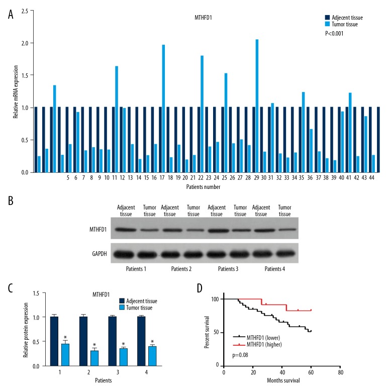
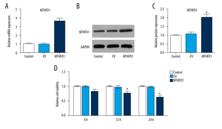
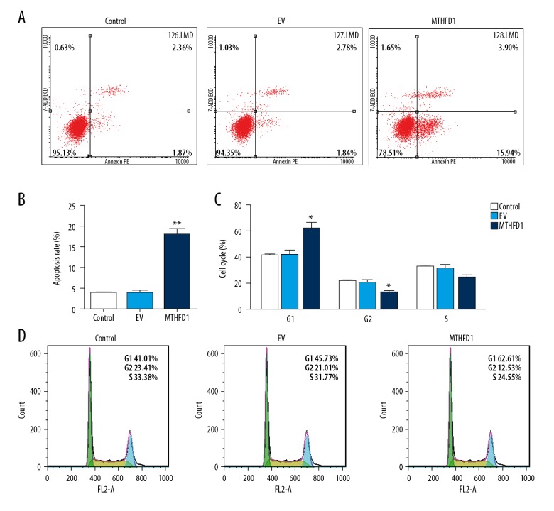
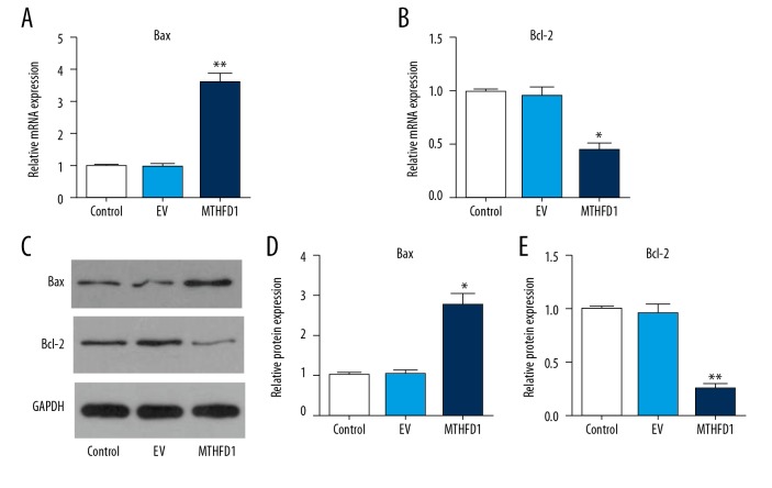
BACKGROUND The aims of this study were to investigate the expression of methylenetetrahydrofolate dehydrogenase 1 (MTHFD1) in human tissue containing clear cell renal cell carcinoma (CCRCC) compared with normal renal tissue, and the effects of upregulating the expression of MTHFD1 in the human CCRCC cell line, Caki-1. MATERIAL AND METHODS Tumor and adjacent normal renal tissue were obtained from 44 patients who underwent radical nephrectomy for CCRCC. Caki-1 human CCRCC cells were divided into the control group, the empty vector (EV) group, and the plasmid-treated group that overexpressed MTHFD1. MTHFD1 mRNA and protein levels were measured by quantitative real-time polymerase chain reaction (qRT-PCR) and Western blot, respectively. The cell counting kit-8 (CCK-8) assay measured cell viability. Flow cytometry evaluated apoptosis and the cell cycle. Western blot measured the protein levels of MTHFD1, Bax, Bcl-2, Akt, p53, and cyclin D1, and qRT-PCR determined the gene expression profiles. RESULTS MTHFD1 mRNA and protein levels in CCRCC tumor tissues were significantly lower compared with adjacent normal renal tissue. MTHFD1 over-expression in Caki-1 cells inhibited cell proliferation, arrested cells in the G1 phase, increased cell apoptosis, and upregulated gene and protein expression of Bax/Bcl-2 and p53 and inhibited p-Akt, and cyclin D1. CONCLUSIONS MTHFD1 was underexpressed in CCRCC tissue when compared with normal renal tissue. MTHFD1 transfection of human CCRCC Caki-1 cells in vitro inhibited cell proliferation and promoted apoptosis, associated with reduced expression of cyclin D1, reduced Akt phosphorylation, and increased expression of Bax/Bcl-2 and p53.
本研究旨在探讨亚甲基四氢叶酸脱氢酶 1(MTHFD1)在人肾透明细胞癌(CCRCC)组织中的表达,并研究上调人 CCRCC 细胞系 Caki-1 中 MTHFD1 的表达的影响。
44 例接受根治性肾切除术的 CCRCC 患者的肿瘤和相邻正常肾组织。将人 CCRCC 细胞系 Caki-1 分为对照组、空载体(EV)组和过表达 MTHFD1 的质粒处理组。通过实时定量聚合酶链反应(qRT-PCR)和 Western blot 分别测量 MTHFD1 mRNA 和蛋白水平。细胞计数试剂盒-8(CCK-8)测定细胞活力。流式细胞术评估细胞凋亡和细胞周期。Western blot 测定 MTHFD1、Bax、Bcl-2、Akt、p53 和细胞周期蛋白 D1 的蛋白水平,qRT-PCR 测定基因表达谱。
与相邻正常肾组织相比,CCRCC 肿瘤组织中的 MTHFD1 mRNA 和蛋白水平明显降低。在 Caki-1 细胞中过表达 MTHFD1 抑制细胞增殖,使细胞停滞在 G1 期,增加细胞凋亡,并上调 Bax/Bcl-2 和 p53 的基因和蛋白表达,抑制 p-Akt 和 cyclin D1。
与正常肾组织相比,CCRCC 组织中 MTHFD1 表达下调。体外转染人 CCRCC Caki-1 细胞的 MTHFD1 抑制细胞增殖并促进凋亡,与 cyclin D1 表达减少、Akt 磷酸化减少以及 Bax/Bcl-2 和 p53 表达增加有关。