• 文献检索
  • 文档翻译
  • 深度研究
  • 学术资讯
  • Suppr Zotero 插件Zotero 插件
  • 邀请有礼
  • 套餐&价格
  • 历史记录
应用&插件
Suppr Zotero 插件Zotero 插件浏览器插件Mac 客户端Windows 客户端微信小程序
定价
高级版会员购买积分包购买API积分包
服务
文献检索文档翻译深度研究API 文档MCP 服务
关于我们
关于 Suppr公司介绍联系我们用户协议隐私条款
关注我们

Suppr 超能文献

核心技术专利:CN118964589B侵权必究
粤ICP备2023148730 号-1Suppr @ 2026

文献检索

告别复杂PubMed语法,用中文像聊天一样搜索,搜遍4000万医学文献。AI智能推荐,让科研检索更轻松。

立即免费搜索

文件翻译

保留排版,准确专业,支持PDF/Word/PPT等文件格式,支持 12+语言互译。

免费翻译文档

深度研究

AI帮你快速写综述,25分钟生成高质量综述,智能提取关键信息,辅助科研写作。

立即免费体验

TGF-β 信号通路与腭裂融合过程中的上皮间质转化

TGF-β Signaling and the Epithelial-Mesenchymal Transition during Palatal Fusion.

机构信息

Department of Orthodontics, Nihon University School of Dentistry, Chiyoda-ku, Tokyo 101-8310, Japan.

Department of Oral Biological and Medical Sciences, Faculty of Dentistry, University of British Columbia, Vancouver, BC V6T 1Z3, Canada.

出版信息

Int J Mol Sci. 2018 Nov 19;19(11):3638. doi: 10.3390/ijms19113638.

DOI:10.3390/ijms19113638
PMID:30463190
原文链接:https://pmc.ncbi.nlm.nih.gov/articles/PMC6274911/
Abstract

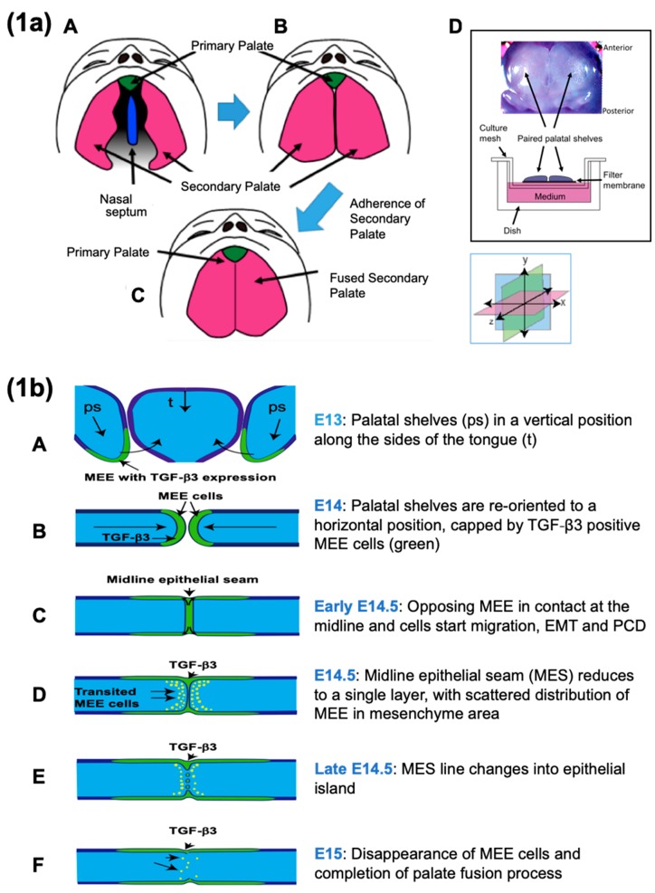
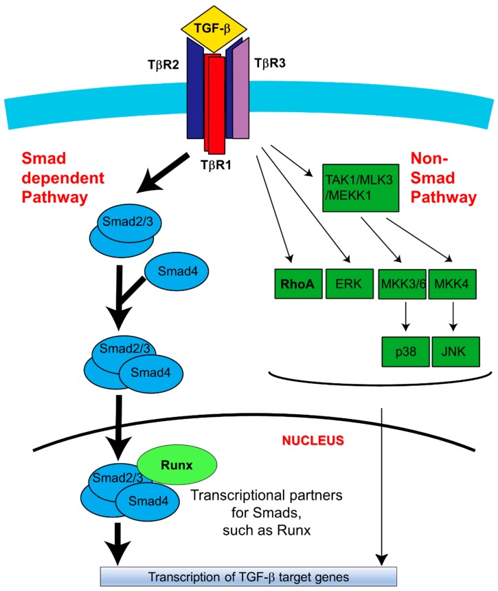
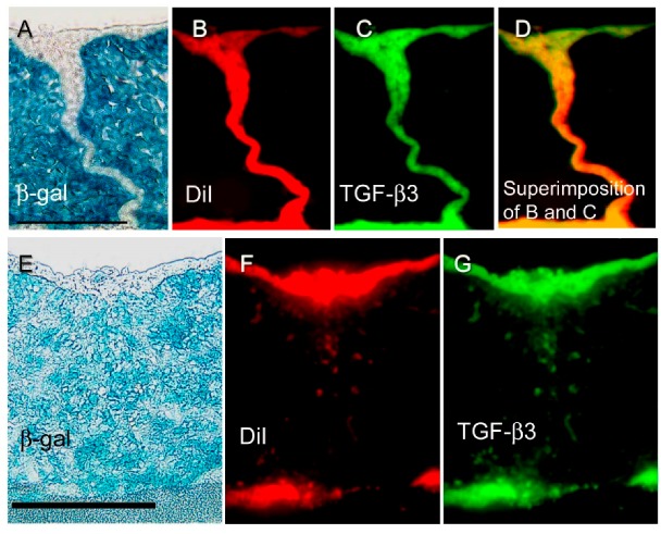
Signaling by transforming growth factor (TGF)-β plays an important role in development, including in palatogenesis. The dynamic morphological process of palatal fusion occurs to achieve separation of the nasal and oral cavities. Critically and specifically important in palatal fusion are the medial edge epithelial (MEE) cells, which are initially present at the palatal midline seam and over the course of the palate fusion process are lost from the seam, due to cell migration, epithelial-mesenchymal transition (EMT), and/or programed cell death. In order to define the role of TGF-β signaling during this process, several approaches have been utilized, including a small interfering RNA (siRNA) strategy targeting TGF-β receptors in an organ culture context, the use of genetically engineered mice, such as Wnt1-cre/R26R double transgenic mice, and a cell fate tracing through utilization of cell lineage markers. These approaches have permitted investigators to distinguish some specific traits of well-defined cell populations throughout the palatogenic events. In this paper, we summarize the current understanding on the role of TGF-β signaling, and specifically its association with MEE cell fate during palatal fusion. TGF-β is highly regulated both temporally and spatially, with TGF-β3 and Smad2 being the preferentially expressed signaling molecules in the critical cells of the fusion processes. Interestingly, the accessory receptor, TGF-β type 3 receptor, is also critical for palatal fusion, with evidence for its significance provided by Cre-lox systems and siRNA approaches. This suggests the high demand of ligand for this fine-tuned signaling process. We discuss the new insights in the fate of MEE cells in the midline epithelial seam (MES) during the palate fusion process, with a particular focus on the role of TGF-β signaling.

摘要

转化生长因子-β(TGF-β)信号在发育过程中发挥着重要作用,包括腭发生。腭融合的动态形态发生过程发生以实现鼻腔和口腔的分离。在腭融合中,重要的是中线上皮(MEE)细胞,它们最初存在于腭中线缝处,并且在腭融合过程中由于细胞迁移、上皮-间充质转化(EMT)和/或程序性细胞死亡而从中线缝中丢失。为了定义 TGF-β信号在这个过程中的作用,已经采用了几种方法,包括在器官培养环境中靶向 TGF-β受体的小干扰 RNA(siRNA)策略、使用基因工程小鼠,如 Wnt1-cre/R26R 双转基因小鼠,以及通过利用细胞谱系标记进行细胞命运追踪。这些方法使研究人员能够区分腭发生事件中某些特定特征的明确细胞群体。在本文中,我们总结了 TGF-β信号作用的最新认识,特别是它与 MEE 细胞命运在腭融合中的关联。TGF-β在时间和空间上受到高度调节,TGF-β3 和 Smad2 是融合过程中关键细胞中优先表达的信号分子。有趣的是,辅助受体 TGF-β类型 3 受体对于腭融合也很重要,Cre-lox 系统和 siRNA 方法提供了其重要性的证据。这表明精细调节信号过程对配体的高需求。我们讨论了在腭融合过程中线间上皮缝(MES)中 MEE 细胞命运的新见解,特别关注 TGF-β信号的作用。

https://cdn.ncbi.nlm.nih.gov/pmc/blobs/16a4/6274911/6fb1984a1a6d/ijms-19-03638-g005.jpg
https://cdn.ncbi.nlm.nih.gov/pmc/blobs/16a4/6274911/dc925789de81/ijms-19-03638-g001a.jpg
https://cdn.ncbi.nlm.nih.gov/pmc/blobs/16a4/6274911/d924241d7744/ijms-19-03638-g002.jpg
https://cdn.ncbi.nlm.nih.gov/pmc/blobs/16a4/6274911/e5d3e2fa9da7/ijms-19-03638-g003a.jpg
https://cdn.ncbi.nlm.nih.gov/pmc/blobs/16a4/6274911/8861c7361e94/ijms-19-03638-g004.jpg
https://cdn.ncbi.nlm.nih.gov/pmc/blobs/16a4/6274911/6fb1984a1a6d/ijms-19-03638-g005.jpg

相似文献

1
TGF-β Signaling and the Epithelial-Mesenchymal Transition during Palatal Fusion.TGF-β 信号通路与腭裂融合过程中的上皮间质转化
Int J Mol Sci. 2018 Nov 19;19(11):3638. doi: 10.3390/ijms19113638.
2
The expression of TGF-β3 for epithelial-mesenchyme transdifferentiated MEE in palatogenesis.TGF-β3 在腭发生中诱导上皮-间充质转分化的 MEE 的表达。
J Mol Histol. 2010 Dec;41(6):343-55. doi: 10.1007/s10735-010-9296-0. Epub 2010 Oct 22.
3
Cell autonomous requirement for Tgfbr2 in the disappearance of medial edge epithelium during palatal fusion.在腭融合过程中内侧边缘上皮消失时,Tgfbr2的细胞自主需求。
Dev Biol. 2006 Sep 1;297(1):238-48. doi: 10.1016/j.ydbio.2006.05.014. Epub 2006 May 19.
4
Medial edge epithelial cell fate during palatal fusion.腭融合过程中内侧边缘上皮细胞的命运
Dev Biol. 2000 Apr 15;220(2):343-57. doi: 10.1006/dbio.2000.9644.
5
Disintegration of the medial epithelial seam: is cell death important in palatogenesis?中缝的解体:细胞死亡在腭发育中重要吗?
Dev Growth Differ. 2011 Feb;53(2):259-68. doi: 10.1111/j.1440-169X.2010.01245.x.
6
The TGF-beta type III receptor is localized to the medial edge epithelium during palatal fusion.转化生长因子βⅢ型受体在腭融合过程中定位于内侧边缘上皮。
Int J Dev Biol. 2000 Jun;44(4):397-402.
7
Pathogenesis of cleft palate in TGF-beta3 knockout mice.转化生长因子β3基因敲除小鼠腭裂的发病机制
Development. 1999 Sep;126(17):3869-79. doi: 10.1242/dev.126.17.3869.
8
Microtubule disassembly prevents palatal fusion and alters regulation of the E-cadherin/catenin complex.微管拆卸会阻止腭融合,并改变E-钙黏蛋白/连环蛋白复合体的调控。
Int J Dev Biol. 2013;57(1):55-60. doi: 10.1387/ijdb.120117yk.
9
Functional role of TGF-β receptors during palatal fusion in vitro.转化生长因子-β受体在体外腭融合过程中的功能作用。
Arch Oral Biol. 2014 Nov;59(11):1192-204. doi: 10.1016/j.archoralbio.2014.07.007. Epub 2014 Jul 24.
10
Transforming growth factor beta (TGFbeta) signalling in palatal growth, apoptosis and epithelial mesenchymal transformation (EMT).转化生长因子β(TGFβ)信号传导在腭部生长、细胞凋亡和上皮-间质转化(EMT)中的作用
Arch Oral Biol. 2004 Sep;49(9):675-89. doi: 10.1016/j.archoralbio.2004.05.007.

引用本文的文献

1
State of omics-based microbial diagnostics of CRC.基于组学的结直肠癌微生物诊断现状。
Gut Microbes. 2025 Dec;17(1):2526132. doi: 10.1080/19490976.2025.2526132. Epub 2025 Jul 2.
2
Identification of Transforming Growth Factor-β3 Signaling by Compressive Force in MC3T3-E1 Cells Reactions.通过压缩力在MC3T3-E1细胞反应中鉴定转化生长因子-β3信号传导。
Cell Biochem Biophys. 2025 Jun 24. doi: 10.1007/s12013-025-01783-6.
3
Molecular Regulation of Palatogenesis and Clefting: An Integrative Analysis of Genetic, Epigenetic Networks, and Environmental Interactions.

本文引用的文献

1
Runx1-Stat3-Tgfb3 signaling network regulating the anterior palatal development.Runx1-Stat3-Tgfb3 信号网络调控前腭发育。
Sci Rep. 2018 Jul 25;8(1):11208. doi: 10.1038/s41598-018-29681-3.
2
IRF6 and AP2A Interaction Regulates Epidermal Development.IRF6 和 AP2A 的相互作用调节表皮发育。
J Invest Dermatol. 2018 Dec;138(12):2578-2588. doi: 10.1016/j.jid.2018.05.030. Epub 2018 Jun 18.
3
Combined Tongue-Palate Fusion With Alveolar Bands in a Patient With Pierre Robin Sequence and Van der Woude Syndrome.Pierre Robin序列和范德伍德综合征患者中合并的舌-腭融合与牙槽嵴带
腭发育及腭裂形成的分子调控:遗传、表观遗传网络及环境相互作用的综合分析
Int J Mol Sci. 2025 Feb 6;26(3):1382. doi: 10.3390/ijms26031382.
4
Unbiased transcriptome analysis of human cleft palate reveals evolutionally conserved molecular signatures of development: experimental study.人类腭裂的无偏转录组分析揭示发育过程中进化保守的分子特征:实验研究
Int J Surg. 2024 Nov 1;110(11):6933-6940. doi: 10.1097/JS9.0000000000001841.
5
A novel gene mutation impacting the regulation of in the TGFβ pathway: A mechanism in the development of Van der Woude syndrome.一种影响转化生长因子β(TGFβ)信号通路中**[此处原文缺失相关内容]**调控的新型基因突变:范德伍德综合征发生的一种机制。
Front Genet. 2024 Jun 5;15:1397410. doi: 10.3389/fgene.2024.1397410. eCollection 2024.
6
Exosomes Derived from Human Palatal Mesenchymal Cells Mediate Intercellular Communication During Palatal Fusion by Promoting Oral Epithelial Cell Migration.人腭中胚层细胞来源的外泌体通过促进口腔上皮细胞迁移在腭裂融合过程中介导细胞间通讯。
Int J Nanomedicine. 2024 Mar 29;19:3109-3121. doi: 10.2147/IJN.S451491. eCollection 2024.
7
Orofacial Clefts: Genetics of Cleft Lip and Palate.口腔颌面部裂隙:唇腭裂的遗传学。
Genes (Basel). 2023 Aug 9;14(8):1603. doi: 10.3390/genes14081603.
8
LncRNA-NONMMUT100923.1 regulates mouse embryonic palatal shelf adhesion by sponging miR-200a-3p to modulate medial epithelial cell desmosome junction during palatogenesis.长链非编码RNA-NONMMUT100923.1通过海绵吸附miR-200a-3p来调节小鼠胚胎腭突黏附,从而在腭发育过程中调控内侧上皮细胞桥粒连接。
Heliyon. 2023 May 19;9(5):e16329. doi: 10.1016/j.heliyon.2023.e16329. eCollection 2023 May.
9
TCDD induces cleft palate through exosomes derived from mesenchymal cells.四氯二苯并二恶英通过间充质细胞来源的外泌体诱导腭裂。
Toxicol Res (Camb). 2022 Oct 8;11(6):901-910. doi: 10.1093/toxres/tfac068. eCollection 2022 Dec.
10
m hypomethylation as a mechanism for non-syndromic cleft lip and palate.甲基化不足作为非综合征性唇腭裂的一种机制。
Epigenetics. 2022 Dec;17(13):2278-2295. doi: 10.1080/15592294.2022.2115606. Epub 2022 Sep 1.
Cleft Palate Craniofac J. 2019 Jan;56(1):123-126. doi: 10.1177/1055665618773192. Epub 2018 Apr 30.
4
A review of FGF signaling in palate development.成纤维细胞生长因子信号在腭发育中的研究进展。
Biomed Pharmacother. 2018 Jul;103:240-247. doi: 10.1016/j.biopha.2018.04.026. Epub 2018 Apr 24.
5
TGF-β signaling inhibits canonical BMP signaling pathway during palate development.TGF-β 信号在腭发育过程中抑制经典 BMP 信号通路。
Cell Tissue Res. 2018 Feb;371(2):283-291. doi: 10.1007/s00441-017-2757-y. Epub 2017 Dec 16.
6
Live Imaging of Mouse Secondary Palate Fusion.小鼠继发腭融合的活体成像
J Vis Exp. 2017 Jul 27(125):56041. doi: 10.3791/56041.
7
Non-Smad Signaling Pathways of the TGF-β Family.转化生长因子-β家族的非Smad信号通路。
Cold Spring Harb Perspect Biol. 2017 Feb 1;9(2):a022129. doi: 10.1101/cshperspect.a022129.
8
Mechanisms of TGFβ-Induced Epithelial-Mesenchymal Transition.TGFβ 诱导的上皮-间充质转化的机制。
J Clin Med. 2016 Jun 29;5(7):63. doi: 10.3390/jcm5070063.
9
EMT: Matter of Life or Death?急救员:关乎生死?
Cell. 2016 Feb 25;164(5):840-2. doi: 10.1016/j.cell.2016.02.024.
10
TGF-β Tumor Suppression through a Lethal EMT.通过致死性上皮-间质转化实现的转化生长因子-β肿瘤抑制作用
Cell. 2016 Feb 25;164(5):1015-30. doi: 10.1016/j.cell.2016.01.009. Epub 2016 Feb 18.