Geisinger Heart and Vascular Institute, Danville, Pennsylvania.
Cardiology Service, Brooke Army Medical Center, Fort Sam Houston, Texas.
Catheter Cardiovasc Interv. 2019 Jul 1;94(1):E37-E43. doi: 10.1002/ccd.27998. Epub 2018 Nov 25.
This study examines the intrapatient variability in peak instantaneous left ventricular outflow tract (LVOT) gradients and aortic pulse pressures during rest, exercise, and after ventricular ectopy.
Although the variability in LVOT gradients in patients with hypertrophic cardiomyopathy (HCM) is well known, the predictors of such variation are not. We hypothesized that quantitative invasive analysis of gradient variation could identify useful predictors of maximal gradients.
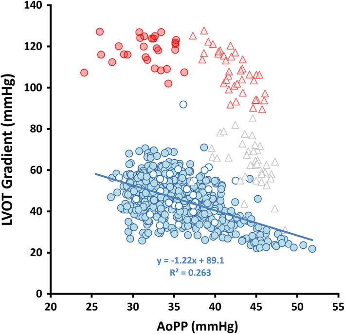
Variability in continuously recorded, high-fidelity left ventricular and aortic pressure waveforms were evaluated by computer-assisted analysis in the resting state (N = 659 beats) and during supine exercise (N = 379 beats) in a symptomatic patient with a resting LVOT gradient >30 mmHg and frequent ventricular ectopy.
At rest, the peak left ventricular and aortic pressures at the time of the peak instantaneous LVOT gradient for all sinus and postectopic beats followed consistent regression slopes characterizing the potential energy loss between the LV cavity and aorta. During exercise, similar regression slopes were identified, and these converged with the resting slopes at the point of the maximal measured LVOT gradient. Component analysis of the LVOT gradient suggests that resting beat-to-beat variability provides information similar to post-ectopic pressures for predicting maximal gradients in obstructive-variant HCM.
Our study suggests that computer-assisted analysis of hemodynamic variability in HCM may prove useful in characterizing the severity of obstruction. Further study is warranted to confirm the reproducibility and utility of this finding in a population with clinically significant exercise-induced gradients.
本研究旨在探讨肥厚型心肌病(HCM)患者在静息、运动和室性早搏期间左心室流出道(LVOT)峰值瞬时梯度和主动脉脉搏压的个体内变异性。
尽管 HCM 患者的 LVOT 梯度变异性众所周知,但这种变化的预测因素尚不清楚。我们假设通过梯度变化的定量有创分析可以识别最大梯度的有用预测因素。
通过计算机辅助分析评估静息状态(N=659 次搏动)和仰卧位运动(N=379 次搏动)期间连续记录的高保真左心室和主动脉压力波形的变异性,该患者静息时 LVOT 梯度>30mmHg,且频发室性早搏。
在静息状态下,所有窦性和室性早搏时的左心室和主动脉峰值压力与峰值瞬时 LVOT 梯度的时间呈一致的回归斜率,特征为 LV 腔和主动脉之间的势能损失。在运动期间,也确定了类似的回归斜率,这些斜率在测量到的最大 LVOT 梯度处与静息斜率趋同。LVOT 梯度的分量分析表明,静息时的逐搏变异性提供了类似于室性早搏后压力的信息,可用于预测梗阻性变异型 HCM 的最大梯度。
我们的研究表明,HCM 血流动力学变异性的计算机辅助分析可能有助于表征梗阻的严重程度。需要进一步研究来证实这一发现在具有临床意义的运动诱导梯度的人群中的重现性和实用性。