Department of Mechanical Engineering, Indian Institute of Technology, Guwahati, 781039, India.
Department of Bioscience and Bioengineering, Indian Institute of Technology, Guwahati, 781039, India.
Sci Rep. 2018 Nov 26;8(1):17357. doi: 10.1038/s41598-018-35646-3.
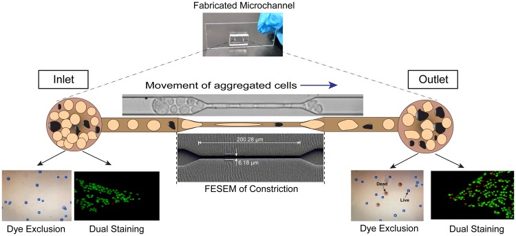
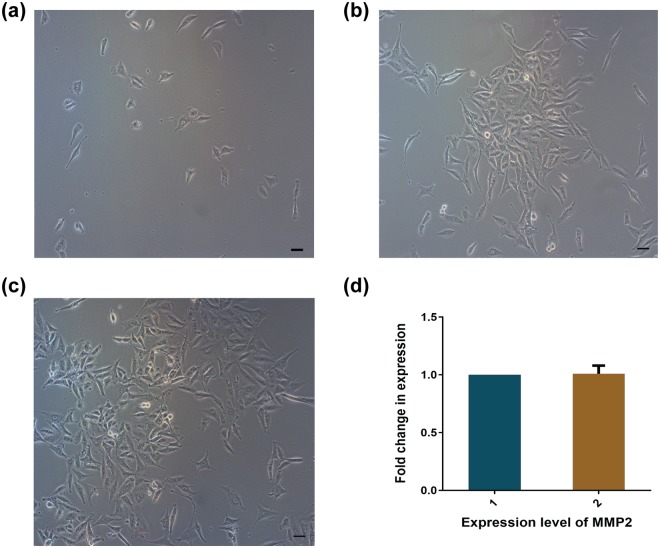
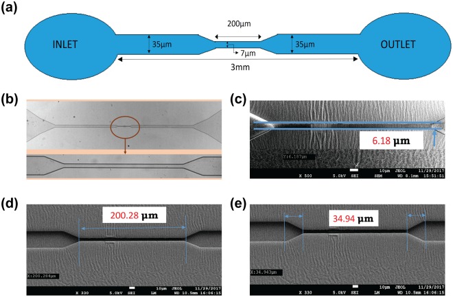
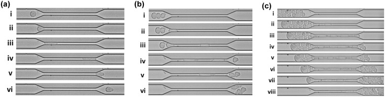
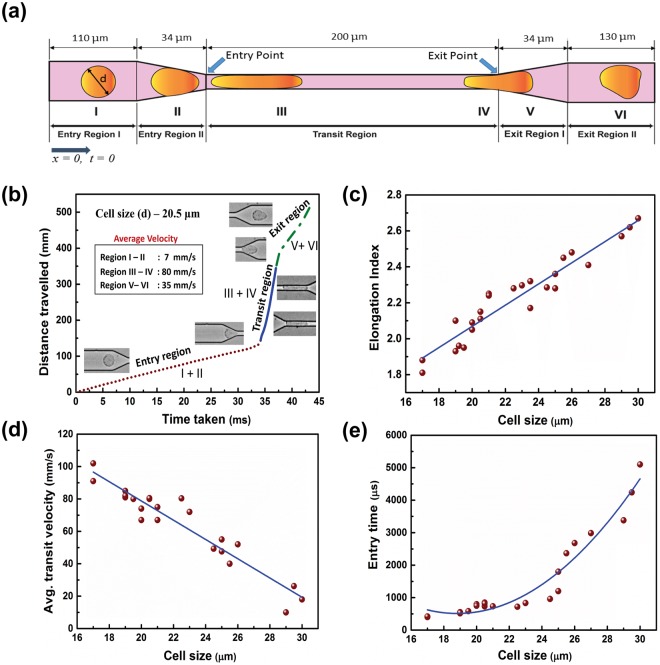
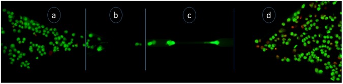
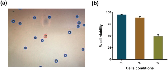
To understand the burgeoning challenges of metastasis, a microchannel of 35 μm diameter, constricted to 7 μm for a distance of 200 μm in a total length of 3 mm, was designed and fabricated using a mask aligner made of polydimethylsiloxane (PDMS) to mimic in vivo capillaries. A thin glass cover-slide was mounted on top to monitor the motion of single or aggregated malignant HeLa cells (size 17-30 μm) microscopically through the constricted microchannel at a constant flow rate of 30 μl/h. Quantitative deconvolution of high-speed videographs of a single cell of 30 μm revealed cellular deformation while passing through constriction, having elongation index, average transit velocity and entry time of 2.67, 18 mm/s and 5.1 ms, respectively. Morphological analysis of live and apoptotic cells by dual staining with Acridine Orange/Ethidium Bromide demonstrated retention of a significant viable cell population after exit through the constriction and a viability index of 50% was quantified by dye exclusion assay. The cumulative data for microfluidic parameters, morphology and relevant metastatic MMP2 gene expression efficiency measured by real-time polymerase chain reaction revealed retention of virulence potency that could possibly cause metastasis, would be beneficial in developing futuristic MEMS device for cancer theranostics.
为了理解转移的新兴挑战,设计并制造了一个 35 μm 直径的微通道,在 3 mm 的总长度内,限制在 7 μm 处 200 μm 的距离,使用由聚二甲基硅氧烷(PDMS)制成的掩模对准器来模拟体内毛细血管。将薄的玻璃盖玻片安装在顶部,通过在 30 μl/h 的恒定流速下通过微通道来显微镜监测单个或聚集的恶性 HeLa 细胞(尺寸为 17-30 μm)的运动。通过对 30 μm 的单个细胞的高速录像进行定量反卷积,揭示了细胞在通过限制时的变形,其伸长指数、平均传输速度和进入时间分别为 2.67、18 mm/s 和 5.1 ms。通过吖啶橙/溴化乙锭双重染色对活细胞和凋亡细胞进行形态分析表明,在通过限制后,仍然保留了大量有活力的细胞群,并且通过染料排除试验定量了 50%的活力指数。通过实时聚合酶链反应测量的微流控参数、形态和相关转移性 MMP2 基因表达效率的累积数据表明,保留了可能导致转移的毒力效力,这可能有益于开发用于癌症治疗的未来 MEMS 设备。